Case Report
Journal of Orthopaedic Case Reports 2016 Sep-Oct: 6(4):77-79
Neuropathic Knee Joint ‑ A Complication of Syrinx Following Spinal Anesthesia: A Rare Case Report and Review of Literature S Swaroop Chandra1, J K Giriraj Harshavardhan1, Ganesan G Ram1, P V Vijayaraghavan1 What to Learn from this Article?
Spinal anesthesia is not without any complication, it might result in complications such as neuropathic joint. Abstract Introduction: Neurological complications due to spinal anesthesia are dysesthesia, paresthesia, cauda equina syndrome, and neuropathic joint. However, neurological complications are rare. We report a case of neuropathic joint of knee as a complication of syrinx following postspinal anesthesia. Case Report: A 33‑year‑old female came with complaints of pain and instability of her left knee and left foot drop. She had developed weakness of her left lower limb following her cesarean section surgery done under spinal anesthesia. Clinically and radiologically, she had features of the neuropathic left knee joint which had occurred as a complication of syrinx following spinal anesthesia. Conclusion: The case is reported for its rarity and to highlight the possible neurological complications of spinal anesthesia. Keywords: Neuropathic joint, syrinx, postspinal anesthesia.
Introduction Most common anesthesia given in day to day practice is spinal due to its cost‑effectiveness, safety, and efficacy, which provides both motor and sensory block with a high success rate. Complications such as hypotension, headache, and urinary retention are relatively common [1, 2]. However, permanent neurological complications are very rare. Neurological complications due to spinal anesthesia are burning sensation over buttocks, dysesthesia, paresthesia, transverse myelitis, anterior spinal artery syndrome, and cauda equina syndrome [1, 3]. We report a case of
the neuropathic left knee joint which developed because of formation of a syrinx following inadvertent dural penetration during spinal anesthesia [2, 3]. Case Report A 33‑year‑old female gravida 2 came with a complaint of weakness of left lower limb and swelling in the left knee. The patient was apparently normal before and underwent lower segment cesarean section under spinal anesthesia in November 2014. She is unaware of the level of
Author’s Photo Gallery
Access this article online Website: www.jocr.co.in
Dr. S Swaroop Chandra
Dr. J K Giriraj Harshavardhan
Dr. Ganesan G Ram
Dr. P V Vijayaraghavan
DOI: 2250-0685.580
1
Department of Orthopaedics, Sri Ramachandra Medical University, Chennai, Tamil Nadu, India.
Address of Correspondence Dr. S Swaroop Chandra, Department of Orthopaedics, Sri Ramachandra Medical University, Chennai, Tamil Nadu, India. E-mail:
[email protected]
77
Copyright © 2016 by Journal of Orthpaedic Case Reports Journal of Orthopaedic Case Reports | pISSN 2250‑0685 | eISSN 2321‑3817 | Available on www.jocr.co.in | doi: 10.13107/jocr.2250-0685.580 This is an Open Access article distributed under the terms of the Creative Commons Attribution Non‑Commercial License (http://creativecommons.org/licenses/by‑nc/3.0) which permits unrestricted non‑commercial use, distribution, and reproduction in any medium, provided the original work is properly cited.
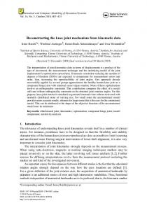
www.jocr.co.in which it was given. Immediately, after introduction of the spinal lumbar puncture needle, she experienced severe shooting pain and tingling sensation over left lower limb. Postoperatively, the patient was not having any bladder/bowel incontinence, but she had weakness of her left lower limb which showed no recovery and was gradually progressive. 2 months before presentation, the patient had a history of slip and fall over her left knee while walking following which she developed swelling over her left knee joint. On examination, diffuse swelling was noted over left knee. Knee flexion was possible up to 90 degrees with some extensor lag. Gross abnormal mobility was present in both sagittal and coronal planes (Fig. 1). Anterior and posterior drawer tests were positive. Valgus and varus instability tests were also positive. The power of left lower limb was reduced (Table 1).
misidentification of Tuffier’s line (line drawn between the highest points of iliac crest in adults which corresponds to L4 spinous process) [6, 7], arachnoid membrane attachment to the conus like a web. A traumatic needle insertion can cause severe disturbance in intramedullary microcirculation, and the direct toxicity of anesthetic agent over the injured cord resulted in syrinx [8, 9]. Neuropathic arthropathy develops in weight bearing joints, and the most common cause is diabetes mellitus [10], other causes being leprosy, meningomyelocele, syringomyelia, spinal cord injury, tabesdorsalis and syphilis [11, 12]. The most common joints affected are the ankle and joints of the feet. Involvement of the knee joint is very uncommon and rarely reported till date [4]. Painless abnormal mobility due to the destruction of afferent proprioceptive fibers, loss of sensation of joint, followed by severe
Knee, ankle, and plantar reflexes were absent. Pain and temperature (crude sensations) were absent involving L4, L5, and S1 dermatomes. Vibration and joint position sensations were intact. Routine blood investigations are done found to be within normal limits except erythrocyte sedimentation rate (90). X‑ray and computerized tomography of knee joint were done which show marked destructive changes, sclerosis, dislocation, and subchondral fractures of knee joint (Fig. 2).
Figure 1: Clinical picture of left knee joint and varus and varus instability test.
Magnetic resonance imaging of the whole spine was done which show syrinx at D7, D9, D10, and D12 and L1 level (Fig. 3). The patient was counseled regarding the possible options of treatment. Arthrodesis could be attempted but with high failure rates and loss of available knee movements. Knee replacement with constrained/hinged custom mega prosthesis (in view of gross instability) could be done with high rates of early loosening and infection. The patient was not willing for any surgical procedure, and hence, she was advised and fitted with orthoses. Figure 2: X-ray and computed tomography films of left knee joint.
Discussion The incidence of permanent neurological injury following spinal anesthesia varies between 0 and 4.2/10000 patients [1]. Preexisting spinal pathology or disease increases the incidence of post‑operative neurological complication following neuraxial blockade [4]. Reynolds reported a series of cases of conusmedullaris injury postspinal anesthesia [5]. The possible reason for this was indicated as misplacement of needle at the lower end of the spinal cord, Table 1: The power of left lower limb was reduced Hip Knee flexion and extension Ankle‑Dorsi flexion Plantar flexion EHL and EDL FHL, FDL
5/5 4/5 1/5 2/5 0/5 2/5
EHL: Extensor hallucis longus, EDL: Extensor digitorum longus, FHL: Flexor hallucis longus, FDL: Flexor digitorum longus
Journal of Orthopaedic Case Reports | Volume 6 | Issue 4 | Sep - Oct 2016 | Page 77-79
78 Figure 3: Magnetic resonance imaging showing syrinx.
www.jocr.co.in degenerative changes, osteophyte formation, and subchondral fractures suggests neuropathic joint. Conclusion Syrinx can occur as a complication of spinal anesthesia. Neurological deficit as a complication of syrinx can be disabling and can lead to the neuropathic joints even in larger joints as illustrated in this case report.
Clinical Message Patients who have severe radicular pain following spinal anesthesia should be carefully followed up for the development of a syrinx and neurological deficit. The neuropathic knee can occur as a complication and is difficult to treat with poor outcome.
References 1. Apan A, Apan OH. Topics in spinal anaesthesia. Whizar-Lugo, V., editor. Complications in Spinal Anaesthesia. InTech; 2014. DOI: 10.5772/58817. Available from: http://www. i ntec h o p en .co m / b o o k s / to p i c s - i n - s p i na l anaest h es i a / complications-in-spinal-anaesthesia. 2. Doherty MJ, Millner PA, Latham M, Dickson RA, Elliott MW. Non‑invasive ventilation in the treatment of ventilator failure following corrective spinal surgery. Anaesthesia 2001;56:235‑247. 3. Kumar N, Patidar SP, Joshi D, Kumar N. Focal myelomalacia and syrinx formation after spinal anaesthesia. J Assoc Physicians India 2010;58:450‑451. 4. Rajakulendran Y, Rahman S, Venkat N. Long‑term neurological complication following traumatic damage to the spinal cord with a 25 gauge whitacre spinal needle. Int J Obstet Anesth 1999;8(1):62‑66. 5. Reynolds F. Damage to the conus medullaris following spinal anaesthesia. Anaesthesia 2001;56(3):238-247. 6. Hogan QH. Tuffier’s line: The normal distribution of anatomic parameters. Anesth Analg 1994;78:194-195. 7. Render CA. The reproducibility of the iliac crest as a marker of lumbar spine level. Anaesthesia 1996;51(11):1070-1071. 8. Waters JH, Watson TB, Ward MG. Conus medullaris injury
Conflict of Interest: Nil Source of Support: None
following both tetracaine and lidocaine spinal anesthesia. J Clin Anesth 1996;8(8):656‑658. 9. Lambert LA, Lambert DH, Strichartz GR. Irreversible conduction block in isolated nerve by high concentrations of local anesthetics. Anesthesiology 1994;80(5):1082‑1093. 10. Rajbhandari SM, Jenkins RC, Davies C, Tesfaye S. Charcot neuroarthropathy in diabetes mellitus. Diabetologia 2002;45(8):1085-1096. 11. Jones EA, Manaster BJ, May DA, Disler DG. Neuropathic osteoarthropathy: Diagnostic dilemmas and differential diagnosis. Radiographics 2000;20:S279-S293. 12. Viens NA, Watters TS, Vinson EN, Brigman BE. Case report: Neuropathic arthropathy of the hip as a sequela of undiagnosed tertiary syphilis. Clin Orthop Relat Res 2010;468(11):3126-3131. 13. Armstrong DG, Peters EJ. Charcot’s arthropathy of the foot. J Am Podiatr Med Assoc 2002;92(7):390-394. 14. Wukich DK, Sung W. Charcot arthropathy of the foot and ankle: Modern concepts and management review. J Diabetes Complications 2009;23:409-426. 15. Gopakumar TS, Rajanish R, Kavitha E, Valsalan R. Neuropathic joint following spinal anesthesia. J Orthop 2008;5(3):e2.
How to Cite this Article Chandra SS, Harshavardhan JKG, Ram GG, Vijayaraghavan PV. Neuropathic Knee Joint - A Complication of Syrinx Following Spinal Anesthesia: A Rare Case Report and Review of Literature. Journal of Orthopaedic Case Reports 2016 Sep-Oct;6(4): 77-79.
79
Journal of Orthopaedic Case Reports | Volume 6 | Issue 4 | Sep - Oct 2016 | Page 77-79