New kidney immobilization method for percutaneous renal biopsy technique in cats. Operational aspects and complications1. Novo método de imobilização do ...
13 – ORIGINAL ARTICLE EXPERIMENTAL UROLOGY
New kidney immobilization method for percutaneous renal biopsy technique in cats. Operational aspects and complications1 Novo método de imobilização do rim para biopsia renal percutânea em gatos. Aspectos operacionais e complicações Daniele Alves SilvaI, Izabela Teodoro OliveiraI, Cecília Braga LaposyII, Cristiane Aparecida Miranda ZacchiI, Juliana Dalarossa AmatuzziIII, Alessandra MelchertIV Graduate student, Veterinary Medicine, UNOESTE, Presidente Prudente-SP, Brazil. Helped with data collection and processing of informations. PhD, Full Professor, Veterinary Medicine, Department of Clinical Pathology, UNOESTE. Presidente Prudente-SP, Brazil. Performed haematological and biochemical analysis of the study. III MD, Resident, Veterinary Medicine, UNOESTE, Presidente Prudente-SP, Brazil. Acquisition of data and technical support. IV PhD, Full Professor, Veterinary Medicine, Department of Clinical Pathology, UNOESTE, Presidente Prudente-SP, Brazil. Main author. Conception, design, intellectual and scientific content of the study. I
II
ABSTRACT PURPOSE: Evaluate a new immobilization kidney method for collecting blind percutaneous renal biopsies (RB) in healthy cats. METHODS: Ten cats were biopsied by a modified blinded percutaneous technique using semi-automated needles. Were evaluated the operational aspects of the technique, its complications, and the quality of the obtained samples. The evaluation included physical examination, hemogram, urinalysis, abdominal ultrasound, renal function, and histopathology of the biopsy specimens. RESULTS: The developed technique was fast and easy to perform; it required two operators, and the right kidney was elected for specimen collection. After the RB, a decrease in hematocrit levels was observed in addition to hematuria and perirenal transient hematoma; however, no clinical consequences were observed, and normal parameters were restored within 48 hrs. There were no major complications or deaths, alterations in the physical examination or renal function, or signs of infection. Of the samples, 95% revealed the presence of renal tissue, and in 100% of the cats the samples were of diagnostic quality. CONCLUSION: The technique was easily performed, provided adequate material for diagnosis with minimal transient complications. Key words: Kidney. Biopsy. Urology. Techniques. Complications. Cats. RESUMO OBJETIVO: Avaliar um novo método de imobilização do rim para coleta de biopsia renal (BR) percutânea às cegas em gatos hígidos. MÉTODOS: Dez gatos foram biopsiados por uma técnica de biopsia percutânea às cegas modificada, com uso de agulha semiautomática. Foram avaliados os aspectos operacionais da técnica, complicações e a qualidade das amostras obtidas. A avaliação incluiu exame físico, hemograma, urinálise, ultrassonografia abdominal, função renal e análise histopatológica do espécime de biopsia. RESULTADOS: A técnica foi de fácil realização; foram requeridos dois operadores, e o rim direito foi eleito para a coleta da BR. Após a BR, diminuição do hematócrito foi observada, em conjunto com hematúria e hematoma peri-renal transitório; entretanto, não foram observadas conseqüências clínicas, e os parâmetros normais foram restabelecidos em 48 horas. Não ocorreram complicações maiores ou óbitos, alterações ao exame físico, função renal ou sinais de infecção. Das amostras obtidas, 95% revelaram presença de tecido renal, e em 100% dos gatos as amostras apresentaram qualidade para diagnóstico. CONCLUSÃO: A técnica foi realizada facilmente, forneceu material adequado para diagnóstico, com complicações clínicas mínimas. Descritores: Rim. Biópsia. Urologia. Técnicas. Complicações. Gatos.
76 - Acta Cirúrgica Brasileira - Vol. 27 (1) 2012
New kidney immobilization method for percutaneous renal biopsy technique in cats. Operational aspects and complications
Introduction Kidney diseases are common in cats1. Accurate assessment of the history, physical examination, laboratory tests, and diagnostic imaging may indicate alterations consistent with renal disease2 but may not determine the specific diagnosis3 or possibility of recovery of renal function2. The renal biopsy is a complementary test to these exams4. Besides providing guidelines for therapeutic aid, the renal biopsy (RB) specimen displays diagnostic and prognostic value. However, this procedure is not frequently performed in cats due to complications associated with the procedure as well as the doubts in relation to the diagnosis1. The biopsy and histopathological evaluation of the renal tissue are valuable diagnostic and prognostic tools5. The RB allows the clinician to establish the histological diagnosis and should be considered when it could alter the clinical approach6 or facilitate the prognosis, including in patients with glomerular desease or acute renal failure (IRA)7. It also justifies the use of specific and expensive therapies, such as dialysis, in those patients with a favorable prognosis2. Several RB techniques have been described in veterinary medicine, and one of these is percutaneous biopsy, which does not require surgical opening of the abdominal cavity5. The percutaneous biopsy can be performed by using imaging such as ultrasound8,9, or “blindly”, which is done when the kidneys are immobilized by abdominal palpation. The blind technique is especially easy to be performed in this species due to the easy palpation of cat kidneys 7. Complications for the RB result from the penetration of the needle in large vessels, which include: severe hemorrhage1 and perirenal bleeding3,10-12. Hydronephrosis due to ureteral or renal pelvis obstruction by blood clots10 and death are uncommon complications1. Hemorrhaging is the most important and most serious complication of the RB procedure10, and the patient should be monitored after the procedure13. To evaluate the quality of the RB sample, it is important to consider if the number of glomeruli is sufficient to represent the renal parenchyma14. There is no standard established for veterinary medicine; however, various references in the literature indicate that the minimum number of glomeruli per sample is five7,14. However, in clinical practice, we observed difficulties in a renal biopsy collection in cats, with the previously described methods1. Thus, the objective of this study was to describe a new method of the blinded percutaneous RB technique and to evaluate its complications and the diagnostic quality of the samples in
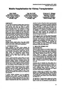
healthy cats. Methods Animal selection and ethical guidelines This study used 10 healthy adult male (n=3) and female (n=7) cats of undefined breed weighing between 3 and 5 kg that came from the cattery of the Institution of origin. In this study, we performed biopsies in the right (n=9) or left (n=1) kidney of these animals. The study was approved by the Research Ethics Committee of the Institution, filed under number 130/06. Kidney immobilization and biopsy collection After adequate liquid and food fasting, the cats, with their abdominal regions properly shaved, were anesthetized with 1 mg/ kg xylazine associated with ketamine 10 mg/kg intramuscularly, preceded by 0.004 mg/kg atropine applied under the skin. The animals were then placed in the left lateral position for palpation of the abdominal cavity and the right and left kidneys by positioning the fingers beneath the transverse apophysis of the lumbar vertebrae and selecting the kidney with better immobilization for biopsy (Figure 1A). The procedure is relatively easy and minimally invasive, and it was performed on an outpatient basis without the need of an operating room. There was adequate antisepsis of the abdominal cavity, and the kidney to be biopsied was immobilized by an assistant who moved the kidney up with one hand and immobilized it sideways using the index and middle fingers of the other hand located in the cranial and caudal poles (Figure 1B). The collection of the RB was done using the blinded percutaneous technique. A small incision was made in the skin (Figure 1C), and the operator of the needle inserted it into the caudal pole of the kidney in the dorsoventral direction using one hand, while the other hand fixed the kidney in its ventral face (Figure 1D) to avoid movement during the procedure. A 16 gauge, 4.5 in, semi-automated Tru-Cut type needle was used, which has automatic gathering of the internal cannula. Two to four punctures were completed per animal, in order to obtain at least two specimens macroscopically viable, taking place however a maximum of four punctures per cat. The needle was inserted into the cavity through the skin incision, surpassing the muscles until its introduction into the renal capsule, which offers resistance to passage of the needle and when the kidney is perforated. The internal cannula of the needle was immediately exposed, and the automatic gathering of the cannula was triggered for the collection of the renal fragment. After the RB, the small
Acta Cirúrgica Brasileira - Vol. 27 (1) 2012 - 77
Silva DA et al.
incision of the skin was sutured with a simple point (Figure 1F) and nylon thread 2-0, which was removed after 7 d. Sanitization of the local area was performed using saline and povidone iodine immediately after and 24 and 72 hrs after the RB.
M2, and M3, respectively); renal ultrasound before and 30 min after the RB to verify the occurrence of hemorrhaging or perirenal hematoma; urea and creatinine levels before and 72 hrs after the RB to evaluate possible alterations in renal function; and coagulation time using the microcapillary method before the RB to exclude hemorrhagic tendencies. The biopsy site was inspected and palpated daily until removal of the suture stitches (7 d). The complications observed in the study were classified as minimal: perirenal hematoma and microscopic hematuria, which spontaneously resolved without the need of a blood transfusion; and more serious: hemorrhages that required blood transfusion15,16. Statistical analysis The profile multivariate statistical analysis was used to analyze the data to compare the evaluation periods. All the data were analyzed using GraphPad Prism (version 1.3). The level of significance for the tests was 5% (CI 95%), and p0.001 X M0, M2 and M3
There was a significant increase in the number of erythrocytes in the urine in M1 and M2 compared to the control time M0; in M3, although there was no statistical difference when compared to M0, it was observed that the average number of erythrocytes was greater (92.1 ± 77.2 in M3 versus 32.6 ± 62.8 in M0). Microscopic hematúria occurred in all cats of this study after RB. The occurrence of macroscopic hematuria was observed in two cats (18.2%) and a significant decrease of the hematocrit (p