NIH Public Access Author Manuscript Nat Genet. Author manuscript; available in PMC 2010 September 1.
NIH-PA Author Manuscript
Published in final edited form as: Nat Genet. 2010 March ; 42(3): 234–239. doi:10.1038/ng.536.
Common variants at 7p21 are associated with frontotemporal lobar degeneration with TDP-43 inclusions
NIH-PA Author Manuscript
Vivianna M. Van Deerlin*,1,2, Patrick M. A. Sleiman*,6, Maria Martinez-Lage*,1,8, Alice ChenPlotkin*,1,3, Li-San Wang1, Neill R Graff-Radford9, Dennis W. Dickson10, Rosa Rademakers10, Bradley F. Boeve11, Murray Grossman3, Steven E. Arnold3,4,5, David M.A. Mann13, Stuart M. Pickering-Brown12,13, Harro Seelaar14, Peter Heutink15, John C. van Swieten14, Jill R. Murrell16, Bernardino Ghetti16,17, Salvatore Spina16,18, Jordan Grafman19, John Hodges20, Maria Grazia Spillantini21, Sid' Gilman22, Andrew P. Lieberman23, Jeffrey A. Kaye24, Randall L. Woltjer25, Eileen H Bigio26,27, Marsel Mesulam27, Safa al-Sarraj28, Claire Troakes29, Roger N. Rosenberg30, Charles L. White III31, Isidro Ferrer32, Albert Lladó33, Manuela Neumann34, Hans A. Kretzschmar35, Christine Marie Hulette36, Kathleen A. Welsh-Bohmer37,38, Bruce L Miller39, Ainhoa Alzualde40, Adolfo Lopez de Munain41, Ann C. McKee42,43, Marla Gearing44,46,47, Allan I. Levey45,46, James J. Lah45, John Hardy48,49, Jonathan D. Rohrer50, Tammaryn Lashley49,51, Ian R.A. Mackenzie52, Howard H. Feldman53,54, Ronald L. Hamilton55, Steven T. Dekosky56, Julie van der Zee57,58, Samir Kumar-Singh57,58, Christine Van Broeckhoven57,58, Richard Mayeux59, Jean Paul G. Vonsattel59,60,61, Juan C. Troncoso62, Jillian J Kril63, John B.J. Kwok64, Glenda M. Halliday64,65, Thomas D. Bird66,67, Paul G. Ince68, Pamela J. Shaw68, Nigel J. Cairns69,70, John C. Morris69,70, Catriona Ann McLean71, Charles DeCarli72, William G. Ellis73, Stefanie H. Freeman74, Matthew P. Frosch74, John H. Growdon74, Daniel P. Perl75, Mary Sano75,76, David A. Bennett77, Julie A. Schneider77, Thomas G. Beach78, Eric M. Reiman79,80, Bryan K. Woodruff81, Jeffrey Cummings82, Harry V. Vinters83,84, Carol A. Miller85, Helena C. Chui85, Irina Alafuzoff86,87,88, Päivi Hartikainen88, Danielle Seilhean89, Douglas Galasko90, Eliezer Masliah90,91, Carl W. Cotman92, M. Teresa Tuñón93,94, M. Cristina Caballero Martínez94,95, David G. Munoz96, Steven L. Carroll97,
NIH-PA Author Manuscript
Users may view, print, copy, download and text and data- mine the content in such documents, for the purposes of academic research, subject always to the full Conditions of use:http://www.nature.com/authors/editorial_policies/license.html#terms Correspondence and requests for materials should be addressed to VVD (
[email protected]) or JQT (
[email protected]).. *Co-first authors with equal contributions Author Contributions VVD, PS, MML, and ACP contributed equally to this manuscript. The overall study was designed and implemented by VMYL, JQT, VVD, HH and MML and discussed with GDS, LSW, and ACP. Coordination, sample handling, DNA extraction, genetic analysis and data management were done primarily by VVD and MML. Genotyping and additional testing, including immunohistochemical analysis, was done by PS, HH, VVD, or MML. Data analysis and quality control were performed by PS, LSW, and ACP and discussed and reviewed by VVD, HH, GS, VMYL, JQT, and MML. Expression analysis was designed and implemented by ACP. The manuscript was prepared by ACP, VVD, and PS and reviewed by MML, GDS, HH, JQT, VMYL, and LSW. The members of the International FTLD Collaboration, which includes all other authors, contributed cases, evaluated pathology, performed genetic studies and reviewed the manuscript. Supplementary Information is available online Accession Numbers GRN: NM_002087.2 TMEM106B: NM_018374.3 Competing Financial Interest A patent application on TMEM106B has been submitted. Dr. James Lah is currently involved in clinical trials involving: Elan, Janssen, Medivation, and Ceregene. Dr. Howard Feldman has been a full time employee in Neuroscience Global Clinical Research and Development at Briston-Myers Squibb.
Van Deerlin et al.
Page 2
Daniel Marson98,99, Peter F. Riederer100, Nenad Bogdanovic101, Gerard D. Schellenberg1,2, Hakon Hakonarson6,7, John Q. Trojanowski1,2, and Virginia M.-Y. Lee1,2
NIH-PA Author Manuscript
1Center
NIH-PA Author Manuscript NIH-PA Author Manuscript
for Neurodegenerative Disease Research, Department of Pathology and Laboratory Medicine, University of Pennsylvania School of Medicine, Philadelphia, PA USA 2Institute on Aging, University of Pennsylvania School of Medicine, Philadelphia, PA USA 3Department of Neurology, University of Pennsylvania School of Medicine, Philadelphia, PA USA 4Department of Psychiatry, University of Pennsylvania School of Medicine, Philadelphia, PA USA 5Penn Memory Center, University of Pennsylvania School of Medicine, Philadelphia, PA USA 6The Center for Applied Genomics, Division of Human Genetics, University of Pennsylvania School of Medicine, Philadelphia, PA USA 7Division of Pulmonary Medicine and Department of Pediatrics, The Children's Hospital of Philadelphia, University of Pennsylvania School of Medicine, Philadelphia, PA USA 8Department of Neurology, Hospital de la Santa Creu i Sant Pau, Universitat Autonoma de Barcelona, Barcelona, Spain 9Department of Neurology Jacksonville, Mayo College of Medicine, Jacksonville, FL, USA 10Department of Neuroscience, Mayo Clinic, Jacksonville, FL, USA 11Department of Neurology, Mayo Clinic, Rochester, MN, USA 12Clinical Neuroscience Research Group, United Kingdom 13University of Manchester, United Kingdom 14Erasmus Medical Centre Rotterdam, The Netherlands 15Section of Medical Genomics, Department of Clinical Genetics, VU University Medical Center Amsterdam, The Netherlands 16Department of Pathology and Laboratory Medicine, Indiana University School of Medicine, Indianapolis, IN, USA 17Indiana Alzheimer Disease Center, Indiana University School of Medicine, Indianapolis, IN, USA 18Department of Neurological, Neurosurgical and Behavioral Sciences, University of Siena, Siena, Italy 19Cognitive Neuroscience Section, National Institute of Neurological Disorders and Stroke, Bethesda, MD, USA 20Prince of Wales Medical Research Institute, New South Wales, Australia 21Cambridge Centre for Brain Repair, Dept of Clinical Neurosciences, University of Cambridge, Cambridge, United Kingdom 22Department of Neurology, University of Michigan, Ann Arbor, MI, USA 23Department of Pathology University of Michigan, Ann Arbor, MI, USA 24Department of Neurology and Biomedical Engineering, Oregon Health and Science University, Portland, OR, USA 25Department of Pathology, Oregon Health and Science University, Portland, OR, USA 26Department of Pathology, Northwestern University Feinberg School of Medicine, Chicago, IL, USA 27Cognitive Neurology and Alzheimer Disease Center Northwestern University Feinberg School of Medicine, Chicago, IL, USA 28Department of Clinical Neuropathology, Institute of Psychiatry, Kings College Hospital, London, United Kingdom 29MRC London Neurodegenerative Diseases Brain Bank, Institute of Psychiatry, Kings College Hospital, London, United Kingdom 30Alzheimer's Disease Center, University of Texas Southwestern Medical Center, Dallas, TX, USA 31Department of Pathology, University of Texas Southwestern Medical Center, Dallas, TX, USA 32Institut de Neuropatologia, IDIBELL-Hospital Universitari de Bellvitge, Barcelona, Spain 33Alzheimer's Disease and Cognitive Disorders Unit, Service of Neurology. ICN. Hospital Clínic Barcelona, Barcelona, Spain 34Institute for Neuropathology, University Hospital Zurich, Zurich, Switzerland 35Center for Neuropathology and Prion Research, Ludwig Maximilians University, Munich, Germany 36Department of Pathology, Duke University Health Sciences Center, Durham, NC, USA 37Dept of Psychiatry, Duke University Health Sciences Center, Durham, NC, USA 38Bryan ADRC, Duke University Health Sciences Center, Durham, NC, USA 39Department of Neurology, University of California at San Francisco, San Francisco, CA, USA 40Neurogenetic Unit, Instituto Biodonostia, San Sebastián, Spain 41Servicio de Neurología, Hospital Donostia, San Sebastián, Spain. 42Departments Neurology and Pathology, Boston University School of Medicine, Boston MA 43Bedford Veterans Administration Medical Center, GRECC, Bedford MA, USA 44Department of Pathology and Laboratory Medicine, Emory University School of Medicine, Atlanta, GA, USA 45Department of Neurology, Emory University School of Medicine, Atlanta, GA, USA 46Alzheimer's Disease Research Center, Emory University School of Medicine, Atlanta, GA, USA 47Center for Neurodegenerative Disease, Emory University School of Medicine, Atlanta, GA, USA 48Reta Lila Laboratories, UCL Institute of Neurology, London, United Kingdom 49Department of Molecular Neuroscience, UCL Institute of
Nat Genet. Author manuscript; available in PMC 2010 September 1.
Van Deerlin et al.
Page 3
NIH-PA Author Manuscript NIH-PA Author Manuscript NIH-PA Author Manuscript
Neurology, London, United Kingdom 50Dementia Research Centre, UCL Institute of Neurology, London, United Kingdom 51Queen Square Brain Bank for Neurological Disorders, UCL Institute of Neurology, London, United Kingdom 52Department of Pathology, University of British Columbia, Canada 53Division of Neurology Vancouver General Hospital and the University of British Columbia, Canada 54Neuroscience, Bristol-Myers Squibb, University of Pittsburgh, Pittsburgh PA, USA 55Department of Pathology, University of Pittsburgh, Pittsburgh PA, USA 56Department of Neurology, University of Virginia School of Medicine, Charlottesville, VA, USA 57Neurodegenerative Brain Diseases Group, Department of Molecular Genetics, VIB, Antwerpen, Belgium 58Institute Born-Bunge and University of Antwerp, Antwerpen, Belgium 59Taub Institute for Research on Alzheimer's Disease, Columbia University, New York, NY, USA 60New York Brain Bank, Columbia University, New York, NY, USA 61Department of Pathology, Columbia University, New York, NY, USA 62Departments of Pathology and Neurology, Johns Hopkins University School of Medicine, Baltimore, MD, USA 63Disciplines of Medicine and Pathology, University of Sydney, Australia 64Prince of Wales Medical Research Institute, Australia 65University of New South Wales, Australia 66GRECC, VA Puget Sound Health Care System, University of Washington, Seattle, WA, USA 67Department of Neurology, University of Washington, Seattle, WA, USA 68Department of Neuroscience, University of Sheffield 69Alzheimer's Disease Research Center, Washington University School of Medicine, St Louis, MO, USA 70Department of Neurology, Washington University School of Medicine, St Louis, MO, USA 71Department of Anatomical Pathology, The Alfred Hospital, Australia 72Alzheimer's Disease Center, Imaging of Dementia and Aging (IDeA) Laboratory, Department of Neurology, Center for Neuroscience, University of California at Davis, Sacramento, CA, USA 73Department of Pathology, University of California at Davis, Sacramento, CA, USA 74C.S. Kubik Laboratory for Neuropathology, Massachusetts General Hospital & Harvard Medical School, Boston, MA, USA 75Department of Pathology, Mount Sinai School of Medicine, New York, NY, USA 76James J Peters VA Medical Center, New York, NY, USA 77Rush Alzheimer's Disease Center, Rush University Medical Center, Sun City, AZ, USA 78Sun Health Research Institute, Sun City, AZ, USA 79Banner Alzheimer's Institute, Translational Genomics Research Institute, University of Arizona, Phoenix, AZ, USA 80Arizona Alzheimer's Consortium, Phoenix, AZ, USA 81Mayo Clinic Arizona, Scottsdale, AZ, USA 82Mary S. Easton Center for Alzheimer's Disease Research, Los Angeles, CA, USA 83Department of Pathology and Laboratory Medicine, Los Angeles, CA, USA 84Department of Neurology David Geffen School of Medicine at UCLA, Los Angeles, CA, USA 85Keck School of Medicine, University of Southern California, Los Angeles, CA, USA 86Department of Genetics and Pathology, Uppsala University, Uppsala, Sweden 87Department of Clinical Medicine, Kuopio University, Kuopio Finland 88Department of Neurology, Kuopio University, Kuopio Finland 89UPMC-Univ Paris 06 and APHP, France 90Department of Neurosciences, University of California, San Diego, San Diego, CA, USA 91Department of Pathology, University of California, San Diego, San Diego, CA, USA 92Department of Neurology, University of California, Irvine, Irvine, CA, USA 93Hospital de Navarra Pathology Department, Spain 94Brain Bank of Navarra, Spain 95Biomedical Research Center, Navarra Health ServiceOsasunbidea, Spain 96Department of Laboratory Medicine and Pathobiology, Li Ka Shing Knowledge Institute of St. Michael's Hospital, University of Toronto, Canada 97Department of Pathology, University of Alabama at Birmingham, Birmingham, AL, USA 98Department of Neurology, University of Alabama at Birmingham, Birmingham, AL, USA 99Alzheimer's Disease Research Center, University of Alabama at Birmingham, Birmingham, AL, USA 100Clinical Neurochemistry Clinic and Policlinic of Psychiatry, Psychosomatic and Psychotherapy of the University of Wuerzburg, Germany 101Geriatric Medicine and Neuropathology at Department of Geriatric Medicine, Karolinska University Hospital, Stockholm Sweden
Abstract
Nat Genet. Author manuscript; available in PMC 2010 September 1.
Van Deerlin et al.
Page 4
NIH-PA Author Manuscript
Frontotemporal lobar degeneration (FTLD) is the second most common cause of presenile dementia. The predominant neuropathology is FTLD with TAR DNA binding protein (TDP-43) inclusions (FTLD-TDP)1. FTLD-TDP is frequently familial resulting from progranulin (GRN) mutations. We assembled an international collaboration to identify susceptibility loci for FTLD-TDP, using genome-wide association (GWA). We found that FTLD-TDP associates with multiple SNPs mapping to a single linkage disequilibrium (LD) block on 7p21 that contains TMEM106B in a GWA study (GWAS) on 515 FTLD-TDP cases. Three SNPs retained genome-wide significance following Bonferroni correction; top SNP rs1990622 (P=1.08×10⁻¹¹; odds ratio (OR) minor allele (C) 0.61, 95% CI 0.53-0.71). The association replicated in 89 FTLD-TDP cases (rs1990622; P=2×10⁻⁴). TMEM106B variants may confer risk by increasing TMEM106B expression. TMEM106B variants also contribute to genetic risk for FTLD-TDP in patients with GRN mutations. Our data implicate TMEM106B as a strong risk factor for FTLD-TDP suggesting an underlying pathogenic mechanism.
NIH-PA Author Manuscript
FTLD manifests clinically with progressive behavioral and/or language deficits with a prevalence of 3.5-15/100,000 in 45 to 64 year olds2-5. The clinical presentation of cases with FTLD pathology varies depending on the referral base6 and among these cases, ~50% are diagnosed as FTLD-TDP1. A family history of a similar neurodegenerative disease may be present in up to 50% of FTLD cases, supporting the existence of a genetic predisposition7. Autosomal dominant GRN mutations occur in ~20% of FTLD-TDP cases8-11. GRN mutations are loss-of-function mutations with most resulting in premature termination of the mutant transcript invoking nonsense-mediated RNA decay and with the ensuing haploinsufficiency causing disease11,12. However, a substantial number of familial FTLD-TDP cases are not explained by GRN mutations. Further, patients with the same GRN mutation show variable clinical phenotypes or ages of disease onset which likely reflect additional genetic and environmental factors13.
NIH-PA Author Manuscript
The GWA phase of the study included 515 cases of FTLD-TDP and 2509 disease-free population controls genotyped on the Illumina HH550 or 610-Quad BeadChips as described14 (Table 1). A large population control cohort was acceptable since the general population incidence of FTLD is low4,5,15. Cases were obtained under institutional review board approval by members of the International FTLD Collaboration consisting of investigators from 45 clinical centers and brain banks representing 11 countries (United States, Canada, United Kingdom, The Netherlands, Belgium, Spain, Germany, Australia, Finland, France, and Sweden). All cases met either pathological (n=499) or genetic (n=16) criteria for FTLD-TDP which was confirmed by detecting TDP-43 inclusions using immunohistochemistry (IHC)1,16. A genetic criterion for inclusion (i.e. presence of a known pathogenic GRN mutation) was used since GRN mutation cases are always diagnosed as FTLDTDP 8,9,17,18. All cases were checked for relatedness using identity by state (IBS). The results confirmed that although some GRN-associated FTLD-TDP cases share the same mutation on chromosome 17 with similarity in the immediate vicinity of GRN, they are no more related in the remainder of the genome than individuals without GRN mutations. Detailed inclusion criteria are provided in Methods; cohort features are in Supplementary Table 1. Cochran-Armitage trend test statistics were calculated at all markers following quality control filtering. In addition to self-reported ancestry, all cases and controls were initially screened at ancestry informative markers (AIM) using the STRUCTURE software package19 to reduce the risk of population stratification from self-reported ancestry alone. Each case was subsequently matched to four controls by ‘genetic matching’ by smartPCA20 as previously described21. The genomic inflation factor (λ) for this study was 1.05 indicating that background stratification was minimal as demonstrated in the quantile-quantile (Q-Q) plots (Supplementary Fig. 1).
Nat Genet. Author manuscript; available in PMC 2010 September 1.
Van Deerlin et al.
Page 5
NIH-PA Author Manuscript
Three SNPs reached genome-wide significance following Bonferroni correction (Figures 1a and b). All three SNPs (rs6966915, rs1020004, and rs1990622) mapped to a 68 kb interval (Supplementary Fig. 2) on 7p21.3 (top marker, rs1990622, minor allele frequency (MAF) 32.1% in cases and 43.6% in controls, OR = 0.61, [95% CI 0.53 – 0.71], P=1.08×10⁻¹¹). For rs1990622, the more common (T) allele confers risk with an OR of 1.64 [95% CI 1.34-2.00]. The interval contained nine additional markers in strong LD (r2 >0.45) that were also associated with FTLD-TDP (P-value range = 8.9×10⁻³ - 7.5×10⁻⁷; OR range 0.63-0.77) (Table 2). All 12 associated SNPs map to a single LD block spanning TMEM106B, which encodes an uncharacterized transmembrane protein of 274 amino acids (Figures 1b and c). SNPs rs1020004 and rs6966915 lie within introns 3 and 5, respectively, of TMEM106B, while rs1990622 is 6.9 kb downstream of the gene. These findings argue strongly for the association of the 7p21 locus, and the gene TMEM106B, with FTLD-TDP.
NIH-PA Author Manuscript
The association with FTLD-TDP in the GWA was replicated by TaqMan SNP genotyping in 89 independent FTLD-TDP cases and 553 Caucasian control samples at two of the genomewide significant SNPs (rs1020004 and rs1990622) (Table 1 and Supplementary Table 1). A polymorphic variation adjacent to rs6966915 interfered with interpretation of TaqMan genotyping therefore precluding its use in the replication. The replication set was selected based on the same pathological criteria and had similar characteristics as the GWA phase cohort (Supplementary Table 1). In this replication cohort, the top SNPs again showed significant association (P=0.004 for rs1020004 and P=0.0002 for rs1990622) with the same directions of association as those found in the GWA phase (Supplementary Table 1). These results suggest that in the 7p21 locus, encompassing the gene TMEM106B, we have identified a common genetic susceptibility factor for FTLD-TDP. Of interest, this association was not confirmed in a cohort of 192 living patients with unselected FTLD (Supplementary Table 2). This likely reflects heterogeneity in neuropathological substrates underlying FTLD, with only ~50% of unselected clinical FTLD cases expected to have FTLD-TDP. Assuming that TMEM106B genetic variants confer risk of FTLD-TDP specifically, the power to detect this association in 192 clinical FTLD cases and 553 controls is ~30% for an alpha-value of 0.05. To have >90% power to detect this association, a clinical FTLD cohort would require more than 1400 clinical FTLD cases and an equal number of controls.
NIH-PA Author Manuscript
We next evaluated TMEM106B gene expression in different human tissues to identify phenotype-associated differential expression and also any potential genetic regulators of expression. We queried the mRNA-by-SNP browser (http://www.sph.umich.edu/csg/liang/asthma/, last accessed June 6, 2009), for genetic regulators of TMEM106B expression (eSNPs) in lymphoblastoid cell lines22. The top SNP, rs1990622, was significantly correlated with TMEM106B average expression levels (LOD 6.32; P=6.9×10⁻⁸), as was SNP rs1020004 (LOD 5.16, P=1.10×10⁻⁶). The risk allele (T) of rs1990622 was associated with a higher level of mRNA expression, indicating that TMEM106B may be under cis-acting regulation by either the FTLD-TDP associated SNPs or another SNP(s) in LD with the associated variants. As the expression data in the publicly available database is derived from lymphoblastoid cell lines from normal individuals22, and the diseased organ in FTLD-TDP is brain, we asked if a similar correlation between genotype and expression phenotype for TMEM106B is also present in tissue types affected by disease, and in diseased individuals themselves. Accordingly, we used total RNA isolated from FTLDTDP postmortem brains (n=18) and neurologically normal control brains (n=7) to evaluate TMEM106B expression in frontal cortex, which is severely affected in FTLD-TDP, by quantitative reverse-transcription PCR (QRT-PCR). All RNA samples used were confirmed to be of equivalent high quality as described23 (Supplementary Table 3). For the same individuals for which we obtained expression data, we genotyped SNPs rs1020004 and rs1990622 using allelic discrimination assays.
Nat Genet. Author manuscript; available in PMC 2010 September 1.
Van Deerlin et al.
Page 6
NIH-PA Author Manuscript
Corroborating results from the cell lines, expression of TMEM106B was significantly correlated with TMEM106B genotype, with risk allele carriers showing higher expression (overall P=0.027, TT vs. TC P=0.017, TT vs. CC P=0.03, for rs1990622, Fig. 2a and Supplementary Fig. 3a). Strikingly, however, expression of TMEM106B was >2.5 times higher in FTLD-TDP cases compared to normal controls (P=0.045, Fig. 2b). In addition, the effects of genotype and TMEM106B expression on risk of developing disease are at least partly independent, as are the effects of genotype and disease status on TMEM106B expression (Supplementary Table 4a and b). Thus, these data suggest that increased TMEM106B brain expression might be linked to mechanisms of disease in FTLD-TDP, and that risk alleles at TMEM106B confer genetic susceptibility by increasing gene expression.
NIH-PA Author Manuscript
The primary criterion for inclusion in the GWAS was a neuropathological diagnosis of FTLDTDP; therefore we studied all cases together regardless of GRN mutation status. Nevertheless, a priori it was difficult to predict whether additional genetic susceptibility loci would be identified in a group with Mendelian inheritance of highly penetrant mutations. We therefore separately evaluated FTLD-TDP cases with (n=89) and without (n=426) GRN mutations. Association to the 7p21 locus persisted in both the GRN negative and positive clusters and there was no significant heterogeneity in the ORs for the disease/SNP association between the clusters (Fig. 3 and Supplementary Tables 2 and 5). Using family history status as a covariate in a logistic regression showed that the 7p21 association is independent of family history (Supplementary Table 6). Thus, TMEM106B variants may act as a modifier locus in the presence of GRN mutations, as the APOE locus has been shown to modify age of onset in patients with PSEN124 or PSEN225 mutations. Additionally, in the whole GWA cohort, we observed a correlation between rs1020004 genotype and disease duration (P=0.03) with homozygotes for the risk allele (AA, wild-type) having shorter duration of disease (i.e. more severe disease) than individuals homozygous for the minor allele (GG, Supplementary Figure 4). These results provide strong confirmatory evidence for association of the 7p21 locus with increased risk for FTLD-TDP in both GRN positive and negative cases.
NIH-PA Author Manuscript
In addition to the 7p21 locus, analysis of the GRN cases alone showed highly significant association with SNPs near the GRN locus on 17q21 (Fig. 3 and Supplementary Table 7). Not unexpectedly, haplotype analysis of the cases indicated that the chromosome 17 association was driven by a shared haplotype among the p.R493X (NM_002087.2:c.1477C>T) mutation carriers which represented 20.2% (18/89) of the GRN mutation cases. To determine if the observed association at the GRN locus was dependent on the association at the TMEM106B locus we carried out a logistic regression analysis conditioning on the most significantly associated SNP at the 7p21 locus, rs1990622, in the patients with GRN mutations. The conditional analysis had no effect on the association at the GRN locus suggesting that the associations with 17q21 and 7p21 are independent. IBS analysis confirms that these individuals are unrelated and therefore the identified association on chromosome 7 cannot be discounted in GRN mutation carriers. Indeed, conditioning on the top SNP at the GRN locus, rs8079488, also had no effect on the TMEM106B association (results not shown). In addition to the 7p21 locus, the GWAS of GRN negative cases showed a trend for association at five other loci (Supplementary Table 8) including a locus on chromosome 9p21.2 that falls within a 7.7 Mb critical interval defined from five previous linkage studies, representing a potential refinement of that region26. We observed no association at the GRN locus in the cases without GRN mutations. We then evaluated mRNA expression of TMEM106B in FTLD-TDP with and without GRN mutations separately, and the GRN mutants showed increased expression (overall P=0.0009), compared to controls (P=0.0005) and FTLD-TDP without GRN mutations (P=0.002) (Figure
Nat Genet. Author manuscript; available in PMC 2010 September 1.
Van Deerlin et al.
Page 7
NIH-PA Author Manuscript
2c). Furthermore, controlling for rs1990622 genotype and focusing on heterozygotes (n=14), the presence of a GRN mutation remained significantly associated with increased TMEM106B expression (P=0.039, Fig. 2d) compared to normal controls. These results are compatible with a model in which mutations in GRN are upstream of increased TMEM106B expression in increasing risk for FTLD-TDP. A mechanistic understanding of the pathogenesis of FTLD has been hampered the heterogeneity in clinical and pathological features. With the discovery of TDP-43 as a major FTLD disease protein, the pathologically-defined entity of FTLD-TDP emerged16. Identification of GRN mutations as a major genetic cause of FTLD-TDP, led to definition of a genetic subgroup of FTLD-TDP. This study identifies TMEM106B as a genetic risk factor for FTLD-TDP. We speculate that the homogeneous pathologically-defined study population used here enabled us to detect a robust signal with relatively small case numbers.
NIH-PA Author Manuscript
Our data suggest a potential disease mechanism in which risk-associated polymorphisms at 7p21 increase TMEM106B expression, and elevated TMEM106B expression increases risk for FTLD-TDP. Additionally, we show that TMEM106B genotypes are a significant risk factor for FTLD-TDP even in GRN mutation carriers implying that GRN mutations may act upstream of TMEM106B in a pathogenic cascade. Future directions of research on this novel genetic risk factor will include a detailed evaluation of the TMEM106B locus by sequencing, collection of more pathologically-defined FTLD-TDP cases for a genome-wide replication, and studies of expression profiles in additional tissues and brain regions. A better understanding of this gene may in turn provide an opportunity to intervene in an otherwise fatal and devastating neurodegenerative disease.
METHODS Inclusion criteria
NIH-PA Author Manuscript
Individuals of European descent with dementia clinically +/− motor neuron disease (MND) and an autopsy diagnosis of FTLD-TDP confirmed by TDP-43 IHC were included. Mixed pathologies were not excluded. Living individuals with a pathogenic GRN mutation were also included18. Only a single proband per family was permitted. Appropriate informed consent was obtained. 598 unique FTLD-TDP cases met inclusion criteria; 515 were used for the GWAS after PCA matching to controls. Characteristics described in Supplementary Table 1. Whole genome amplification (WGA), performed in duplicate and pooled, was used for 15 (Repli-g Mini, Qiagen), but only 6 cases ultimately passed quality control parameters for the GWAS. The replication, using SNP genotyping, included cases of insufficient quality or quantity for the GWA phase (n=27), cases available only as formalin-fixed paraffin-embedded tissue (n=6), and cases randomly not used for GWA phase (n=56). Three FTLD-TDP cases with mutations in valosin-containing protein (VCP) gene were included (two in GWA and one in replication)18. Controls GWAS controls consisted of 2509 samples, including 1297 self-reported Caucasian children of European ancestry recruited from CHOP Health Care Network and 1212 samples from the 1958 birth cohort genotyped by the WTCCC27. Although the controls were not selected for absence of neurodegenerative disease, the large size of the cohort relative to the low population frequency of FTLD overrides this potential concern. Furthermore, the minor allele frequencies at the 7p21 loci are very similar ( 60 years from the UPenn Center for Neurodegenerative Disease Research (CNDR), and 95 population controls from CHOP. DNA extraction and quality assessment Samples sent as DNA from external sites were extracted using different methods. Remaining samples (376) were extracted at UPenn from frozen brain tissue or blood. Genomic DNA was extracted from frozen brain tissue (50 mg) by the Qiagen MagAttract DNA Mini M48 Kit on the M48 BioRobot. Genomic DNA was purified from whole blood using FlexiGene kit (Qiagen). High quality DNA was required for the Illumina genotyping. All DNA samples were evaluated for purity by spectrophotometric analysis (Nanodrop) and for degradation by 1% agarose gel electrophoresis (Invitrogen). TDP-43 IHC
NIH-PA Author Manuscript
Autopsy cases were confirmed to have TDP-43 pathology by IHC performed by the sending institution or at UPenn CNDR as previously described16. TDP-43 negative cases were excluded. GRN sequencing To stratify the analysis according to GRN mutation status, exons 1-13 (with exon 1 representing exon 0 in Gass et al.10) and adjacent intronic regions were sequenced as described13 in cases not previously evaluated. GRN sequencing was not possible due to limited sample quantity in a few cases (n=13 in GWA, n=15 in replication). Novel variants identified in this study not predicted to cause a frameshift or premature termination and previously described variants of uncertain significance were grouped with GRN mutation negative cases. The most common mutations identified are given in Supplementary Table 1. Illumina genotyping and quality control
NIH-PA Author Manuscript
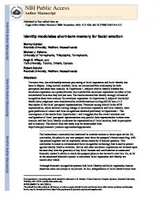
The FTLD-TDP cases and CHOP control samples were genotyped on either the Illumina HH550 BeadChip or the Illumina human610-quad BeadChip at the Center for Applied Genomics at CHOP as previously described14. The 1958 birth cohort samples were genotyped on the HH550 BeadChip by the WTCCC27. Sixteen individuals, 13 cases and 3 controls, were excluded from GWA phase for low genotyping (T (p.R493X) GRN mutation carriers representing ~20% of mutation positive cases, however the chromosome 7 association is not related to any single GRN mutation and remains when the cases with c.1477C>T are removed (P=1.446×10⁻¹⁰). The same locus on chr 7 identified in the GRN mutation cases is also the strongest signal in the GRN negative cases, although it does not reach genome-wide significance. A list of the SNPs with the highest signals in b is given in Supplementary Table 8.
NIH-PA Author Manuscript Nat Genet. Author manuscript; available in PMC 2010 September 1.
NIH-PA Author Manuscript
NIH-PA Author Manuscript Control Numbers
2509
553
Case Study Source International FTLD Consortium International FTLD Consortium
λ
1.05
Method of Testing Illumina HH550 or 610-Quad BeadChips TaqMan genotyping of 2 SNPs
1297 CHOP EuropeanCaucasian, 1212 WTCCC Penn Autopsy, Penn ADC, Coriell Neurologically Normal panel, CHOP European-Caucasian
Control Study Source
CHOP, Children's Hospital of Philadelphia; Penn ADC, University of PennsylvaniaAlzheimer's Disease Center; WTCCC, Wellcome Trust Case Control Consortium; λ,genomic control inflation factor.
89
515
GWA
Replication
Case Numbers
Phase
Summary of samples and controls used for GWA and replication phases
NIH-PA Author Manuscript
Table 1 Van Deerlin et al. Page 17
Nat Genet. Author manuscript; available in PMC 2010 September 1.
NIH-PA Author Manuscript
NIH-PA Author Manuscript
12182087
12194417
12222303
12232513
12243606
12250312
12252934
rs12671332
rs1468915
rs1020004
rs6966915
rs10488192
rs1990622
rs6945902
A
C
T
T
G
C
C
T
G
C
G
G
Minor Allele
0.183
0.321
0.139
0.321
0.233
0.171
0.173
0.154
0.287
0.154
0.133
0.148
MAF case
0.230
0.436
0.204
0.435
0.338
0.242
0.245
0.207
0.351
0.191
0.166
0.184
MAF cont OR 0.77 0.77 0.77 0.75 0.70 0.64 0.65 0.60 0.61 0.63 0.61 0.75
CA P-val 5.82×10⁻³ 8.97×10⁻³ 4.90×10⁻³ 9.45×10⁻⁵ 9.88×10⁻⁵ 7.50×10⁻⁷ 9.49×10⁻⁷ 5.00×10⁻¹¹ 1.63×10⁻¹¹ 1.46×10⁻⁶ 1.08×10⁻¹¹ 7.17×10⁻⁴ 0.63
0.53
0.52
0.53
0.51
0.54
0.54
0.58
0.64
0.64
0.64
0.64
Lower 95% CI
0.89
0.71
0.76
0.71
0.70
0.77
0.77
0.84
0.86
0.93
0.94
0.92
Upper 95% CI
Chr, chromosome; BP, base pairs (NCBI build 36); MAF, minor allele frequency; cont, controls; CA P-val, Cochrane-Armitage P-value; OR, odds ratio; CI, confidence interval.
SNPs are listed in genomic order based on location on chromosome 7. SNPs in bold text have the lowest P-values.
12166585
12101859
rs10226395
rs6952272
12088321
rs1990602
12130625
12071795
rs1006869
rs1003433
BP
SNP rs ID
SNPs on chromosome 7 in region with highest association in the GWAS
NIH-PA Author Manuscript
Table 2 Van Deerlin et al. Page 18
Nat Genet. Author manuscript; available in PMC 2010 September 1.