Communicated by Paul A. Marks, December 1, 1975. ABSTRACT ..... ed [Rabbits, T. H., Jarvis, J. M. & Milstein, C. (1975) Cell 6,5-12]. We acknowledge the ...
Proc. Nat. Acad. Sci. USA Vol. 73, No. 3, pp. 727-731, March 1976 Biochemistry
No detectable reiteration of genes coding for mouse MOPC 41 immunoglobulin light-chain mRNA (antibody diversity/RNA'DNA hybridization/hydroxylapatite/gene reiteration)
M.-G. FARACE, M.-F. AELLEN, P.-A. BRIAND, C. H. FAUST*, P. VASSALLI, AND B. MACH Department of Pathology, University of Geneva Faculty of Medicine, Geneva, Switzerland
Communicated by Paul A. Marks, December 1, 1975
ABSTRACT
RNA fractions rich in immunoglobulin light
sary for such hybridizations in DNA excess, the mRNA was labeled chemically with '25I (7), a procedure that preserves the specificity and the rate of hybridization of RNA (8). The probe used contains, therefore, the entire V and C sequences, as well as the untranslated regions (9, 10). So far, there have been two limitations of such studies: (i) the lack of control of the fate of the [125I]mRNA probe during the course of hybridization and the possibility that the observed extensive RNA breakdown could be nonrandom; and (ii) the finding of two distinct transitions in the hybridization profile, corresponding to components with different gene reiteration frequencies (11-17). In this paper, an extension of preliminary reports (18), we present data obtained under hybridization conditions allowing the entire sequence of the L-chain mRNA probe, including, therefore, the V region, to remain available for hybridization throughout the reaction. Moreover, as the result of further purification of the mRNA, only a single transition is now observed in the hybridization kinetics, corresponding to two to four genes.
(L)chain mRNA were isolated from mouse myeloma MOPC 41 by procedures previously described, and chemically labeled with 125I. These RNA fractions were hybridized with MOPC 41 DNA under conditions of DNA excess. Hybridization conditions were chosen under which the entire sequence of the L-chain mRNA probe, thus including the variable region, remains available for hybridization throughout the reaction. The hybridization (Cot) curve showed double transition kinetics, with one component corresponding to about 250 gene copies and the other to about two to four copies. In contrast, when MOPC 41 L-chain mRNA was further purified as a single band by gel electrophoresis in 99% formamide, the hybridization curve showed only a single transi-
tion, corresponding to about two to four genes, with the disappearance of the "reiterated" component. That component resulted therefore from contaminating RNA species. The data indicate that no reiteration can be detected by RNase or by hydroxylapatite for the genes corresponding to the entire sequence of MOPC 41 L-chain mRNA, including the untranslated segments, within the limits of detectability of short reiterated segments. It thus appears that there is only one or very few genes corresponding to the 41 L-chain variable region "subgroup" in MOPC 41 DNA. The possibility that the variable genes of plasmocytes might result from a combination of several nonreiterated germline genes is discussed.
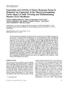
MATERIALS AND METHODS Purification and Iodination of L-Chain mRNA. Mouse myeloma tumors were obtained originally from Dr. M. Potter (N.I.H.). Preparation of the polysomes, extraction of polysomal RNA, fractionation of RNA by oligo(dT)-cellulose chromatography, and isolation of 14S mRNA coding for the L chain of MOPC 41 myeloma were performed as described earlier (3, 9). 14S RNA, representing the peak fraction of the two successive sucrose gradients, was further fractionated by preparative gel electrophoresis using 4.5% polyacrylamide gels in 99% formamide (19). After elution of the RNA from individual 1-mm slices (see legend of Fig. 1), aliquots were assayed in a wheat-germ cell-free system (21) and RNA from the main band in Fig. 1, where L-chain mRNA is located, was used for iodination. Iodination of RNA was performed according to the procedure of Prensky et al. (7) with slight modifications: optimal iodination was obtained in the presence of 0.01 mM KI, in addition to Na'25I (Amersham, IMS 30), 0.8 mCi in a volume of 20 ,l. Iodinated RNA was passed through a column of Sephadex G-75 (0.6 X 7 cm) in 2 mM EDTA, heated at 600 for 20 min as described (22), and passed through a second G-75 column. After ethanol precipitation, ['"I]RNA was centrifuged through a sucrose gradient (3), where it sedimented as a peak of 13-14 S. The peak fraction was precipitated and stored in ethanol at -200. The specific activity of [125I]RNA was between 5 and 10 X 107 cpm/,tg. After alkali treatment (10) and sucrose gradient sedimentation, the [125I]RNA fragments were fractionated on oligo(dT)-cellulose (20). Purification of Mouse Hemoglobin 9S mRNA. Polysomes were isolated from mouse reticulocytes (23). RNA was
The different models that have been proposed to explain the generation of diversity of antibodies have different implications as to the number of different genes required to code for the constant (C) and variable (V) regions of immunoglobulin (Ig). According to the germline hypothesis (1), each of the different V region specificities of antibodies is coded for by a distinct V gene, all of them being present in germline DNA. According to the somatic models (2), only a few "basic" genes are present in the genome and the diversity of the different V sequences is acquired de novo during differentiation by somatic mutations and/or recombinations. An evaluation of the number of genes coding for the V and the C regions of Ig chains in the genome could allow a distinction between the two models. By following the kinetics and reannealing of enzymatically synthesized DNA, complementary to mouse myeloma Lchain mRNA (cDNA), with mouse DNA, we and others (3-6) have estimated as 1 to 3 the number of genes coding for the sequence studied, which corresponded mainly to the C region of the L chain and the 3'-end untranslated region of the mRNA. An alternative approach makes use of the mRNA itself. To obtain the high specific radioactivity necesAbbreviations: L chain, light chain of Ig; V, variable; C, constant; SSC standard saline citrate (0.15 M NaCl-0.015 M Na citrate, pH 7.0); x X SSC, concentration is x times that of SSC; HAP, hydroxylapatite; Cot, initial concentration (moles of nucleotide/liter) X time
(sec); Hb, hemoglobin. * Present address: University of Oregon Medical School, Portland, Oreg.
727
728
Biochemistry: Farace et al.
Proc. Nat. Acad. Sci. USA 73 (1976)
x
E Cl
10
4
8
FIG. 1. Acrylamide gel electrophoresis of 14S L-chain mRNA from.MOPC 41 myeloma tumor (16 jig of 14S RNA) obtained after the second sucrose gradient step was loaded on a 4.5% polyacrylamide gel (0.6 X 12 cm) in 99% deionized formamide (19) and the electrophoresis was run at 28 V/cm for 3 hr, with recirculation of the electrophoresis buffer (20 mM Tris-acetate, pH 7.4). Scale is cm of migration. The gel was scanned in a Beckman Acta II spectrophotometer at 260 nm, and ink marks were placed on either side of the major RNA band, 12 mm apart. The gels were then cut into 1-mm thick slices, and the position corresponding to each individual slice was identified on the absorbance tracing of the RNA peak. RNA was eluted from selected slices by diffusion in 0.1 M Na acetate, 0.5% sodium dodecyl sulfate at room temperature with gentle shaking. RNA of the peak fraction was obtained by pooling the 1-mm slice corresponding to this peak in four gels run in parallel. After ethanol precipitation, the RNA was chromatographed through a column of oligo(dT)-cellulose (20) (6 X 2 mm) to eliminate soluble acrylamide. The stained gel (Pyronine G, Serva) (19) was obtained from another electrophoretic run (inset).
extracted and chromatographed on oligo(dT)-cellulose (20). The 9S RNA peak obtained after sedimentation through a sucrose gradient was assayed in a wheat-germ system and iodinated (specific activity: 5 to 10 X 107 cpm/,ug). Preparation of Myeloma DNA. DNA was prepared as described (3). After sonication, it was passed through a column of Sephadex-Chelex (Bio-Rad) (24). Its average length,
estimated by sedimentation through alkaline sucrose gradients (3, 25), was about 300 to 400 nucleotide pairs.
RESULTS Isolation of L-Chain mRNA. After two sucrose gradient fractionations, the poly(A)-containing RNA prepared from the membrane-bound polysomes of myeloma MOPC 41 sediments as a symmetrical peak ("14S RNA"), which has been shown to be translated into MOPC 41 "Pro-L chain" by sodium dodecyl sulfate-acrylamide gel analysis and tryptic fingerprints (9). The peak fraction of "14S RNA" was further fractionated by electrophoresis on acrylamide gels, using denaturing conditions in 99% formamide. This system allows a clear distinction between the mRNAs for the and f, chains of hemoglobin (Hb), which differ by about 75 nucleotides (26). Under these conditions, 14S RNA is resolved into a single major band, containing the L-chain mRNA activity (see below), and a smear of minor RNA species with a slightly faster mobility (Fig. 1). After scanning, the gels were sliced into 1-mm slices, and RNA was eluted (Fig. 1). Aliquots were assayed in a wheat-germ system (21), and the product of synthesis was analyzed by gel electrophoresis and radioautography (9) with and without prior immunoprecipia
10 20 FRACTION NUMBER
20
FIG. 2. Analysis of 1251-labeled L-chain mRNA on sucrose gradients. Samples of MOPC 41 125I-labeled L-chain mRNA (see Materials and Methods) were sedimented through 5-20% sucrose gradients (3) directly (O in A and B), after melting at 900 for 5 min in 62.5% formamide (A in A), or after incubation in 50% formamide, 4 X SSC at 420 for 26 hr ( Cot value of 4000 in Fig. 3) (A in B) and for 3 days (Cot value of 12,000) (o in B). The gradients were fractionated, and radioactivity of the samples was determined directly in a gamma counter. Arrows indicate the position of 18S and 4S RNA markers.
tation. Pro-L-chain-mRNA activity coincides with the main RNA band seen in Fig. 1. RNA eluted from this major band will be referred to as "L-chain mRNA", even though the presence of contaminant RNA species of the same size cannot be ruled out. Hybridization Conditions. Since iodination is a random process (27), when L-chain mRNA or hemoglobin 9S mRNA is iodinated, all molecules in these preparations will be labeled. Therefore, in spite of possible contaminants, the RNA eluted from the major band and iodinated can be used as a probe for studies of L-chain sequences. However, a crucial point in RNA-DNA hybridization concerns the fate of the RNA probe during the reaction. Preliminary observations had shown that under standard conditions of hybridization [phosphate buffer or 0.15 M NaCI-0.015 Na citrate (SSC) at 650 or 700], [125I]RNA was rapidly degraded. We therefore explored the effect of different conditions of buffer and temperature on the fate of a freshly iodinated mRNA preparation. At 650 in 2 X SSC, after 24 hr of incubation (corresponding to a Cot value of 10,000 in Fig. 3; Cot is mol of nucleotide/liter X sec), the size of the remaining [125I]RNA was mostly smaller than 4 S, as analyzed on sucrose gradient. In contrast, when incubation was performed at 42° in 50% formamide and 4 X SSC (28), there was no detectable change in the size of [125I]RNA after short periods of incubation (Cot value of 10), and even after 26 hr or 3 days (Cot value of 4000 and 12,000, respectively), the average size of the [1251]mRNA probe remained in the range of 9-12 S (Fig. 2). This represents, on the average, less than one break per molecule. Furthermore, after 24 hr of incubation at 650 in phosphate buffer or in 2 X SSC, only 30% of the input [125I]RNA radioactivity could be recovered as trichloroacetic acid-precipitable material, whereas, when the incubation was performed at 420 or 500 in 50% formamide, 4 X SSC, 95-100% of the input [125I]RNA counts remained acid-precipitable after 3 days of incubation (Fig. 3). Under these milder conditions of hybridization, therefore, the entire [1251]mRNA sequence, thus including the V region, remains in a physical state that allows hybridization. A large excess of DNA was used such that the ratio of L-
Biochemistry:
Proc. Nat. Acad. Sci. USA 73 (1976)
Farace et al.
Lu N
30-
50%i
z
20-
C~~~~~~~o 10
lo10
o3
1
cot
FIG. 3. Kinetics of hybridization of MOPC 41 1251-labeled L chain mRNA from different stages of purification, with MOPC 41 DNA. RNA and DNA were melted in 62.5% formamide for 5 min at 900. This completely separates the DNA strands, as assayed by chromatography on hydroxylapatite (29). After melting, samples containing 10-4 gg of [125I]RNA (specific activity 5 X 10⁷ cpm/ 4g) and 0.9 mg of MOPC 41 sonicated DNA in a total volume of either 0.3 ml (DNA concentration = 1.5 mg/ml), for Cot values from 1 to 103, or 60 Ml (DNA concentration = 15 mg/ml), for Cot values from 102 to 2 x 10⁴, were incubated in 4 X SSC, 50% formamide at 42° for the times required to achieve the desired Cot values. Samples were diluted with 2 X SSC to 1% formamide and divided into two parts, which were incubated for 30 min at 370, one with 20 ,g/ml of pancreatic RNase (Worthington), the other without enzyme. RNase-treated and control samples were precipitated with cold 10% trichloroacetic acid, and radioactivity was determined in a Packard gamma counter. RNase-resistance of the 0 time controls (2%) was subtracted from each point. The % RNA hybridized was calculated from the acid-precipitable radioactivity of the control and RNase-treated aliquot of each point. "Co" is the initial DNA concentration (mol of nucleotide/liter) and "t" is time of incubation in sec. Cot1/2: Cot value for 50% hybridization. (03 o) The % of input [1251]RNA recovered as trichloroacetic acid-precipitable radioactivity. (0-0) 14S [125I]RNA from the second sucrose gradient. (0-0) L-chain [125I]mRNA eluted from the peak fraction (1-mm slice) of the acrylamide gel (Fig. 1 and Materials and Methods). On the right-hand ordinate, the results are normalized to 100%. (A-A) Samples containing 10-4 Mg of 9S mouse Hb [I25I]mRNA and 1.8 mg of MOPC 41 DNA at the same DNA concentrations indicated above. In three other experiments, the percent hybridization of Hb [125I]mRNA at saturation varied from 42 to 45%. -
chain sequences in DNA to those in [125I]mRNA would be about 2:1, if there were only one single gene for the L-chain sequences. The effect of the difference of rate between DNA-DNA reannealing (Kd) and RNA-DNA hybridization (Kh) on the percent of the cDNA or RNA probe that can hybridize at saturation has been recently studied, as a function of the ratio of the specific labeled and unlabeled sequences (30, 31). Accordingly, since Kh is lower than Kd, especially in formamide (32), the percentage of mRNA hybridized should be below that expected on the basis of the sequence ratio alone (66% for a DNA/probe ratio of 2:1). As expected, the observed percent [125I]RNA hybridized in 50% formamide was found to be 55-60% at 500 and 40-45% at the suboptimal temperature of 420. To maximize the chances of detecting DNA sequences with a slight degree of divergence from the mRNA probe (different V sequences, see Discussion), we have used the less stringent conditions of 420 (33). At this temperature, the percent hybridization of hemoglo-
729
bin 9S [125I]mRNA at saturation varied between 42 and 45% in three experiments (Fig. 3). This indicates that such a rather low plateau of hybridization is not a special feature of Lchain mRNA. Hybridization of [125I~mRNA. When 14S MOPC 41 RNA, obtained from the peak of the second gradient but without further purification by gel electrophoresis, was used for hybridization with homologous DNA, the resulting hybridization curve showed two distinct transitions as a function of Cot (Fig. 3), whose reiteration frequencies were calculated to be about 250 copies and two to four copies per haploid genome (see below: standard curve with Hb mRNA). The "reiterated" RNA fraction represented about 15% of the RNA hybridized. A similar pattern of double transition has been observed by other authors, and different interpretations have been proposed for the "reiterated" component (11-17, 34, 38) (see Discussion). When hybridization was performed with L-chain mRNA obtained from the major RNA band (1-mm slice) observed after gel electrophoresis of 14S RNA (Fig. 1), the hybridization profile (Fig. 3) showed no detectable reiterated component and only a single transition (Cot 1/2 value of 1700). Furthermore, when L-chain [125I]mRNA was treated mildly with alkali (10) and the RNA fragments fractionated on oligo(dT)-cellulose, the poly(A)-minus fraction (enriched in V region sequences) gave an identical hybridization profile as the unfractionated RNA (data not shown). It must be pointed out that the finding of a Cot curve with only a single transition with purified L-chain mRNA does not exclude the possibility that a small length of the entire mRNA sequence might correspond to reiterated genes. Such a situation, without a distinct additional transition in the curve, might escape detection when the hybrids are analyzed by RNase sensitivity alone, since the [125I]RNA segment actually in hybrid form could represent a percentage of radioactivity too small for accurate detection and close to RNase-resistance background values (about 2%). An alternative assay for RNA-DNA hybrids is chromatography on hydroxylapatite (HAP) under conditions where, if only a segment of an RNA molecule is in hybrid form, the totality of that molecule is retained on HAP and therefore scored as "hybridized" RNA (35, 36). During hybridization (Fig. 2), as well as during the HAP chromatography procedure (data not shown and ref. 36), the size of the [125I]mRNA is either unaffected or only reduced to the extent of less than one break per molecule on average. Therefore, if L-chain mRNA contained a short segment complementary to reiterated genes, a much higher percentage of the [125I]RNA would be scored as hybrids with the HAP assay than with the RNase assay at low Cot values. The results of hybridization experiments performed over a range of Cot values (10104) indicate that this is not the case. Fig. 4 illustrates the results obtained at Cot 10 and at Cot 12,000. Only 2.5% of the [125I]RNA was scored as hybrid with HAP and 2.6% with RNase at Cot 10 (Fig. 4). At Cot value of 12,000, 60% of the [125I]RNA is found in a hybridized form by the HAP assay and 45% with RNase. In contrast, in an another experiment, the RNA side fraction from the formamide gel gave, at a Cot of 10, 18% hybridization by the HAP assay and only 5% by the RNase assay, thus indicating that the HAP procedure can detect RNA chains hybridized over only a portion of their length. Finally, hybridization was performed under the same conditions with Hb [125I]mRNA used as kinetic marker (Fig. 3). From other studies it has been concluded that the hemoglobin gene is detected in the mouse genome as one or two
730
Biochemistry: Farace et al. cot
Proc. Nat. Acad. Sci. USA 73 (1976) Cot 12000
10 A
B
97.5%
6
~~~~~~~~60%
X
~~~~40%
0L 2-
-I 0
12.5%~
[ 5
10
15 5 FRACTIONS
10
15
FIG. 4. Fractionation of hybridized samples on HAP. Samples prepared in parallel from those described in the legend of Fig. 3 (gel-eluted MOPC 41 L-chain [125I~mRNA) were hybridized to a Cot value of 10 (A) and 12,000 (B) as described, and then diluted to 1% formamide in 0.12 M NaPO4 buffer (equimolar Na2HPO4 and NaH2PO4). They were applied to a column of HAP (1 X 8 cm, BioRad) equilibrated with 0.12 M NaPO4 buffer and maintained at 650. Stepwise elution was then performed with 0.12 M, 0.14 M (first arrow) and 0.4 M NaPO4 buffer (second arrow), the respective buffer being started as indicated by the arrows. Controls performed with the same batch of HAP had shown that all singlestranded RNA elutes in the 0.12 M fraction and that all RNaseresistant RNA (hybridized) elutes in the 0.4 M fraction (35). The percent [125I1RNA found at 0 time of incubation in the 0.4 M fraction ( = 0.7%) has been subtracted.
copies (37). The Cot curve obtained with mouse Hb mRNA also shows a single transition with a C0t1/2 value of 2700, which should, therefore, correspond along the Cot scale to one or two gene copies. It can now be estimated that the single transition observed with MOPC 41 L-chain mRNA (Cotl/2 value = 1700) corresponds to twice that number of genes. Within experimental errors, it can therefore be concluded that all sequences in MOPC 41 L-chain mRNA, detectable by hybridization by either HAP or RNase assay, correspond to no more than twice the number of Hb genes, i.e., two to four genes.
DISCUSSION A first transition in the hybridization kinetic curve (Fig. 3), analogous to that observed with 14S mRNA, has been reported for various preparations of L-chain mRNA (11-15), as well as for fractions containing Ig heavy chain mRNA (16, 17). With a single exception (16), this rapidly hybridizing RNA corresponds to DNA sequences reiterated 200 to 300 times. Several interpretations of these "reiterated sequences" have been considered. They cannot be attributed to the C region, or to the 3'-end untranslated region (10) of the Lchain mRNA, since these have been shown to correspond to only one or very few genes (3-6). It has been suggested that they correspond to the genes coding for the variable part of the Ig molecules (11, 14, 16). According to an alternative explanation, it is the untranslated region located at the 5' end of L-chain mRNA which correspond to reiterated genes. Indeed, hybridization experiments performed with cDNA molecules long enough to include sequences located beyond the V region coding sequences themselves, and assumed to be transcripts of L-chain mRNA, have shown a rapidly hy-
bridizing component (34, 38). This led to the suggestion that the 5' end of L-chain mRNA could be a feature common to all genes coding for the V region sequence, in all types of heavy and light chains of Ig (38). Components corresponding to reiterated DNA sequences have also been observed for mRNA
unrelated to immunoglobulin, such as fibroblast
(12)
myoblast (24) mRNA; furthermore, in amphibian oocyte mRNA, a 5'-end RNA segment has been shown to corre-
or
spond to reiterated sequences in DNA (35). According to a third interpretation, the RNA sequences that hybridize rapidly might represent distinct RNA molecules, contaminating the preparation of L-chain mRNA (18). In the present experiments, in fact, the further purification of L-chain mRNA as a single band on acrylamide gels in formamide has revealed that the RNA sequences that hybridize at low Cot values are not a part of the L-chain 41 mRNA molecules. The reiterated sequences correspond to one or several contaminating RNA species, distinct from the main band detected in the formamide gels. This main RNA band, which is homogeneous in size, contains the L-chain mRNA activity. Its exact degree of purity is not known, but the 14S RNA preparation from which it is purified has been estimated to be about 50% homogeneous (3). The finding that L-chain mRNA hybridizes with a single transition kinetics, with a Cot1/2 value of about 1700 suggests that, in the MOPC 41 genome, the number of gene copies corresponding to each of the various parts of the mRNA molecules (C and V regions as well as untranslated regions) is less than 5. This value is obtained by comparison with the hybridization kinetics of Hb mRNA (Fig. 3), assuming one or two genes for Hb (37). It is in agreement with the values obtained with hybridization of cDNA, and which concerned mainly the C region and the untranslated region of the 3' end (3). The hybridization Cot curve of 41 L-chain mRNA, however, does not exclude the possibility that one or several small segments of this RNA correspond to reiterated genes. If such segments represent only a small percentage of the RNA submitted to hybridization, the detection of their hybridization might be difficult with the RNase assay. Therefore, an alternative assay of the hybrids has been used to explore this possibility, using a procedure which scores as hybrids the entire RNA molecule, even when only a segment of RNA is in duplex form (35, 36). In these experiments with hydroxylapatite, no hybridization to reiterated genes could be detected. This reduces the length of the segment of Lchain mRNA that might still correspond to reiterated genes to the minimal length required for discrimination of hybrids by hydroxylapatite. This length is about 20 nucleotides in the case of DNA-DNA duplexes (29, 36), and has not been determined precisely for DNA-RNA hybrids. The V region of MOPC 41 mRNA is expected to hybridize with all the possible genes corresponding in the genome to this L-chain "subgroup". Amino-acid sequence data have indeed indicated that V region sequences fall into subgroups, or prototype sequences, characterized by an almost complete sequence identity over about two-thirds of their length ("framework sequence"), with amino-acid differences responsible for the specificity of the antibody combining site restricted to three "hypervariable regions" of about 10 to 12 amino acids each (39). The V region subgroups are clearly defined in man, where there are three for K chains and four for X chains. In mice, the situation appears more complex; if the same pattern of subgroups exists, the number of K-chain subgroups is much larger, perhaps larger than 20 (40). Consequently, there is no large collection of known sequences within these subgroups. In one such subgroup, seven V sequences are known, five of which differ by less than 10 amino acids out of 110 (40). There is no reason to suspect that the range of diversity of V sequences in the hypervariable regions should differ markedly in different subgroups. The existence in the genome of reiterated genes for the "framework sequence" of the V gene of the 41 L-chain subgroup would have been detected by hybridization, at least with the HAP assay. This is likely, even consid-
Biochemistry:
Farace et al.
ering the possible use of synonymous codons or the preewce of silent mutations in these sequences. The conditions of low stringency used for the hybridization (420 versus 50°) were indeed chosen to maximize the possibility of detecting even partially mismatched hybrids. One is therefore led to conclude that MOPC 41 DNA contains only one or very few genes for the "framework" V sequence of the 41 L-chain subgroup. Similar conclusions have been reached, although under different experimental conditions, in the case of the L-chain of several myelomas (13, 15, 34). This suggests the existence of a single gene for the "framework sequence" of each V subgroup. Such a conclusion renders very unlikely the classical "germ-line theory" of the generation of antibody diversity, which postulates the existence of one distinct V gene for each different V sequence. It is more compatible with any of the various "somatic models", which require the existence of a small number of "basic V genes" in germ-line DNA (one for each subgroup) which are modified in each individual during differentiation as the result of somatic mutations or recombinations affecting the "hypervariable" sequences. We are now confronted with an interesting paradox, with on one hand the hybridization data, favoring the existence of only one gene per V subgroup, and on the other hand recent data which suggest that the spectrum of different antibody specificities (idiotypes) might be inherited and not generated de novo in each individual animal (41). These seemingly contradictory requirements may be explained if one considers that different portions of the V region might be represented, in germ-line DNA, by different genes. The organization of germ-line V genes could thus include at least one gene for the "framework" (or "unvariable") region of each V subgroup, into which would be inserted, during lymphocyte differentiation, small DNA segments specifying the hypervariable regions, as suggested by Wu and Kabat (39). Evidence in favor of this model has also recently come from the observation that distinct genes might exist for the "framework part" and for the "idiotypic determinant" ("combining site") of a given antibody molecule (42). Many such segments, different from one another, would exist in germ-line DNA, perhaps not as individual episomes as originally suggested, but rather as part of chromosomal DNA. These segments could be clustered and organized as a "hypervariable segment gene", with appropriate spacer regions, from which they could be specifically excised, perhaps in a manner similar to the selective cleavage of DNA segments by restriction endonucleases. An interesting prediction of any kind of model involving DNA "insertion" in the formation of a given V gene in a given cell is that the hybrids formed between a V region probe (cDNA or mRNA) and the V gene sequence from either plasma cell DNA or germline DNA should be different. In the latter case, hybridization to the "framework" V genes would produce hybrids with specific "deletion loops", which could be identified experimentally. Note Added in Proof. Since submission of this paper, similar conclusions concerning the "reiterated component" have been presented [Rabbits, T. H., Jarvis, J. M. & Milstein, C. (1975) Cell 6,5-12]. We acknowledge the skillful technical assistance of Miss Dubant, and the advice of M. Crippa and D. Dina on HAP chromatography. This work was supported by the Swiss National Foundation (Grant no. 3.1310.73). 1. Hood, L., Gray, W. R., Sanders, B. G. & Dreyer, W. J. (1967) Cold Spring Harbor Symp. Quant. Biol. 32, 133-146. 2. Gally, J. A. & Edelman, G. M. (1972) Annu. Rev. Genet. 6, 1-46.
Proc. Nat. Acad. Sci. USA 73 (1976)
731
3. Faust, C. H., Diggelmann, H. & Mach, B. (1974) Proc. Nat. Acad. Sci. USA 71, 2491-2495. 4. Rabbits, T. H. (1974) FEBS Lett. 42,323-326. 5. Honjo, T., Packman, S., Swan, D., Nau, M. & Leder, P. (1974) Proc. Nat. Acad. Sci. USA 71, 3659-3663. 6. Stavnezer, J., Huang, R. C. C., Stavnezer, E. & Bishop, J. M. (1974) J. Mol. Biol. 88, 43-63. 7. Prensky, W., Steffensen, D. M. & Hughes, W. L. (1973) Proc. Nat. Acad. Sci. USA 70, 1860-1864. 8. Tereba, A. & McCarthy, B. J. (1973) Biochemistry 12, 46754679. 9. Mach, B., Faust, C. H. & Vassalli, P. (1973) Proc. Nat. Acad. Sci. USA 70,451-455. 10. Milstein, C., Brownlee, G. G., Cartwright, E. M., Jarvis, J. M. & Proudfoot, N. J. (1974) Nature 252,354-359. 11. Delovitch, T. L. & Baglioni, C. (1973) Proc. Nat. Acad. Sci. USA 70, 173-178. 12. Rabbits, T. H., Bishop, J. O., Milstein, C. & Brownlee, G. G. (1974) FEBS Lett. 40, 157-160. 13. Tonegawa, S., Steinberg, C., Dube, S. & Bernardini, A. (1974) Proc. Nat. Acad. Sci. USA 71, 4027-4031. 14. Storb, U. (1974) Biochem. Biophys. Res. Commun. 57,31-38. 15. Leder, P., Honjo, T., Packman, S., Swan, D., Nau, M. & Morman, B. (1974) Proc. Nat. Acad. Sci. USA 71,5109-5114. 16. Premkumar, E., Shoyab, M. & Williamson, A. R. (1974) Proc. Nat. Acad. Sci. USA 71, 99-103. 17. Bernardini, A. & Tonegawa, S. (1974) FEBS Lett. 41, 73-77. 18. Mach, B., Farace, M. G., Faust, C. H., Vassalli, P. & Diggelmann, H. (1974) Fed. Eur. Biochem. Soc. Meet. Symp. 9th 7, 294. 19. Pinder, J., Staynov, D. Z. & Gratzer, W. (1974) Biochemistry 13,5373-5378. 20. Faust, C. H., Diggelman, H. & Mach, B. (1973) Biochemistry
12,925-931. 21. Roberts, B. E. & Paterson, B. M. (1973) Proc. Nat. Acad. Sci. USA 70,2330-2334. 22. Knochel, W. (1974) Mol. Biol. Rep. 1, 311-320. 23. Lingrel, J. B. (1972) in Methods in Molecular Biology, eds. Last, J. A. & Laskin, E. (Dekker, New York), Vol. 2, pp. 231262. 24. Campo, S. & Bishop, J. 0. (1974) J. Mol. Biol. 90, 649-663. 25. Studier, F. W. (1965) J. Mol. Biol. 11, 373-390. 26. Gould, H. J. & Hamlyn, P. H. (1973) FEBS Lett. 30,301-304. 27. Dube, S. J. (1973) Nature 246,483. 28. Neiman, P. E., Wright, S. E., McMillia, C. & McDonnell, D. (1974) J. Virol. 13, 837-846. 29. Britten, R. J., Graham, D. E. & Neufeld, B. R. (1974) in Methods in Enzymology, eds. Grossman, L. & Moldave, K. (Academic Press, New York) Vol. 29, pp. 363-418. 30. Straus, N. A. & Bonner, T. J. (1972) Biochim. Biophys. Acta 277,87-95. 31. Gambino, R., Kacian, D., O'Donnell, J., Ramirez, F., Marks, P. A. & Bank, A. (1974) Proc. Nat. Acad. Sci. USA 71, 39663970. 32. Bishop, J. 0. (1972) Biochem. J. 126, 171-185. 33. McCarthy, B. J. & Church, R. B. (1970) Annu. Rev. Biochem. 39, 131-150. 34. Rabbits, T. H. & Milstein, C. (1975) Eur. J. Biochem. 52, 125-133. 35. Dina, D., Meza, J. & Crippa, M. (1974) Nature 248,486-490. 36. Klein, W. H., Murphy, W., Attardi, G., Britten, R. J. & Davidson, E. H. (1974) Proc. Nat. Acad. Sci. USA 71, 1785-1789. 37. Harrison, P. R., Birnie, G. D., Hell, A., Humphries, S., Young, B. D. & Paul, J. (1974) J. Mol. Biol. 84,539-554. 38. Rabbits, T. H., Brownlee, G. G. & Milstein, C. (1974) in Proc. 9th FEBS Meeting (North Holland, Amsterdam), Vol. 36, pp. 141-149. 39. Wu, T. T. & Kabat, E. (1970) J. Exp. Med. 132, 211-250. 40. Hood, L., McKean, D., Farnsworth, V. & Potter, M. (1973) Biochemistry 12, 741-749. 41. Eichmann, K. (1973) J. Exp. Med. 137, 603-621. 42. Kindt, T. J., Seide, R. K., Bokisch, V. A. & Krause, R. M. (1973) J. Exp. Med. 138,522-536.