humans, autoimmune diabetes is a polygenic disorder in which more than 15 ..... tion increases circulating corticosterone and progesterone concentrations (in ...
Neuroendocrine Immuno-ontogeny of the Pathogenesis of Autoimmune Diabetes in the Nonobese Diabetic (NOD) Mouse Franc¸oise Homo-Delarche
Abstract Type 1 diabetes (T1D) is a T cell-mediated autoimmune disease in which insulin-producing beta cells of the pancreatic islets of Langerhans are destroyed. The nonobese diabetic (NOD) mouse is one of the rare spontaneous models that enable the study of prediabetic pancreatic events. The etiology of the autoimmune attack in human and animal T1D is still unknown, but genetic and environmental factors are involved in both cases. Although several autoantigens have been identified and defective immune-system regulation is implicated, this information does not satisfactorily explain the generally accepted beta-cell specificity of the disease or how so many and diverse environmental factors intervene in its pathogenesis. Based on data obtained from evaluating glucose homeostasis in a variety of situations, particularly stress and cytokine administration, in young prediabetic NOD mice, the author hypothesizes that the islet of Langerhans is a major actor, and its altered regulation through environmentally induced insulin resistance might reveal latent T1D. It is also postulated that T1D pathogenesis might be linked to abnormal pancreas development, probably due to disturbances of glutamic acid decarboxylase (GAD)+ innervation phagocytosis by defective macrophages during the early postnatal period. Also discussed is the role of defective presentation of pancreatic hormones and GAD in the thymus, and its potential repercussion on T-cell tolerance. Observations have demonstrated that the diabetogenic process in the NOD mouse is extremely complex, involving neuroendocrine immune interaction from fetal life onward. Key Words: androgens; beta-cell hyperactivity; cytokines; glucocorticoids; inflammation; insulin resistance; nonobese diabetic mouse; type 1 diabetes
Introduction
T
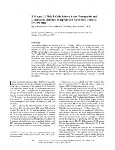
ype 1 diabetes mellitus (T1D1), observed predominantly in children and young adults, is a multigenic disease strongly dependent on numerous environmental factors, as schematized in Figure 1 (Atkinson and EisenFranc¸oise Homo-Delarche, M.D., Ph.D., is an Institut National de la Sante´ et de la Recherche Me´dicale ( INSERM) researcher at the Centre National de la Recherche Scientifique (CNRS), Universite´ Paris 7/D.Diderot, Paris, France. 1 Abbreviations used in this article: APC, antigen-presenting cell; BB, biobreeding; CBG, corticosteroid-binding globulin; DC, dendritic cell; EFAD,
Volume 45, Number 3
2004
barth 2001; Dahlquist et al. 1999; Homo-Delarche 1997; Todd and Wicker 2001). T1D results from the autoimmune destruction of insulin-producing beta cells in pancreatic islets of Langerhans. Despite extensive genetic and immunological investigations, mainly conducted in spontaneous animal models (particularly the nonobese diabetic [NOD1] mouse but also the bio-breeding [BB1] rat), the etiology of the disease remains unknown (Atkinson and Leiter 1999; Mathis et al. 2001; Notkins and Lernmark 2001). As in humans, autoimmune diabetes is a polygenic disorder in which more than 15 susceptibility loci have been described (Todd and Wicker 2001). Because genetic and immunological data have been abundantly reviewed (Atkinson and Eisenbarth 2001; Atkinson and Leiter 1999; Bach 1994; Beyan et al. 2003; Moriyama and Eisenbarth 2002; Todd 1995; Todd and Wicker 2001), the focus in this article is on the less known neuroendocrine aspects of diabetes development in the prediabetic NOD mouse. In NOD mice, the first immune cells, macrophages (M⌽s1) and dendritic cells (DCs1), also known as antigenpresenting cells (APCs1), accumulate around NOD pancreatic islets and ducts at weaning (3 wk of age) (Figure 2) (Jansen et al. 1994; Jansen et al. 1996; Rosmalen et al. 2002a). Subsequently, T cells migrate to the pancreas and home (or accumulate) around ducts (periductulitis) and islets (peri-insulitis), followed by M⌽, DC, and T-cell infiltration into the islets (insulitis). Destructive insulitis coincides with scavenger M⌽ influx and finally leads to beta-cell destruction and overt symptoms of T1D (Jansen et al. 1994; Rosmalen et al. 2002a). Generally, NOD females, whose islets are precociously infiltrated, become diabetic earlier (as of 3 mo of age) and at greater frequency than males. In the Necker Hospital colony (Paris, France), 80% and 40% of females and males, respectively, generally become diabetic at 6 mo of age. Because of the existence of NOD sexual dimorphism and the impact of some environmental factors (e.g., stress, infections, and diet) on the clinical onset of T1D, colleagues
essential fatty acid deficient; GABA, ␥-aminobutyric acid; GAD, glutamic acid decarboxylase; GFAP, glial fibrillary acidic protein; HPA, hypothalamuspituitaryadrenal; IAA, insulin autoantibody; IFN␥, interferon-␥; IGF1, insulin growth factor-1; IL, interleukin; LADA, latent autoimmune diabetes of adults; MHC, major histocompatibility complex; M⌽, macrophage; NOD, nonobese diabetic; NON, nonobese nondiabetic; NOR, nonobese resistant; Sc, Schwann cell; T1D, type 1 diabetes; T2D, type 2 diabetes; TNF, tumor necrosis factor; TZD, thiazolidinedione; VNTR, variable number of tandem repeats.
237
Figure 1 The complexity of one or more genotype−environment interactions in type 1 diabetes (T1D). From Todd JA, Wicker LS. 2001. Genetic protection from the inflammatory disease type 1 diabetes in humans and animal models. Immunity 15:387-395. Copyright © 2001 Elsevier. Reprinted with permission.
and I2 examined more closely the impact of some of these factors on NOD mice. The data obtained led us to study glucose homeostasis in young prediabetic NOD mice, and we advanced the hypothesis that the islet of Langerhans and its innervation are major actors in T1D progression (HomoDelarche 1997; (Homo-Delarche and Boitard 1996; HomoDelarche 1997). Moreover, in the context of the possible role of defective presentation of pancreatic hormones in the thymus with one or more potential repercussions on T-cell tolerance, we also evaluated the thymic expression of pancreatic hormones. Our own data presented in this article are interwoven with those of other investigators obtained in similar or close domains to yield a coherent working hypothesis of T1D pathogenesis. Furthermore, as often as possible, we compare the neuroendocrine events that take place during the progression of the NOD disease with human T1D.
Effects of Hormones and Environment on NOD Insulitis and Diabetes Incidence
anti-inflammatory and immunosuppressive actions but also act as counterregulatory hormones inducing hyperglycemia (Homo-Delarche et al. 1991; Porte and Woods 1983). Therefore, in T1D, glucocorticoids might have both potentially beneficial and deleterious effects. Moreover, NOD diabetes can be modulated not only by multiple immunotherapeutic agents (Bach 2002b), but also by various other factors, including melatonin, ambient temperature, variations of protein and carbohydrate intake, insulin and drugs modulating its secretion or sensitivity, insulin growth factor-1 (IGF-11), and leptin.
Sex Steroids, Pregnancy, Prolactin, and Vitamin D As mentioned above, NOD mice show sexual dimorphism, with diabetes appearing earlier and more often in females than males in most colonies. Orchidectomy soon after weaning significantly increases the prevalence of the disease,
Hormones and environmental factors modulate immune and autoimmune responses (Homo-Delarche and Dardenne 1993; Homo-Delarche and Durant 1994). Sex steroids are part of the mechanism underlying the well-recognized immune sexual dimorphism, which is particularly evident in autoimmune diseases (Ansar Ahmed et al. 1985; Talal and Ansar Ahmed 1987). Moreover, clinical and experimental observations link stressful life events and the onset of these diseases (Homo-Delarche et al. 1991). As part of the endocrine response to stress, glucocorticoids exert well-known
2
Allusions in this article to the author’s work with others relate to research with colleagues in their laboratory at CNRS UMR 8603, Hospital Necker/ Universite´ Paris 5, Paris, France.
238
Figure 2 Sequential appearance of antigen-presenting cells (APCs), both dendritic cells and macrophages, and lymphocytes in nonobese diabetic (NOD) type 1 diabetes mouse pancreata. Clinical onset of diabetes appears when more than 80% of the islets evidence insulitis. ILAR Journal
whereas ovariectomy reduces it in young adults (Fitzpatrick et al. 1991; Hawkins et al. 1993; Makino et al. 1981; Verheul et al. 1995). Generally in those studies, the degree of insulitis coincided with progression to diabetes. Androgen (dihydrotestosterone) administration, at pharmacological but also near physiological doses, prevents the disease in females, whereas a physiological or near physiological, nontoxic dose of estrogens does not affect disease symptoms, raising the possibility that the NOD sexual dimorphism is linked to the protective immunosuppressive effects of androgens (Fox 1992; Hawkins et al. 1993; Verheul et al. 1995). In terms of circulating sex steroids, when compared with NOD females, NOD males appear, as expected, to differ only in androgen levels (Durant et al. 1998). Intriguingly, although the incidence of diabetes is lower in NOD females treated with androgens from birth onward, ovariectomy + testosterone increases it to 100%, suggesting that the neonatal hormonal environment strongly influences diabetes incidence in NOD mice (Hawkins et al. 1993). To what extent this effect on diabetes incidence results from the alteration of the hypothalamus-pituitary-adrenal (HPA1) axis due to ovariectomy (and also sham surgery) performed during the early postnatal period, as demonstrated in rats, is unknown (Vallette et al. 1982). In this setting, glucocorticoid effects all along the HPA axis might be downregulated, thereby modifying the postnatal increase and functioning of their receptors (Sakly and Koch 1981). Of note, at variance with what is observed in NOD mice, no sexual dimorphism exists in the BB rat (Rossini et al. 1995). In human T1D, a slight male preponderance can be observed, at least in some countries (Homo-Delarche 1997). The percentages of thymus and spleen cell subpopulations, mitogen reactivity, lymphokine production, and in vivo response to a thymus-dependent antigen (e.g., sheep red blood cells) are dependent on or independent of the sex-steroid environment in adult prediabetic NOD mice. For example, although castration in both sexes does not affect splenic T-cell populations, splenic B cells and M⌽s are significantly increased (Fitzpatrick et al. 1991). Moreover, the sexual dimorphism in NOD diabetes incidence (females being more diabetes prone than males) is reflected by a difference in thymocyte apoptosis (Casteels et al. 1998). Apoptosis is decreased 24 hr after incubation with dexamethasone in both sexes of NOD compared with C57BL/6 mice, but was lower in NOD females than in NOD males. Moreover, orchidectomy (which increases this incidence) diminishes thymocyte apoptosis, and androgen treatment of NOD females (which lowers diabetes incidence) restores thymocyte apoptosis to male levels. The active form of vitamin D3, 1,25-dihydroxyvitamin D3, which prevents diabetes in NOD mice, was shown to restore apoptosis levels to those of C57BL/6 controls (Casteels et al. 1998; Gregori et al. 2002; Zella and DeLuca 2003). At the pancreas level, sexual dimorphism in cytokine production is also found during the early stages of insulitis at 4 wk of age (Bao et al. 2002). The level of the TH1 inflammatory cytokine, interferon-␥ (IFN␥1), is signifiVolume 45, Number 3
2004
cantly higher in females, in contrast to the TH2 antiinflammatory cytokine, interleukin (IL1)-4, which is higher in males. Moreover, estrogens enhance the production of IL-12, an inflammatory cytokine, by CD4+ T cells, whereas androgens lower IFN␥-induced IL-12 production. It is pertinent that mega-islet formation, which takes place from 6 to 10 wk of age in NOD mice, also shows sexual dimorphism (Rosmalen et al. 2001; Rosmalen et al. 2002a). NOD females spontaneously develop more megaislets than NOD males, in which orchidectomy further increases their number. More mega-islets in castrated NOD males are associated with more infiltrating immune cells. These mega-islets are always only weakly insulin positive compared with smaller islets, suggesting their lower insulin content, as if they were actively secreting insulin or, by contrast, completely exhausted. It should be noted that castrated lymphocyte-deficient NODscid mice also have enhanced mega-islet formation and DC infiltration, indicating that lymphocytes are not required for these castrationinduced effects. The latter data suggest that the sex difference in mega-islet numbers in NOD mice reflects, in part, an organ-specific androgen effect that is independent of lymphocytes. Pregnancy is associated with depressed inflammatory and immune responses, and increased growth and function of islets of Langerhans (Aerts and Assche 1975; HomoDelarche and Durant 1994). NOD mice treated for 13 wk with a cocktail of pregnancy hormones, including a glucocorticoid (dexamethasone), progesterone, estradiol, growth hormone, and prolactin, are protected from diabetes and show less insulitis compared with the control group (saline) (Atwater et al. 2002). Intriguingly, prolactin alone appears to have only a partial protective effect on diabetes progression, despite this hormone being known for its immunostimulatory and deleterious properties in various autoimmune diseases, and its mild counterregulatory effect on insulin, particularly during normal pregnancy (Atwater et al. 2002; Durant et al. 1995; Homo-Delarche and Durant 1994). In NOD mice, pituitary implants on day 35 of age enhance prolactin levels and increase the incidence of diabetes in intact males and ovariectomized females. Similarly, metoclopramide, which counteracts the effect of bromocriptine, known to inhibit prolactin secretion, aggravates diabetes. Inversely, bromocriptine protects NOD females when administered daily subcutaneously, but not intraperitoneally (Durant et al. 1995; Hawkins et al. 1994). This difference results from the hyperglycemic effect of intraperitoneal administration of bromocriptine. Finally, prolactin and IL-2 receptors in splenic and pancreatic immunocompetent cell populations appear to be similarly expressed in NOD females and males (Hawkins et al. 1996).
Behavior, Stress, Glucocorticoids, and Melatonin Based on behavioral testing of the numbers of squares crossed and rearings in an open field, Amrani and col239
leagues (1994) concluded that 2-mo-old prediabetic NOD mice of both sexes are more active than their control C57BL/6 counterparts, which were described as a very active mouse strain. It is generally held that the most active mice in the open field are the least emotional. Moreover, the less emotional females become diabetic earlier and the more diabetes-resistant NOD males are more emotional than NOD females (Ader et al. 1991). NOD mice also exhibit increased sensitivity to the behavioral effects (social exploration, locomotor activity, and rearings) of IL-1 injections compared with outbred ICR mice from which the inbred NOD strain originated (Bluthe et al. 1999). These data suggest that emotionality via neuroendocrine changes, possibly linked to the degree of physical activity, may be a mediating factor that modulates diabetes expression (Ader et al. 1991). NOD mice have been subjected to various stressors prenatally, neonatally, or during early adulthood. Prenatal stress induced by immobilization of the mothers during the last week of gestation accelerates diabetes onset and increases its prevalence in NOD females, whereas it has no effect in NOD males (Saravia-Fernandez et al. 1996a). Neonatal separation of NOD progeny from their mothers has been reported to increase the risk of developing diabetes in both sexes significantly, but to have no effect on insulitis (Dahlquist and Kallen 1997). It has been suggested that the heightened risk could be associated with the induction of metabolic alterations leading to increased peripheral insulin requirements, perhaps in relation to insulin resistance. Short acute stress, induced by a triad of stressors (overcrowding + repeated restraint + anesthesia) applied during the week immediately after weaning reportedly does not affect diabetes expression (Durant et al. 1993). However, when applied later, from the 6th to the 8th week of age, the same combination of stressors has protected NOD mice, particularly males, against diabetes, (Saravia-Fernandez et al. 1996a). Long-term chronic stress, obtained by restraint once a week or overcrowding, significantly protects NOD females (Durant et al. 1993). However, the stress effect on insulitis does not parallel the effects on diabetes incidence for all of the groups described above. It should also be noted that long-term injection of vehicle (0.9% saline) alone is able to delay the appearance and/or decrease the incidence of diabetes in both sexes of NOD mice (Saravia-Fernandez et al. 1996a). A stressful condition such as immobilization increases circulating corticosterone and progesterone concentrations (in both sexes of NOD mice) concomitantly with testosterone decrease in males (Durant et al. 1998). Moreover, rodents, particularly NOD mice, exhibit marked sexual dimorphism in stress-induced corticosterone (Amrani et al. 1996; Durant et al. 1998; Homo-Delarche et al. 1991). Although NOD females that have been subjected to repeated restraint remain able to respond with a dramatic increase of serum corticosterone, various immune parameters, including spleen B- and T-lymphocyte proliferation, are unaffected (Durant et al. 1993; Fitzpatrick et al. 1992). However, in vivo, NOD mice appear to be resistant to thymus 240
cell depletion caused by acute and chronic stress and/or infection, and in vitro, as mentioned above, NOD thymocytes are resistant to glucocorticoid-induced apoptosis (Casteels et al. 1998; Durant et al. 1993; Martins and Aguas 1999). Although up-regulated expression of the bcl-2 gene has been postulated, evidence suggests that the molecule responsible for this resistance acts up-stream from the bcl-2 gene (Garchon et al. 1994; Martins and Aguas 1996). It has also been shown that in NOD mice, the glucocorticoid response involves a prolonged exposure to high levels of the survival signal represented by the Myc protein, which might delay thymocyte and splenocyte apoptosis (Martins and Aguas 1998). Adrenalectomy, which reduces corticosterone secretion (albeit not completely in mice), tends to accelerate diabetes onset, particularly in NOD males (Durant et al. 1993; Saravia-Fernandez et al. 1996a). The number of islet glucocorticoid receptors in all types of endocrine pancreatic cells decreases appreciably at 3 wk of age, but with no sexual dimorphism (Thompson et al. 2002). This glucocorticoidreceptor loss might contribute to the onset of insulitis and potentially to the ontogeny of diabetes in NOD mice as a result of loss of immunosuppressive glucocorticoid effects, particularly on cytokine production (Perretti et al. 1989). Pertinently, in prediabetic NOD islets, IL-1 receptors are expressed with the same density, affinity, and specificity as in control strains (Jafarian-Tehrani et al. 1995). Researchers have also assessed the effect of melatonin, a hormone produced by the pineal gland and known to counteract glucocorticoid-induced thymus atrophy and immunosuppression in NOD females. Conti and Maestroni (1996) have reported that neonatal pinealectomy accelerates diabetes development, and exogenously administered melatonin protects the animals, in spite of its enhancement of insulin autoantibodies (IAAs1).
Infections Infections generate opposite effects on T1D, as described above for other environmental factors. First, they have an apparent protective effect against both NOD and human T1D (Bach 1994). For example, in susceptible strains of mice or rats, autoimmune diseases, particularly T1D, develop earlier and more frequently among animals bred in a specific pathogen-free environment. Indeed, infecting young NOD mice with various pathogens can prevent diabetes (Bach 2002a). It is unknown to what extent this protection might be the partial result of long-term immunosuppression of glucocorticoids, whose secretion is triggered by cytokines (Bateman et al. 1989; Besedovsky and del Rey 1996; Fukata et al. 1994; Tilders et al. 1994). Second, among environmental factors triggering T1D, much attention has been focused on viruses (Jun and Yoon 2003). To date, 13 different viruses have been reported to be associated with T1D development in humans and various animal models. The most clear and unequivocal evidence ILAR Journal
that a virus induces diabetes in animals comes from studies on the encephalomyelitis virus in mice and the Kilham rat virus in rats (Jun and Yoon 2003). In human diabetes, several other viruses in addition to the enteroviruses have been considered potentially causal agents (Hyoty and Taylor 2002). With the exception of congenital rubella, their role is not clear. Enteroviral infections appear to accompany or precede the onset of diabetes in children. As with rubella, enteroviral infection may induce beta-cell autoimmunity even in utero (Dahlquist et al. 1999; Otonkoski et al. 2000). The following several mechanisms have been advanced to explain how viruses might trigger T1D: (1) Sequence similarities between islet-cell GAD and coxsackievirus could cause an immune attack against coxsackievirus and also beta cells. (2) Enteroviral infections could sustain autoimmunity until a final “hit” results in beta-cell loss. (3) Acute or chronic enteroviral infections in peri-insular tissue, because of excess production of free radicals, could cause beta-cell destruction (Graves et al. 1997; Jun and Yoon 2003). However, there is also evidence in humans that increased insulin resistance during infection (e.g., a flu-like viral infection) might cause insulin deficiency in individuals with widely fluctuating residual beta-cell activity (Wasmuth et al. 2000). Even moderate infections can be responsible for severe insulin resistance, with striking plasma insulin increases to maintain normal glycemia (Sammalkorpi 1989; Yki-Jarvinen et al. 1989). Pertinently, after a single injection of IL-1, an inflammatory cytokine, NOD females mount a huge corticosterone response, which induces a transient state of insulin resistance that prevents the IL-1elicited hypoglycemia that occurs normally (Amrani et al. 1996). When mice are adrenalectomized before IL-1 injection, hypoglycemia characterizes only the NOD females, thus demonstrating the role of glucocorticoids in their IL1-induced insulin resistance. NOD males, despite their higher insulin secretion and lower corticosterone response, compared with females, are resistant to the IL-1-induced hypoglycemic effect. Furthermore, their strong IL-1induced insulin secretion is androgen dependent, because it is suppressed by orchidectomy (Amrani et al. 1996). In normal rats, testosterone has a direct effect on pancreatic islet function by favoring insulin gene expression and secretion (Moriyama and Eisenbarth 2002). Hence, androgens appear to be intricately involved in the IL-1-induced insulin resistance of NOD males. The androgen-induced insulin resistance has been described in other mouse strains (Leiter 1989).
Ambient Temperature Low ambient temperature may be considered a stressful condition that alters glucose homeostasis (MacDonald et al. 1987). In this context, NOD mice placed at 23.7°C upon weaning have a lower diabetes incidence at 30 wk of age than those placed at 21.1°C, yet the degree of insulitis does not differ (Williams et al. 1990). Animals housed at the lower temperature have a transiently higher food intake beVolume 45, Number 3
2004
tween 13 and 15 wk of age that stimulates beta cells. Pertinently, children’s overeating has recently been cited as partly explaining the increasing incidence of juvenile T1D, as for type 2 diabetes (T2D1) (Pundziute-Lycka et al. 2003). Cold temperature and overeating, and their potential relationship, should be investigated more thoroughly as factors favoring diabetes expression along the so-called T1D North-South gradient, just as cold temperature and infections might explain increased seasonal (autumn/winter) T1D incidence, perhaps concomitantly with overeating (Bach 2002b; Dahlquist et al. 1999). Moreover, a cold environment is also associated with low exposure to sunlight, which might lead to vitamin D deficiency (Bach 2002b). As mentioned above, vitamin D has been shown to protect against T1D.
Proteins, Free Fatty Acids, and Carbohydrates Epidemiological and animal studies in BB rats and NOD mice have shown that diabetes is a food-influenced disease. Notably, the timing of dietary changes is important, because the maternal diet influences diabetes in NOD offspring (Funda et al. 1999; Reddy et al. 1995). Although the data concerning the diabetogenic effects of milk proteins have become very controversial, wheat flour and soybean proteins are still thought to be the major identified diabetogenic foods (Hoorfar et al. 1993; Paxson et al. 1997; Scott and Marliss 1991; Scott et al. 1988). NOD mice fed a glutenfree diet, while maintaining the same potentially diabetogenic protein (e.g., soybean and milk proteins) consumption and never exposed to gluten even in utero because NOD breeding pairs ate same diet, evidence a much lower T1D incidence (Funda et al. 1999). The mechanisms by which the different diabetogenic components act on diabetes development in BB rats and NOD mice are still unknown, but the involvement of the mucosal immune system (e.g., in celiac disease), which is often associated with T1D, is not excluded. Essential fatty acid deficiency (EFAD1) has an antidiabetogenic effect in multiple low-dose streptozotocin-treated mice and BB rats (Lefkowith et al. 1990; Wright et al. 1991). NOD females fed an EFAD diet from weaning have a sharply decreased cumulative T1D incidence, but the insulitic process was unaffected (Benhamou et al. 1995). The results of studies on various immune parameters suggested a heightened activity of TH2-like cells, concomitantly with an effect on antigen-presenting cells (APCs1), which is linked to less eicosanoid metabolism. Pertinently, we demonstrated that peritoneal M⌽s of prediabetic NOD mice have enhanced production of prostaglandins and leukotrienes (Lety et al. 1992). A final important dietary component that influences diabetes is that of carbohydrates. As for some other environmental factors, the timing of exposure to glucose appears to be important. Although NOD mice rarely become diabetic during their first gestation (indeed, they are slightly hypoglycemic because of hyperinsulinemia; our unpublished ob241
servations), the risk is greatly increased during their second and third pregnancies. According to the degree of maternal hyperglycemia, the consequences can be mild (macrosomia, organomegaly, increased insulin content) or severe (intrauterine death, malformations) (Formby et al. 1987). At birth, beta cells, which are immature, produce only basal amounts of insulin and are unresponsive to glucose (Asplund 1973). Exposure to a high level of glucose in utero might accelerate the beta-cell maturation process and enhance the expression of different beta-cell antigens (Bock et al. 1991; Buschard 1991). The results of two studies have shown that neonatal beta-cell stimulation by intraperitoneal injection of glucose, associated or not with glucagon or arginine, twice a day for the first 6 days of life attenuates diabetes in BB rats and NOD mice. However, in those studies, controls were left untreated, therefore underestimating the protective effect of vehicle injection alone, as mentioned above (Bock et al. 1991; Buschard 1991; Saravia-Fernandez et al. 1996a). By contrast, in a third study in which control NOD mice received two daily injections of vehicle (saline) for the first 6 days of life, neonatal injections of glucose and arginine aggravated insulitis and accelerated diabetes only in NOD females (Senecat et al. 1994). Concomitantly, pancreatic expression of antigens recognized by islet cell antibodies and mRNA for the potential autoantigen GAD67 were increased by glucose-arginine treatment, and the immune parameters evaluated were not affected. The authors concluded that the effects observed might reflect accelerated beta-cell maturation and overexpression of islet antigens during the completion of self-tolerance or amplification of the destructive process due to the existence of more target cells for effector cells. In humans, insulin secretion may differ in response to mother’s milk or cow’s milk formula (Lucas et al. 1981). Infants fed formula (from birth) mounted a stronger insulin response than breast-fed infants, suggesting beta-cell hyperactivity in the former. To what extent this effect might be linked to the extensively debated protective effect of breast milk versus formula in T1D is unknown. Glucose-enriched water given ad libitum after weaning (from 4 wk of age) to NOD mice does not increase diabetes incidence, as expected, but instead lowers blood glucose levels and leads to delayed onset and lower diabetes incidence, particularly in females. This effect has been attributed to improved insulin release and/or lower intake of chow diet with its diabetogenic catalysts (Granot et al. 1994). Also contrary to expectations, a fructose-enriched diet, which causes insulin resistance and hyperinsulinemia in normal and diabetes-prone rats, protects NOD mice against developing diabetes when administered from weaning (Orban et al. 2001). NOD mice fed fructose have increased glucose tolerance and insulin secretion, elevated liver insulin-receptor substrate, and preserved beta-cell mass. The effects of diazoxide, which inhibits insulin secretion, and tolbutamide, which stimulates insulin secretion, were examined in NOD mice treated from 3 wk of age 242
(Williams et al. 1993). Only tolbutamide lowers their diabetes incidence, but high dose levels of tolbutamide aggravate insulitis. Monosodium glutamate administered to neonatal NOD females results in obesity associated with stunting, hyperinsulinemia, and elevated corticosteronemia (Nakajima et al. 1985). However, insulitis and cumulative incidence are lower than for the control group. Prophylactic insulin administration prevents insulitis and diabetes in NOD mice and BB rats, but available clinical trial data are not convincing (Pozzilli 2002). Both NOD and NODscid mice receiving this therapy develop fewer mega-islets (Jansen et al. 1996; Rosmalen et al. 2002a). Moreover, treated NODscid mice have less APC infiltration, indicating that the protective effect is not solely the induction of immunological tolerance to insulin, which is considered to be a major autoantigen (Karounos et al. 1997). These observations suggest one or more important links between the functional status of the islet, leukocyte infiltration, mega-islet formation in NOD and NODscid mice, and—only in NOD mice—between insulitis and diabetes (Rosmalen et al. 2002a). Thiazolidinediones (TZDs1) are drugs that act as receptor agonists to reduce insulin resistance. They are currently used in combination with other hypoglycemic agents to treat T2D, and their effects on NOD mice have been evaluated. TZDs are also of interest because they possess antiinflammatory properties that may have the potential to limit immune-cell inflammation that occurs in the islets of T1D. Both rosiglitazone and troglitazone, given by gavage from 3 wk of age, lower diabetes incidence. However, troglitazone has marked hepatotoxicity at the dose used (Beales and Pozzilli 2002). By down-regulating IL-1-induced intercellular molecule adhesion-1 expression on islet cells, troglitazone modulates the ability to target autoimmune beta-cell destruction. By modifying IFN␥ production, it also acts on T cells, the major effectors of the autoimmune process (Augstein et al. 2003).
Insulin Growth Factors IGF-1, which shares structural homology with insulin and has an insulin-like metabolic effect, has been given to NOD females (Kaino et al. 1996; Yakar et al. 2001). Only early short-term (4-9 wk of age) but not long-term (4-34 wk of age) treatment delays diabetes onset and limits insulitis. IGF-1 also lowers the T1D incidence after transfer of autoreactive T cells from diabetic mice to nondiabetic NOD recipients (Bergerot et al. 1995). In these transfer experiments, IGF-1 treatment contains insulitis and T-cell trafficking to lymphoid organs. Moreover, beta-cell mass, as assessed by insulin immunolabeling, is better preserved in IGF-1-treated mice than in controls, but pancreatic insulin contents and amounts of mRNA proinsulin transcripts are similar. Pertinently, IGF-1 is known to stimulate proliferation and prevent apoptosis of pancreatic beta cells (Hill et al. 1999; Hugl et al. 1998). ILAR Journal
IGF-2 might be implicated in biological functions relevant to autoimmune diabetes, including inhibition of stimulation of beta-cell proliferation and apoptosis (Christofori et al. 1995; Halminen et al. 1996; Hill and Hogg 1991). That IGF-2 is produced by lymphocytes and expressed in the thymus might be important in the control of the autoimmune response (Geenen and Lefebvre 1998; Nyman and Pekonen 1993; Vafiadis et al. 1998). The defective thymus IGF-2 expression observed in BB rats led to the hypothesis that this defect might be detrimental to insulin tolerance and favor diabetes development (Geenen and Lefebvre 1998).
increased risk observed (1) for maternal-child blood group incompatibility, which is associated with beta-cell dysfunction; (2) with older maternal age, excess weight gain during pregnancy, and amniocentesis (Vaarala et al. 1999); and, as mentioned above, (3) with some viral infections during pregnancy.
Characteristics of NOD Glucose Homeostasis and Its Regulation During the Prediabetic Stage Perinatal Pancreas Abnormalities in Autoimmune Diabetes
Leptin Finally, the impact of leptin, the product of the obese gene, has also been examined in NOD mice. Initial evidence suggests that its main function is to regulate body weight, energy balance, and endocrine functions (Lo¨ nnqvist and Schalling 1997). However, leptin also influences immune and inflammatory responses, particularly, exerting proinflammatory effects (Fantuzzi and Faggioni 2000; Howard et al. 1999; Lord et al. 1998). In NOD females, serum leptin levels, which are already higher than those of NOD males or control strain females at 6 wk of age, increase sharply soon after the onset of hyperglycemia (Matarese et al. 2002). Intraperitoneal injections of recombinant leptin, when administered early in life, accelerate the development of autoimmune diabetes only in NOD females, enhance immune-cell infiltration, and augment IFN␥ production by T cells, suggesting that leptin favors proinflammatory T-cell responses. Thus, T1D is very sensitive to numerous hormonal and environmental factors. For this reason, it is not surprising that the diabetes incidence can vary among NOD colonies worldwide (Pozzilli et al. 1993). Moreover, it is worth noting that in spontaneous animal models of T1D and in human disease, factors such as infections, dietary modifications, stressful events, puberty, and pregnancy, which increase insulin resistance and beta-cell activity thereby lowering resistance of T1D-prone individuals to microenvironmental factors involved during the progression of insulitis, could exacerbate the insidious progression to T1D. The effect of puberty, which intervenes immediately after weaning in rodents, has not been evoked until now, except indirectly in terms of castration in NOD mice. However, in humans, puberty, which induces insulin resistance, is well recognized as a factor triggering the clinical onset of both T1D and T2D (Amiel et al. 1991; Bergstrom et al. 1996; Caprio et al. 1989). One important feature of puberty is rapid growth, and growth hormone is known to increase peripheral insulin resistance (Amiel et al. 1986). Finally, in addition to its transmissible genetic origin, it has been suggested that human T1D diabetes is a congenital disorder (Bruining and Batstra 1999; Todd and Wicker 2001). Evidence related to human T1D has accumulated to indicate that the pathogenetic process may start early in life, even in utero, with Volume 45, Number 3
2004
Some of the data presented above would seem to indicate that the clinical expression of T1D is “programmed” as early as intrauterine life in NOD mice. Additional NOD and human T1D data presented below reinforce the hypothesis. NOD neonates born to mildly hyperglycemic mothers have greater birth weights than control newborns (Formby et al. 1987). Maternal metabolic anomalies lead to high glucose levels in utero, which stimulate fetal beta cells and increase birth weight, due to the pleiotropic effects of insulin. We also studied various parameters that affect the pancreas and glucose homeostasis of NOD and NODscid neonates born to young nondiabetic mothers (7 wk old at mating) and compared them with those of C57BL/6 newborns (Homo-Delarche et al. 2003). Pancreatic insulin contents and mean islet size, assessed by insulin immunolabeling, are similar in the three strains during the first month of life. Antiglucagon labeling of NOD and NODscid pancreata reveals high percentages of small glucagon+ islets that decline within the first 2 wk of age, and is suggestive of islet neogenesis (Pelegri et al. 2001). Concomitantly, compared with control C57BL/6 mice, both NOD strains are histologically characterized at birth by higher numbers of large and irregularly shaped islets that also progressively disappear (S.Geutkens, F. Homo-Delarche, J.M Ple´ au, S.Durant, H.A Drexhage, and W. Savino, manuscript in preparation). Reverse transcriptase-polymerase chain reaction detected enhanced expression of preproinsulin I and II in both NOD and NODscid mice between 1 and 2 wk of life. Notably, in all mouse strains investigated, including controls, pancreatic preproinsulin II expression is 10-fold higher than that of preproinsulin I. In situ hybridization revealed that 1-day-old NOD mice express significantly higher levels of primary preproinsulin II transcripts than age-matched C57BL/6 mice, but this difference disappeared during the first week of life. When we treated NOD and NODscid mothers with insulin (Ultratard, NovoNordisk, 1 U/100 g BW) during the last 2 wk of gestation, insulin transcriptional activity at birth was down-regulated in female NOD mice but not in males or both sexes of NODscid. These findings demonstrate that signs of beta-cell stimulation exist in NOD and NODscid neonates. We cannot exclude that insulin resistance, possibly linked to HPA-axis disturbances, might play a role in both NOD and NODscid 243
mice (our preliminary results). Moreover, in newborn NOD mice, sexual dimorphism is already manifest, reflected in a lack of pancreas preproinsulin II down-regulation in males after maternal prophylactic insulin administration. Such a lack is perhaps due to insulin resistance induced by androgens, which are known to increase during the few days before birth in males, exclusively (Weisz and Ward 1980). In humans, some evidence supports a weak, albeit significant, association of higher birth weight and increased risk for T1D, with the possibility of sexual dimorphism (Podar et al. 1999; Stene et al. 2001). Moreover, results of a retrospective study have indicated that children who later developed T1D gained more weight (body mass index) during the first year of life than their healthy siblings (Bruining and Batstra 1999). Taken together, these data suggest that perinatal beta-cell hyperactivity is associated with a higher risk of developing diabetes.
Postweaning Anomalies of the Pancreas and Glucose Homeostasis in T1D The accumulation of DCs and M⌽s, particularly at the duct and islet periphery, which is generally thought to be the first sign of the autoimmune process in NOD mice and BB rats, usually starts soon after weaning (Figure 2). We investigated glucose homeostasis in prediabetic mice because of reported heightened beta-cell sensitivity to glucose observed in perifused inflamed islets from prediabetic BB rats compared with noninflamed islets and transient hyperinsu-
linemia accompanying insulin resistance during the prediabetic phase of T2D (Figure 3) (DeFronzo 1997; Teruya et al. 1993). We detected transient hyperinsulinemia in both sexes of NOD and NODscid mice compared with several control strains (C57BL/6, BALB/c, DBA/2), indicating that their beta cells are hyperactive during the prediabetic period (Amrani et al. 1998; Homo-Delarche 1997). Others have confirmed the beta-cell hyperactivity in NOD, compared with BALB/c mice (Orban et al. 2001). This mild hyperinsulinemia develops between 2 and 4 wk of age, or about the time of weaning, when the shift from maternal milk to laboratory chow takes place. Even in normal mouse strains, the sharply increased dietary glucose at this time awakens beta cells, which have been at rest because of the low glucose content of maternal milk (Girard et al. 1992). At 8 wk of age, basal nonfasting blood insulin levels are still higher in NOD and NODscid mice than in controls (Amrani et al. 1998). Mild hyperinsulinemia in NOD and NODscid mice is associated with decreased glycemia, as expected, compared with sex-matched C57BL/6, DBA/2 and BALB/c mice (Homo-Delarche 1997). Because glucose has been shown to protect beta cells, this low glycemia level might lower their resistance to the various inflammatory factors produced in the islet microenvironment (Hoorens et al. 1996). It is pertinent that transiently elevated insulinemia has also been observed in the BB rat a few days before the onset of T1D, and, as mentioned above, perifused inflamed, but not uninflamed, islets exhibit beta-cell hyperactivity (Nakhooda et al. 1978; Teruya et al. 1993). A defect in the
Figure 3 (A) Markedly greater beta-cell hyperactivity of inflamed versus noninflamed islets from prediabetic diabetes-prone (DP)-BioBreeding (BB) rats. In diabetic (D)-BB rats, the second phase of insulin secretion is decreased. WF, insulin secretion in the control strain. From Teruya M, Takei S, Forrest LE, Grunewald A, Chan EK, Charles MA. 1993. Pancreatic islet function in nondiabetic and diabetic BB rats. Diabetes 42:1310-1317. Copyright © 1993 American Diabetes Association. Reprinted with permission from The American Diabetes Association. (B) Chronological appearance of insulin resistance, insulin secretion, and fasting blood glucose in type 2 diabetes. From Sacks DB, McDonald JM. 1996. The pathogenesis of type 2 diabetes mellitus. Am J Clin Pathol 105:149-156. Copyright ©1996 American Society of Clinical Pathologists. Reprinted with permission. 244
ILAR Journal
first phase of glucose-induced insulin secretion is observed only as of 10 wk of life, when the beta-cell mass begins to decrease, probably due to the progression of insulitis (Kano et al. 1986). Pertinently, less insulin is secreted by NOD islets as long as lymphocytes are present in the culture (Strandell et al. 1992). In human T1D, some indications of transient beta-cell hyperactivity during the prediabetic phase also exist. Circulating proinsulin levels (combined or not with insulin and C-peptide levels) have been evaluated in nondiabetic twins, siblings, or first-degree relatives of T1D patients under basal fasting and stimulated conditions and compared with control subjects. Despite similar fasting blood glucose concentrations, fasting proinsulin levels were higher in twins, siblings, and first-degree relatives than in controls (Hartling et al. 1989; Heaton et al. 1988; Spinas et al. 1992). After oral and/or intravenous glucose challenge, proinsulin levels were also higher in identical twins and siblings (Heaton et al. 1988; Lindgren et al. 1991; Roder et al. 1994). Increased insulin and C-peptide secretions have been described after glucose administration to nondiabetic identical ICA+ twins who were followed for more than 11 yr after one of them had been diagnosed with T1D (Heaton et al. 1987). The results of another study suggest that mild insulin resistance responsible for beta-cell hyperactivity might take place during the prediabetic period (Hollander et al. 1982). Recently, 3773 first-degree relatives of T1D probands were screened for antibodies to islets. Relatives confirmed as having one or more such antibodies were followed with antibody and metabolic testing for a median of 4 yr (Fourlanos et al. 2003). Indeed, antibody-positive relatives who progressed to T1D had elevated fasting insulin, fasting glucose, and insulin resistance at least several years before its onset. These data led the authors to conclude that this metabolic profile in a population “at risk” for T1D is thought to be related to disease progression and is important in understanding the pathophysiology and prediction of T1D. Finally, patients with latent autoimmune diabetes of adults (LADA1), characterized by anti-islet antibodies and suspected of having a slow autoimmune assault on their islets, present some features of insulin resistance and early islet dysfunction (Juneja et al. 2001). LADA is also characterized by an increase of the proinsulin:insulin ratio (Rodriguez-Villar et al. 1997). These observations, which are well recognized in T2D (DeFronzo 1997; Rhodes and Alarcon 1994), suggest that periods of hyperinsulinemia and insulin resistance might occur very early during the prediabetic period in both human T1D and rodent models of the disease.
Potential Beta-cell Stimuli During the Early, Relatively Long, Prediabetic Period in Autoimmune Diabetes High in utero glucose levels in NOD mice can result in relatively greater insulin secretion at weaning, because of Volume 45, Number 3
2004
the existence of the so-called “glucose-priming effect” (Formby et al. 1987; Fujimoto et al. 2000). This effect predicts that the more beta cells have previously been stimulated, the higher their insulin response to a rise in glucose levels will be. Thus, postweaning islet abnormalities might be the consequence of earlier altered beta-cell activity that spontaneously normalizes after birth but reappears at weaning together with beta-cell awakening induced by diet change and islet microenvironment linked to the onset of the inflammatory reaction. However, signs of beta-cell stimulation are seen in NOD pups, whose young mothers (7 wk of age at mating) were not diabetic before and after gestation. Intriguingly, as described above, signs of molecular beta-cell stimulation are also observed in lymphocytedeficient NODscid neonates, whose mothers never become diabetic. Pertinently, NODscid mice have an unexpected pattern of higher-level basal glycemia before weaning than C57BL/6 and NOD mice, with higher insulinemia but low glucagonemia (Pelegri et al. 2001). To our knowledge, it is presently unknown whether insulin resistance exists in mice with the scid mutation. However, athymic BALB/c nude mice develop spontaneous hyperglycemia, impaired glucose tolerance and insulin insensitivity early in life, which makes them a good model of T2D (Zeidler et al. 1991, 1992). They also exhibit HPA-axis disturbances at the hypothalamus level (Spinedi et al. 1997). On the basis of these data and the fact that the NOD background derives from a common ICR progenitor shared with the nonobese nondiabetic (NON1) strain, a model of pre-T2D, some insulin resistance on the NOD background might indeed occur (Atkinson and Leiter 1999). Moreover, with regard to glucose homeostasis, NODscid mice might share some similarities with nude mice, perhaps related to HPA-axis perturbations. We have evidence of HPA-axis hypersensitivity in NOD and NODscid mice during gestation and of the abnormal response of this axis to IL-1 in NOD pups before weaning (unpublished results). With regard to postweaning hyperinsulinemia, other factors (e.g., infiltrating cells and their cytokine products) might also be involved, either affecting the endocrine cells of the islet directly or altering their innervation. As to whether such factors act directly on beta cells, because low concentrations of cytokines (e.g., IL-1) stimulate insulin secretion in vivo and in vitro, inflammatory cells might indeed contribute to beta-cell hyperactivity (Wogensen et al. 1990; Zumsteg et al. 1993). Pertinently, we have shown that DCs from young adult prediabetic NOD mice stimulate in vitro insulin secretion by NOD prediabetic islets (Durant et al. 2002). Mild hyperglucagonemia has been observed simultaneously with hyperinsulinemia in NOD and NODscid mice (Ohneda et al. 1984; Pelegri et al. 2001). The causes of this hyperglucagonemia could again be linked to cytokines (e.g., IL-1), which also stimulate alpha cells that also appear to contain IL-1 receptors (Jafarian-Tehrani et al. 1995; Wogensen et al. 1990; Zumsteg et al. 1993). Another nonmutually exclusive explanation for hyper245
glucagonemia might be defective regulation of glucagon secretion, which is normally tightly controlled by the ␥-aminobutyric acid (GABA1ergic) glutamic acid decarboxylase ([GAD1]-containing) innervation (Sorenson et al. 1991). In NOD and control mouse pancreata, as in rats, neurons and beta cells express GAD (Saravia-Fernandez et al. 1996b; Sorenson et al. 1991). Moreover, lymphocytes infiltrate around GABAergic fibers in 1-mo-old NOD pancreata (Saravia-Fernandez et al. 1996b). The fibers then disappear rapidly in NOD females but persist in control mice. After weaning, when transient beta-cell hyperactivity starts, autoantibodies to peripherin, a marker of developing innervation, and T-cell reactivities against peripherin and GAD67 appear in NOD mice (Boitard et al. 1992; Kaufman et al. 1993; Saravia and Homo-Delarche 2003; Tisch et al. 1993). Schwann cells (Scs1), which accompany axons, envelop the islet at its periphery, as nerves do, as if delimiting the islet and exocrine parenchyma (Honjin 1956; Saravia and HomoDelarche 2003; Winer et al. 2003). Pertinently, in 1-mo-old NOD females, T cells are attracted to the endocrineexocrine junction, where they accumulate (Winer et al. 2003). T cells then penetrate into the Sc mantle and usually concentrate contiguous to it, rather than moving into the central islet beta cells. Scs progressively disappear, before beta-cell deficiency appears, and 1-mo-old NOD females already have autoreactive T cells and autoantibodies specific to glial fibrillary acidic protein (GFAP1). In contrast, Scs are not destroyed in control mice, NODscid, or after chemically induced beta-cell damage (Winer et al. 2003). Injection of GFAP-specific T-cell lines into NODscid mice alters the Sc envelope but does not induce beta-cell destruction and diabetes. Therefore, peri-islet nerves might be a precocious actor in T1D pathogenesis, as we recently suggested on the basis of their resemblance to beta cells (common molecule expression and cellular processes (Saravia and Homo-Delarche 2003). On a genetic basis, polymorphisms in the NeuroD/BETA2 gene may be linked to T1D (Hansen et al. 2000; Iwata et al. 1999; Owerbach et al. 1997). NeuroD/BETA2 is essential in the mouse not only for beta-cell morphogenesis and differentiation, but also for neurons (Naya et al. 1997).
Gene-determined Alterations of Pancreatic Hormone Expression at the Pancreas and Thymus Levels in Autoimmune Diabetes Specific genes or alleles unique to diabetes have not been identified in either human or NOD mouse mice. Instead, the risk of disease development is determined by an unfortunate combination of permissive alleles that, by themselves, are also found in the genomes of nondiabetes-prone individuals and that culminate in diabetes only when combined with specific environmental factors (Pugliese and Miceli 2002). To date, genetic studies in NOD mice have mainly identified chromosomal loci associated with immune system ab246
normalities, and the pancreas has received little attention (Todd and Wicker 2001). In human patients with T1D, there are also indications of a genetic basis underlying aberrant islet function. Among the T1D susceptibility loci, the IDDM2 locus maps to a variable number of tandem repeats (VNTR1), which are minisatellites of the insulin gene. The short class I VNTR alleles predispose to T1D, whereas the long class III alleles have a protective effect (Pugliese and Miceli 2002; Todd and Wicker 2001). Predisposing class I VNTR alleles are associated with higher pancreatic but lower thymus preproinsulin-mRNA levels than protective class III VNTR alleles, which are therefore associated with low pancreatic preproinsulin expression. By contrast, human T2D is associated with the class III VNTR (Huxtable et al. 2000; Ong et al. 1999) and low pancreatic mRNA preproinsulin expression (Bennett et al. 1995; Vafiadis et al. 1996). In human T1D, it is unknown whether these different levels of expression in thymus (via tolerance induction) and/ or in pancreas (via sensitivity to autoimmunity) are responsible for the difference in diabetes susceptibility. Nevertheless, specific polymorphisms in the insulin gene region do appear to be significantly more frequent in concordant monozygotic twins than discordant twins (She and Marron 1998). Epidemiological studies have revealed that the T1D incidence is lower for children of diabetic mothers than for those with diabetic fathers (Todd and Wicker 2001). These observations may be explained by imprinting, a mechanism that regulates gene expression by silencing either the maternal or the paternal allele (Pugliese and Miceli 2002). The IGF2 gene, which is adjacent to the insulin gene, was the first human gene found to be imprinted, and it is expressed exclusively on the paternal chromosome (Polychronakos et al. 1995). Hence, imprinting and parent-of-origin effects appear to be important modulators of insulin gene (INS) expression and, in turn, susceptibility to both T1D and T2D (Pugliese and Miceli 2002). Finally, as for NOD mice, epidemiological studies have shown that parental T2D increases the risk for T1D in their offspring, suggesting that T1D and T2D thereby share some common genetic factors (Kajio et al. 1995; Rich et al. 1991). In NOD mice, attempts have been made to assess the role of thymic expression of potential pancreatic autoantigens, particularly insulin. The thymus, as a primary lymphoid organ, promotes the proliferation and differentiation of a specific T-cell repertoire through which the immune system distinguishes “self” antigens from invading pathogens and mutagens (Mondino et al. 1996; Rosmalen et al. 2002b). The thymus produces various cytokines and humoral factors, which influence thymocyte differentiation in an autocrine and/or paracrine manner, as well as neuroendocrine and peripheral hormones, which also modulate thymus physiology (Savino and Dardenne 1995). It is thought that intrathymic presentation of neuroendocrine selfantigens by or in close association with major histocompatibility complex (MHC1) proteins is responsible for the establishment of central immune tolerance of neuroendoILAR Journal
crine molecules (Geenen and Brilot 2003). For this reason, attention has been focused on thymic expression of various factors, some of which have been considered to act as autoantigens, particularly in T1D (Geenen and Lefebvre 1998; Throsby et al. 1998). We and others have described the expression in the mouse thymus of (1) pancreatic endocrine hormones such as preproinsulin II, which is considered together with insulin major potential T1D autoantigens, proglucagon, prosomatostatin, and propancreatic polypeptide; (2) exocrine hormones, such as amylase, elastase, and trypsin; and (3) an isoform of GAD67, which is considered to be a major potential autoantigen in T1D (Chentoufi and Polychronakos 2002; Jolicoeur et al. 1994; Throsby et al. 1998; Throsby et al. 1999). Moreover, all members of the insulin gene family are expressed in the thymic stroma according to the following precise hierarchy and cell topography: IGF-2 (thymic epithelial cells) > IGF-1 (thymic M⌽) > insulin (thymic epithelial cells and/or DC) (Geenen and Brilot 2003). M⌽s were reported to express proglucagon, prosomatostatin, and propancreatic polypeptide (Throsby et al. 1998).
Thus, investigators have attempted to correlate autoantigen expression levels with induction and/or maintenance of tolerance in humans, rats, or transgenic mice (Adams et al. 1987; Heath et al. 1998; Pugliese et al. 1997; Smith et al. 1997; Vafiadis et al. 1997). However, until fine quantification at the level of the various, well-defined cell types susceptible to express these hormones becomes available, comparison will be difficult (Pleau et al. 2001). Unlike humans, lower mammals show no VNTR upstream from the gene encoding insulin. However, rodents express two proinsulin isoforms, encoded by distinct genes that are located on chromosomes 7 and 19 in the mouse. The two isoforms differ by two amino acids in the B-chain, three amino acids in C-peptide, and several differences in the leader peptide sequence (Dubois-Lafforgue et al. 2002; Philippe 1991). One isoform, proinsulin I, is expressed exclusively in islets. The second isoform, proinsulin II, is abundantly expressed in islets and also, at very low levels, in selected other tissues, particularly the thymus. In adult rodent pancreas, proinsulin II is more abundantly expressed than proinsulin I, whereas an inversed ratio is observed during organogenesis (Deltour et al. 1993) . T cells specific to four proinsulin epitopes were identified in the mononuclear cell infiltrate of prediabetic NOD females (Halbout et al. 2002). Immunogenic epitopes are found on both isoforms and particularly proinsulin II, which is the isoform expressed in the thymus. To elucidate the role of proinsulin in the development of T1D, NOD transgenic mice were generated for the mouse proinsulin II gene driven by an MHC class II promoter to direct expression of the transgene to MHC class II-bearing cells, including those of the thymus (French et al. 1997). Insulitis is almost completely absent and diabetes is preVolume 45, Number 3
2004
vented in these transgenic mice. Salivary gland infiltration (sialitis) and immune response to ovalbumin are not altered, indicating that the protective effect of the transgene is specific to islet pathology and not due to general immunosuppression. Inversely, homozygote proinsulin II−/− NOD mice develop accelerated insulitis and diabetes (ThebaultBaumont et al. 2003). These mice were characterized by the absence of preproinsulin II-mRNA expression but the presence of preproinsulin I (normally present in the pancreas but not in thymus) in both glands (Dubois-Lafforgue et al. 2002). Although IAAs are increased in proinsulin II NOD−/− mice, anti-GAD autoantibodies and sialitis remain unchanged (Moriyama et al. 2003; Thebault-Baumont et al. 2003). The detection of IAAs in proinsulin II−/− NOD mice confirms that proinsulin I is also an autoantigen in this model. Moreover, that study provided evidence that proinsulin II expression silences T cells specific to an epitope shared by proinsulin I and II (Thebault-Baumont et al. 2003). These data on proinsulin II−/− NOD mice have been confirmed by others, who showed that by contrast, insulitis and diabetes are much lower in heterozygote proinsulin I+/− NOD mice and completely absent in most homozygote proinsulin I−/− mice (Moriyama et al. 2003). In that case, proinsulin II is expected to be expressed in both pancreas and thymus, but it is unknown to what extent. Despite the absence of insulitis, proinsulin I−/− NOD mice express similar levels of IAAs than proinsulin I+/− and wild-type NOD mice. Moreover, like the wild-type, proinsulin I+/− and proinsulin I−/− mice are not protected against sialitis. Finally, insulin I is not the only lymphocytic target of NOD mice, because proinsulin I−/− NOD islets, when transplanted into recently diabetic wild-type NOD mice, become infiltrated and only transiently reverse diabetes. These observations indicate that (1) loss of either insulin gene influences progression to diabetes; (2) thymic preproinsulin II expression silences T cells specific to an epitope common to proinsulin I and II, and (3) islets can be destroyed by lymphocytes targeting multiple molecules with evidence of epitope spreading. The immune events thought to take place in the thymus during tolerance induction, their repercussions on the peripheral immune system, and their potential modulation by environmental factors are complex (Todd and Wicker 2001). Taken together, these data at the thymus level and those described above at the islet level underline the complexity of the alterations of the neuroendocrine-immune circuit that might lead to T1D.
Hypothetical Scenario of the Diabetogenic Process in NOD Mice Endocrine/neuroendocrine and immune events in the order of their appearance at the NOD islet level and culminating in T1D are shown in Figure 4. Weaning, when there is a dietary change, is characterized by APC infiltration and the 247
Figure 4 Chronological appearance of endocrine/neuroendocrine and immune events in nonobese diabetic mice, focusing on pancreas and glucose homeostasis. APCs, antigen-presenting cells; GABA, ␥-aminobutyric acid.
onset of hyperinsulinemia, concomitantly with the decline of islet glucocorticoid receptors, which reduces the antiinflammatory effects of glucocorticoids, as described above. On an immunological basis, weaning is considered to be the first “checkpoint” in NOD T1D, where peri-insulitis begins and thereafter progresses to severe insulitis (Andre et al. 1996)). The second checkpoint occurs at the onset of clinical disease, which corresponds to the change of the autoimmune status from “benign” to “malignant,” manifested by destructive insulitis and loss of insulin secretion (Andre et al. 1996; Wegmann 1996). This second checkpoint might also have an endocrine basis, due to the diminishing levels of insulin, also known for its anti-inflammatory effects and, concomitantly, the proinflammatory effects of higher glucose and leptin levels (Dandona et al. 2003; Matarese et al. 2002). A neuroendocrine-immune approach to T1D pathogenesis might explain the different disease evolutions observed between NOD and T-cell receptor-transgenic NOD mice, in which both checkpoints also exist but are more clearly delineated. Some puzzling data raise doubts concerning the currently acknowledged scheme of T1D pathogenesis, particularly that beta cells are considered the only targets in the context of, successively, MHC-class II and I interactions with CD4+ and CD8+ lymphocytes, respectively. First, as mentioned above, GFAP+ Scs and neurons surrounding the islet of Langerhans are destroyed before beta cells and antibodies and T-cell reactivities against the former appear early, about the time of weaning (Kaufman et al. 1993; Saravia and Homo-Delarche 2003; Tisch et al. 1993; Winer et al. 2003). Second, the interaction of beta-cell class I with MHC class I-restricted CD8+ T cells is not necessary for the initiation or early progression of insulitis, but it is important for the late switch from benign to malignant insulitis, sug248
gesting that beta-cell MHC class I might be a late “requirement” for diabetes. Third, diabetes is accelerated in neonatal NOD mice transgenic for rat insulin promoter-tumor necrosis factor (TNF1), independent of the NOD allele at idd1 and the expression of functional MHC class II (Rajagopalan et al. 2003). These observations suggest that TNF expression in the pancreas might bypass the requirement of class II molecules for antigen presentation, and might lead to a direct effector pathway in the context of class I and CD8+ cells, independent of CD4+ cells, unless this TNF expression is deleterious for beta-cell viability. In light of these recent findings, one might ask whether it is possible to envision a coherent pathogenetic model of NOD T1D that intercalates all of the different target cells (e.g., Scs, neurons, and beta cells) in a logical progression. We recently proposed a hypothetical scenario that attempts to solve the “murder mysteries” of autoimmune diabetes in NOD mice (Figure 5) (Kaufman et al. 1993; Saravia and Homo-Delarche 2003). Physiologically, perinatal neuronal loss occurs, especially in the sympathetic nervous system. The loss explains the presence of Fas+ nerves, which are observed perinatally in pancreata of all mouse strains (Durant et al. 2003). Sympathetic innervation, which is GAD+, is known to inhibit insulin secretion (Saravia and Homo-Delarche 2003; Sorenson et al. 1991). One can therefore envision that the NOD and NODscid beta-cell hyperactivity observed at birth might stimulate the fetal sympathetic innervation to down-regulate insulin secretion. After birth, as in control mice, this innervation will regress by apoptosis and will be eliminated by M⌽s, because of the dietary change that induces beta-cell rest (Girard et al. 1992; Homo-Delarche et al. 2003; Saikumar et al. 1999). Normally, apoptosis does not trigger an inflammatory response, but in certain circumstances, apoptotic cells induce an imILAR Journal
Figure 5 Hypothetical scenario attempting to solve the murder mysteries of autoimmune diabetes in nonobese diabetic (NOD) mice, based on recent evidence that Schwann cells and neurons surrounding the islet of Langerhans are destroyed before beta cells. During normal perinatal development, macrophages, which are already present at birth in NOD pancreata, are involved in neuron apoptosis. Their possible abnormal control of nerve apoptosis together with transient beta-cell hyperactivity and lymphocyte anomalies might conjointly participate in type 1 diabetes pathogenesis. APCs, antigen-presenting cells; ␣, glucagon-producing alpha cells; , insulin-producing beta cells; ␦, somatostatin-producing delta cells; pp, pancreatic polypeptide-producing cells. Reprinted from Saravia FE, Homo-Delarche F. 2003. Is innervation an early target in autoimmune diabetes? Trends Immunol 24:574-579. Reprinted with permission from Elsevier.
mune reaction that can increase susceptibility to or development of autoimmunity (Fadok et al. 2001). In the context described above, NOD mouse M⌽s present several defects in T1D—particularly defective phagocytosis of apoptotic beta cells, which could induce an inflammatory reaction adjacent to neurons and Scs, in turn stimulating cytokine production (IL-1, IL-6, TNF) (Durant et al. 2003; O’Brien et al. 2002). Damaged neurons and Scs are abnormally phagocytized by M⌽s, and processed antigen- and MHC class II-expressing APCs would activate T cells in peripancreatic lymph nodes. At this point, the scenario rejoins that of Kaufman and colleagues (Kaufman et al. 1993; Saravia and Homo-Delarche 2003): Once activated, lymphocytes migrate to and accumulate around the islet periphery (peri-insulitis). T cells synthesize cytokines, particularly IFN␥, and propagate the autoimmune reaction that intensifies GAD-containing neuron and Sc destruction before that of endocrine cells. Breaches then form in the islet basement membrane, allowing lymphocyte infiltration (insulitis) and beta-cell targeting. Hyperactive endocrine cells are more susceptible to autoimmune reactions because of higher expression of autoantigens and adhesion and MHC molecules, and also because of their greater sensitivity to cytokine-induced damage (Homo-Delarche and Boitard 1996; Rosmalen et al. 2002a). In Figure 6, the last part of the scenario that we advanced for the progression of the diabetogenic process and Volume 45, Number 3
2004
in which beta-cell hyperactivity plays a major role is illustrated. NOD beta-cell hyperactivity reaches its maximum between 6 and 8 wk (Amrani et al. 1998). Because beta cells have a limited life span, their hyperactivity in NOD mice should be accompanied by beta-cell renewal (Finegood et al. 1995). As a consequence, structurally normal islets of 4-wk-old NOD mice contain more somatostatin-pancreatic duodenal homeobox gene 1 (SOM+/PDX-1+) endocrine precursor cells than control ICR mice, and their number increases with age concomitantly with that of APCs (Fernandes et al. 1997). Evidence for ductal cell proliferation that is characteristic of neogenesis in 6-wk-old NOD mice has also been reported (O’Reilly et al. 1997). IAAs peak sharply in NOD mice 8 to 16 wk old (Yu et al. 2000). Moreover, IAA positivity at 8 wk of age (when beta-cell hyperactivity reaches its maximum) is strongly associated with early diabetes development in NOD mice but, curiously, not in the insulitis-resistant, diabetes-free nonobese resistant [NOR1] strain, which shares 85% of its genome with NOD mice, and in which a transient IAA peak is also observed (Abiru et al. 2001). Beta-cell hyperactivity also leads to secretion of a potential autoantigen, proinsulin, which is normally not secreted (Rosmalen et al. 2002a). From 10 wk of age onward, NOD mice have a high percentage of mega-islets, which are specifically associated with infiltrating immune cells, and concomitantly enhanced beta-cell proliferation, suggesting 249
Figure 6 Hypothetical scenario for the progression of the diabetogenic process in nonobese diabetic mice taking into account potential maternal, fetal, and perinatal disturbances of glucose homeostasis, insulin resistance, and hypothalamus−pituitary−adrenal (HPA) axis. GABA, ␥-amino butyric acid; GAD, glutamic acid decarboxylase.
a compensatory proliferative response to ongoing autoimmune beta-cell destruction (Rosmalen et al. 2002a). Finally, beta-cell damage and subsequent decreased beta-cell mass induced by leukocyte infiltration lead to greater demands for insulin from the remaining beta cells, which also become hyperactive. Thus, a vicious cycle begins and ultimately results in the development of clinical symptoms of diabetes. In Figure 7, the hypothetical successive steps of NOD beta-cell functional activity from the perinatal period to diabetes onset are briefly summarized. The potential causes underlying these different phases of beta-cell activity are indicated. In addition, insulin resistance from fetal life through adulthood might fluctuate, particularly if the mother is diabetic (Homo-Delarche 1997). Finally, this neuroendocrine-immune interpretation is able to incorporate environmental events known to influence T1D expression because most of them induce insulin resistance and compensating beta-cell hyperactivity.
Concluding Remarks Despite their highly inbred characteristic, which therefore inadequately reflects the complexity of the human disease, 250
the use of NOD mice makes it possible for scientists to study spontaneous evolution of T1D. The use of spontaneous animal T1D models allows sequential in situ studies at the pancreas level. Obviously, such studies are impossible in prediabetic patients. Notably, one of the problems encountered in studying animal models is the choice of “control” strains. The most widely used controls (e.g., NOR or NODscid mice) are closely related strains. However, closely related strains might be inappropriate controls, inasmuch as several of the diabetes-predisposing factors could be missed because they are also potentially present in them. More precisely, the NOD strain shares a common origin with the NON strain, a pre-T2D model that offers the possibility of some common features, such as insulin resistance. Nevertheless, NODscid mice, which share several endocrine anomalies with NOD mice although to a lesser degree, are highly informative because their data suggest that these abnormalities are not triggered by lymphocytes. In humans, although genetic risk markers appear to be necessary, they alone are not sufficient for the development of T1D, and the interactions of different genes with different environmental risk factors may be required (Dahlquist et al. 1999). Clearly, this situation holds true for the NOD mouse, ILAR Journal
Figure 7 Evolution of the beta-cell compartment, from fetal life to overt type 1 diabetes, and potential causes of each stage in the nonobese diabetic mouse as a function of age.
and elucidation of how its glucose homeostasis is regulated has improved our understanding of the possible mechanisms by which some of these environmental factors might be involved (Leslie et al. 1997). Indeed, we demonstrated a transient period of hyperinsulinemia in NOD and NODscid mice. These mice, particularly NODscid, exhibit mild insulin resistance, at least early in life. It should be noted that this sequence of events appears to exist also in prediabetic T1D patients, as recently reported (Fourlanos et al. 2003). Based on these endocrinological observations, T1D might be considered an “accelerated” form of T2D, whose evolution is complicated by defective cells of the innate and acquired immune systems on a T1D background, probably associated with genetic predisposition at the HLA or MHC in humans and rodents, respectively (Beyan et al. 2003; Delovitch and Singh 1997). This perspective might lead to a better integration of LADA patients, who present insulin resistance and slow autoimmune islet aggression. Furthermore, inflammation is increasingly thought to be linked to the pathogenesis of T2D and its complications (Dandona et al. 2003; Fernandez-Real and Ricart 1999; Pickup et al. 1997). As recently suggested, T1D might be a disease linked to abnormal pancreas development, particularly its innervation, which is in close contact with islets and regulates their hormonal secretion (Durant et al. 2003; Homo-Delarche 2001; Saravia and Homo-Delarche 2003; SaraviaFernandez et al. 1996b; Winer et al. 2003). Both T1D and T2D are modulated by environmental factors during prenatal life (Barker 1998; Dahlquist et al. 1999; Hattersley and Tooke 1999). One way for these events to intervene during the prenatal period is glucocorticoid “programming” (Clark 1998; Seckl 2001). In rodents and other species, antenatal exposure to glucocorticoids causes low birth-weight Volume 45, Number 3
2004
offspring and produces permanent hypertension, hyperglycemia, hyperinsulinemia, altered behavior, and neuroendocrine responses throughout their lives. A priori, these findings would appear to be more closely linked to T2D pathogenesis. However, glucocorticoid programming might also be considered in T1D pathogenesis. Indeed, processes underlying fetal programming include determination of the “set point” of the HPA axis and glucocorticoid expression (Seckl 2001). As mentioned above, we have obtained evidence, in gestating NOD mothers and at least in their female newborns, of early HPA-axis abnormalities evidenced by hyperactivity to various stressors. Such hyperactivity might down-regulate glucocorticoid receptors in some organs, as documented in the NOD pancreas as early as 3 wk of age (e.g., when the first immune cells, particularly M⌽s and DCs, accumulate around ducts and islets). It is therefore possible that HPA-axis hyperactivity, regardless of its origin during fetal or perinatal life, might later diminish the natural anti-inflammatory and immunosuppressive effects of glucocorticoids. In the NOD T1D model, this effect begins about the time of weaning, when diet changes and pancreas growth and islet neogenesis can still be observed. The numerous M⌽ defects that exist in NOD mice, and particularly defective phagocytosis, might also trigger an inflammatory response in the early postnatal developing pancreas (Durant et al. 2003; Saravia and Homo-Delarche 2003). Indeed, it has been shown that both defective control of apoptosis and inflammatory signals can lead to organ autoimmunity (Saikumar et al. 1999; Vezys and Lefrancois 2002). Diminished glucocorticoid efficacy might further enhance the local M⌽-induced inflammatory response. ACTH levels in NOD mice decrease with age, although no corresponding change is detected in circulating corticosterone but adrenalitis is seen (Beales et al. 2002). Finally, this 251
proinflammatory-prone profile will evolve toward autoimmunity because of additional defects of other APCs such as dendritic cells and lymphocytes (Delovitch and Singh 1997; Lam-Tse et al. 2002). To what extent this NOD polyendocrine disease characterized by insulitis, adrenalitis, sialitis, parathyroiditis, and thyroiditis might be linked to defective glucocorticoid activity deficiency remains to be determined. Notably, several other animal and human autoimmune diseases are associated with HPA-axis disturbances and diminished glucocorticoid efficacy. Examples include obese strain chickens with spontaneous thyroiditis; murine models of lupus; and the BB rat, whose corticosteroid-binding globulin (CBG1) contains an amino acid substitution and has lower affinity for glucocorticoids than Wistar CBG (Besedovsky and del Rey 1996; Smith and Hammond 1991). Such a CBG disturbance, already present during fetal life, results in less effective glucocorticoids and might be responsible for the neonatal mortality of and frequent respiratory infections in BB rats (Like et al. 1982). It is worth noting that bipolar (manic depressive) patients have a high prevalence of autoimmune thyroiditis (Lam-Tse et al. 2002). They also have HPA-axis disorders responsible for hypercortisolism and glucocorticoid insensitivity. Pertinently, this glucocorticoid insensitivity exists at the T-cell level in these patients and favors development of autoimmunity (Lam-Tse et al. 2002). Taken together, these observations suggest that defective anti-inflammatory effects of glucocorticoids, whether they are consequences of environmental, prenatal, or perinatal factors, should be considered in both T1D and T2D. In conclusion, the NOD mouse, although not duplicating all of the clinical features of human T1D, appears nevertheless to be a good model for the understanding and analysis of the complex neuroendocrine-immune interactions that underlie the onset and progression of T1D and potential relationships with environmental factors. Primarily, the model has made it possible for scientists to elucidate events—particularly those concerning islet innervation— that occur during the perinatal period and the early transient stages of beta-cell hyperactivity (Rosmalen et al. 2002a; Saravia and Homo-Delarche 2003). Information obtained with the NOD model prompts us to wonder whether a given autoantigen is required to initiate autoimmune diabetes. Rather, as emphasized elsewhere, “it remains possible that a still unrecognized combination of genes and environmental factors triggers the destruction of beta cells and that the autoimmune response is secondary to this process” (Notkins and Lernmark 2001, p. 1251). That possibility might explain the plethora of autoantigens present in T1D and facilitate the understanding of the immune mechanisms.
Acknowledgments The author thanks the following individuals: Dr Christian Boitard (INSERM U561, Hoˆ pital Saint-Vincent-de-Paul, 252
Paris, France), Dr. Mireille Dardenne (CNRS FRE 2444, Hoˆ pital Necker, Paris, France), Prof. Dr. Hemmo A. Drexhage (Department of Immunology, Erasmus MC, Rotterdam, The Netherlands), Prof. Alejandro De Nicola (Instituto de Biologia y Medicina Experimental, Buenos Aires, Argentina) and Dr. Wilson Savino (Department of Immunology, Oswaldo Cruz Foundation of Rio de Janeiro) for their continued support and/or long-term collaboration; Ve´ ronique Alves, Josiane Coulaud, Sylvie Durant and JeanMarie Ple´ au for their daily assistance; Aziz Amrani, Sacha Geutkens, Anna-Marie Jansen, Carme Pelegri, Judith Rosmalen, Flavia Saravia, and Marc Throsby, for their enthusiastic work as doctoral and postdoctoral fellows; He´ le`ne Feillet and the staff of the Jean Hamburger-Pierre Royer library of Hospital Necker for bibliographical help; Janet Jacobson for editorial assistance; and Martine Netter for the artwork. This research was supported by grants from CNRS, Universite´ Paris V, Fondation de France, Alfediam (associated with Lilly Laboratories), INSERM-NWO, INSERMCONICET, INSERM-CNPq, BIOMED “Betimmune,” 5th PCRD “Monodiab,” Dutch Diabetes Research, and the University of Buenos Aires.
References Abiru N, Yu L, Miao D, Maniatis AK, Liu E, Moriyama H, Eisenbarth GS. 2001. Transient insulin autoantibody expression independent of development of diabetes: Comparison of NOD and NOR strains. J Autoimmun 17:1-6. Adams TE, Alpert S, Hanahan D. 1987. Non-tolerance and autoantibodies to a transgenic self antigen expressed in pancreatic beta cells. Nature 325:223-228. Ader DN, Johnson SB, Huang SW, Riley WJ. 1991. Group size, cage shelf level, and emotionality in non-obese diabetic mice: Impact on onset and incidence of IDDM. Psychosom Med 53:313-321. Aerts L, Assche FA. 1975. Ultrastructural changes of the endocrine pancreas in pregnant rats. Diabetologia 11:285-289. Amiel SA, Caprio S, Sherwin RS, Plewe G, Haymond MW, Tamborlane WV. 1991. Insulin resistance of puberty: A defect restricted to peripheral glucose metabolism. J Clin Endocrinol Metab 72:277-282. Amiel SA, Sherwin RS, Simonson DC, Lauritano AA, Tamborlane WV. 1986. Impaired insulin action in puberty: A contributing factor to poor glycemic control in adolescents with diabetes. N Engl J Med 315:215219. Amrani A, Chaouloff F, Mormede P, Dardenne M, Homo-Delarche F. 1994. Glucose, insulin, and open field responses to immobilization in nonobese diabetic (NOD) mice. Physiol Behav 56:241-246. Amrani A, Durant S, Throsby M, Coulaud J, Dardenne M, Homo-Delarche F. 1998. Glucose homeostasis in the nonobese diabetic mouse at the prediabetic stage. Endocrinology 139:1115-1124. Amrani A, Jafarian-Tehrani M, Mormede P, Durant S, Pleau JM, Haour F, Dardenne M, Homo-Delarche F. 1996. Interleukin-1 effect on glycemia in the non-obese diabetic mouse at the pre-diabetic stage. J Endocrinol 148:139-148. Andre I, Gonzalez A, Wang B, Katz J, Benoist C, Mathis D. 1996. Checkpoints in the progression of autoimmune disease: Lessons from diabetes models. Proc Natl Acad Sci U S A 93:2260-2263. Ansar Ahmed S, Penhale WJ, Talal N. 1985. Sex hormones, immune responses, and autoimmune diseases: Mechanisms of sex hormone action. Am J Pathol 121:531-551.
ILAR Journal
Asplund K. 1973. Effects of glucose on insulin biosynthesis in foetal and newborn rats. Horm Metab Res 5:410-415. Atkinson MA, Eisenbarth GS. 2001. Type 1 diabetes: New perspectives on disease pathogenesis and treatment. Lancet 358:221-229. Atkinson MA, Leiter EH. 1999. The NOD mouse model of type 1 diabetes: As good as it gets? Nat Med 5:601-604. Atwater I, Gondos B, DiBartolomeo R, Bazaes R, Jovanovic L. 2002. Pregnancy hormones prevent diabetes and reduce lymphocytic infiltration of islets in the NOD mouse. Ann Clin Lab Sci 32:87-92. Augstein P, Dunger A, Heinke P, Wachlin G, Berg S, Hehmke B, Salzsieder E. 2003. Prevention of autoimmune diabetes in NOD mice by troglitazone is associated with modulation of ICAM-1 expression on pancreatic islet cells and IFN-gamma expression in splenic T cells. Biochem Biophys Res Commun 304:378-384. Bach JF. 1994. Insulin-dependent diabetes mellitus as an autoimmune disease. Endocr Rev 15:516-542. Bach JF. 2002a. Immunotherapy of type 1 diabetes: Lessons for other autoimmune diseases. Arthritis Res 4(Suppl 3):S3-S15. Bach JF. 2002b. The effect of infections on susceptibility to autoimmune and allergic diseases. N Engl J Med 347:911-920. Bao M, Yang Y, Jun HS, Yoon JW. 2002. Molecular mechanisms for gender differences in susceptibility to T cell-mediated autoimmune diabetes in nonobese diabetic mice. J Immunol 168:5369-5375. Barker DJ. 1998. In utero programming of chronic disease. Clin Sci (Lond) 95:115-128. Bateman A, Singh A, Kral T, Solomon S. 1989. The immunehypothalamic-pituitary-adrenal axis. Endocr Rev 10:92-112. Beales PE, Castri F, Valiant A, Rosignoli G, Buckley L, Pozzilli P. 2002. Adrenalitis in the non-obese diabetic mouse. Autoimmunity 35:329333. Beales PE, Pozzilli P. 2002. Thiazolidinediones for the prevention of diabetes in the non-obese diabetic (NOD) mouse: Implications for human type 1 diabetes. Diabetes Metab Res Rev 18:114-117. Benhamou PY, Mullen Y, Clare-Salzler M, Sangkharat A, Benhamou C, Shevlin L, Go VL. 1995. Essential fatty acid deficiency prevents autoimmune diabetes in nonobese diabetic mice through a positive impact on antigen-presenting cells and Th2 lymphocytes. Pancreas 11:26-37. Bennett ST, Lucassen AM, Gough SC, Powell EE, Undlien DE, Pritchard LE, Merriman ME, Kawaguchi Y, Dronsfield MJ, Pociot F, Nerup J, Bouzekri N, Cambon-Thomsen A, Rønningen KS, Barnett AH, Bain SC, Todd JA. 1995. Susceptibility to human type 1 diabetes at IDDM2 is determined by tandem repeat variation at the insulin gene minisatellite locus. Nat Genet 9:284-292. Bergerot I, Fabien N, Maguer V, Thivolet C. 1995. Insulin-like growth factor-1 (IGF-1) protects NOD mice from insulitis and diabetes. Clin Exp Immunol 102:335-340. Bergstrom E, Hernell O, Persson LA, Vessby B. 1996. Insulin resistance syndrome in adolescents. Metabolism 45:908-914. Besedovsky HO, del Rey A. 1996. Immune-neuro-endocrine interactions: Facts and hypotheses. Endocr Rev 17:64-102. Beyan H, Buckley LR, Yousaf N, Londei M, Leslie RD. 2003. A role for innate immunity in type 1 diabetes? Diabetes Metab Res Rev 19:89100. Bluthe RM, Jafarian-Tehrani M, Michaud B, Haour F, Dantzer R, HomoDelarche F. 1999. Increased sensitivity of prediabetic nonobese diabetic mouse to the behavioral effects of IL-1. Brain Behav Immun 13:303-314. Bock T, Kjaer TW, Jorgensen M, Josefsen K, Rygaard J, Buschard K. 1991. Reduction of diabetes incidence in NOD mice by neonatal glucose treatment. Apmis 99:989-992. Boitard C, Villa MC, Becourt C, Gia HP, Huc C, Sempe P, Portier MM, Bach JF. 1992. Peripherin: An islet antigen that is cross-reactive with nonobese diabetic mouse class II gene products. Proc Natl Acad Sci U S A 89:172-176. Bruining GJ, Batstra MR. 1999. Recent advances in immunology of type 1 diabetes mellitus: A congenital disorder? Diabetes Nutr Metab 12: 68-74.
Volume 45, Number 3
2004
Buschard K. 1991. The functional state of the beta cells in the pathogenesis of insulin-dependent diabetes mellitis. Autoimmunity 10:65-69. Caprio S, Plewe G, Diamond MP, Simonson DC, Boulware SD, Sherwin RS, Tamborlane WV. 1989. Increased insulin secretion in puberty: A compensatory response to reductions in insulin sensitivity. J Pediatr 114:963-967. Casteels KM, Gysemans CA, Waer M, Bouillon R, Laureys JM, Depovere J, Mathieu C. 1998. Sex difference in resistance to dexamethasoneinduced apoptosis in NOD mice: Treatment with 1,25(OH)2D3 restores defect. Diabetes 47:1033-1037. Chentoufi AA, Polychronakos C. 2002. Insulin expression levels in the thymus modulate insulin-specific autoreactive T-cell tolerance: The mechanism by which the IDDM2 locus may predispose to diabetes. Diabetes 51:1383-1390. Christofori G, Naik P, Hanahan D. 1995. Deregulation of both imprinted and expressed alleles of the insulin-like growth factor 2 gene during beta-cell tumorigenesis. Nat Genet 10:196-201. Clark PM. 1998. Programming of the hypothalamo-pituitary-adrenal axis and the fetal origins of adult disease hypothesis. Eur J Pediatr 157(Suppl 1):S7-S10. Conti A, Maestroni GJ. 1996. Role of the pineal gland and melatonin in the development of autoimmune diabetes in non-obese diabetic mice. J Pineal Res 20:164-172. Dahlquist GG, Boman JE, Juto P. 1999. Enteroviral RNA and IgM antibodies in early pregnancy and risk for childhood-onset IDDM in offspring. Diabetes Care 22:364-365. Dahlquist G, Kallen B. 1997. Early neonatal events and the disease incidence in nonobese diabetic mice. Pediatr Res 42:489-491. Dahlquist GG, Patterson C, Soltesz G. 1999. Perinatal risk factors for childhood type 1 diabetes in Europe. The EURODIAB Substudy 2 Study Group. Diabetes Care 22:1698-1702. Dandona P, Aljada A, Chaudhuri A, Bandyopadhyay A. 2003. The potential influence of inflammation and insulin resistance on the pathogenesis and treatment of atherosclerosis-related complications in type 2 diabetes. J Clin Endocrinol Metab 88:2422-2429. DeFronzo RA. 1997. Pathogenesis of type 2 diabetes: Metabolic and molecular implications for identifying diabetes genes. Diabetes Rev 5:177269. Delovitch TL, Singh B. 1997. The nonobese diabetic mouse as a model of autoimmune diabetes: Immune dysregulation gets the NOD. Immunity 7:727-738. Deltour L, Leduque P, Blume N, Madsen O, Dubois P, Jami J, Bucchini D. 1993. Differential expression of the two nonallelic proinsulin genes in the developing mouse embryo. Proc Natl Acad Sci U S A 90:527-531. Dubois-Lafforgue D, Mogenet L, Thebault K, Jami J, Krief P, Boitard C. 2002. Proinsulin 2 knockout NOD mice: A model for genetic variation of insulin gene expression in type 1 diabetes. Diabetes 51(Suppl 3):S489-S493. Durant S, Alves V, Coulaud J, El Hasnaoui A, Dardenne M, HomoDelarche F. 1995. Attempts to pharmacologically modulate prolactin levels and type 1 autoimmune diabetes in the non-obese diabetic (NOD) mouse. J Autoimmun 8:875-885. Durant S, Alves V, Coulaud J, Homo-Delarche F. 2002. Nonobese diabetic (NOD) mouse dendritic cells stimulate insulin secretion by prediabetic islets. Autoimmunity 35:449-455. Durant S, Christeff N, Coulaud J, Nunez EA, Dardenne M, Homo-Delarche F. 1998. Basal concentrations of various steroids in the nonobese diabetic (NOD) mouse and effect of immobilization stress. Autoimmunity 28:249-258. Durant S, Coulaud J, Amrani A, el Hasnaoui A, Dardenne M, HomoDelarche F. 1993. Effects of various environmental stress paradigms and adrenalectomy on the expression of autoimmune type 1 diabetes in the non-obese diabetic (NOD) mouse. J Autoimmun 6:735-751. Durant S, Geutskens S, Van Blokland SC, Coulaud J, Alves V, Pleau JM, Versnel M, Drexhage HA, Homo-Delarche F. 2003. Proapoptosis and antiapoptosis-related molecules during postnatal pancreas development in control and nonobese diabetic mice: Relationship with innervation. Lab Invest 83:227-239.
253
Fadok VA, Bratton DL, Henson PM. 2001. Phagocyte receptors for apoptotic cells: Recognition, uptake, and consequences. J Clin Invest 108: 957-962. Fantuzzi G, Faggioni R. 2000. Leptin in the regulation of immunity, inflammation, and hematopoiesis. J Leukoc Biol 68:437-446. Fernandes A, King LC, Guz Y, Stein R, Wright CV, Teitelman G. 1997. Differentiation of new insulin-producing cells is induced by injury in adult pancreatic islets. Endocrinology 138:1750-1762. Fernandez-Real JM, Ricart W. 1999. Insulin resistance and inflammation in an evolutionary perspective: The contribution of cytokine genotype/ phenotype to thriftiness. Diabetologia 42:1367-1374. Finegood DT, Scaglia L, Bonner-Weir S. 1995. Dynamics of beta-cell mass in the growing rat pancreas: Estimation with a simple mathematical model. Diabetes 44:249-256. Fitzpatrick F, Christeff N, Durant S, Dardenne M, Nunez EA, HomoDelarche F. 1992. Glucocorticoids in the nonobese diabetic (NOD) mouse: Basal serum levels, effect of endocrine manipulation and immobilization stress. Life Sci 50:1063-1069. Fitzpatrick F, Lepault F, Homo-Delarche F, Bach JF, Dardenne M. 1991. Influence of castration, alone or combined with thymectomy, on the development of diabetes in the nonobese diabetic mouse. Endocrinology 129:1382-1390. Formby B, Schmid-Formby F, Jovanovic L, Peterson CM. 1987. The offspring of the female diabetic “nonobese diabetic” (NOD) mouse are large for gestational age and have elevated pancreatic insulin content: A new animal model of human diabetic pregnancy. Proc Soc Exp Biol Med 184:291-294. Fourlanos S, Narendran P, Colman P, Harrisson L. 2003. Fasting insulin, fasting glucose and insulin resistance is increased in pre-clinical type 1 diabetes. Diabetologia 46(Suppl 2):A14. Fox HS. 1992. Androgen treatment prevents diabetes in nonobese diabetic mice. J Exp Med 175:1409-1412. French MB, Allison J, Cram DS, Thomas HE, Dempsey-Collier M, Silva A, Georgiou HM, Kay TW, Harrison LC, Lew AM. 1997. Transgenic expression of mouse proinsulin II prevents diabetes in nonobese diabetic mice. Diabetes 46:34-39. Fujimoto S, Tsuura Y, Ishida H, Tsuji K, Mukai E, Kajikawa M, Hamamoto Y, Takeda T, Yamada Y, Seino Y. 2000. Augmentation of basal insulin release from rat islets by preexposure to a high concentration of glucose. Am J Physiol Endocrinol Metab 279:E927-940. Fukata J, Imura H, Nakao K. 1994. Cytokines as mediators in the regulation of the hypothalamic-pituitary-adrenocortical function. J Endocrinol Invest 17:141-155. Funda DP, Kaas A, Bock T, Tlaskalova-Hogenova H, Buschard K. 1999. Gluten-free diet prevents diabetes in NOD mice. Diabetes Metab Res Rev 15:323-327. Garchon HJ, Luan JJ, Eloy L, Bedossa P, Bach JF. 1994. Genetic analysis of immune dysfunction in non-obese diabetic (NOD) mice: Mapping of a susceptibility locus close to the Bcl-2 gene correlates with increased resistance of NOD T cells to apoptosis induction. Eur J Immunol 24: 380-384. Geenen V, Brilot F. 2003. Role of the thymus in the development of tolerance and autoimmunity towards the neuroendocrine system. Ann N Y Acad Sci 992:186-195. Geenen V, Lefebvre PJ. 1998. The intrathymic expression of insulinrelated genes: Implications for pathophysiology and prevention of type 1 diabetes. Diabetes Metab Rev 14:95-103. Girard J, Ferre P, Pegorier JP, Duee PH. 1992. Adaptations of glucose and fatty acid metabolism during perinatal period and suckling-weaning transition. Physiol Rev 72:507-562. Granot M, Perlman R, Sprecher E, Tenenbaum G, Barzilai D, Vardi P. 1994. The role of beta cell maturity in the development of type I diabetes mellitus. Isr J Med Sci 30:22-25. Graves PM, Norris JM, Pallansch MA, Gerling IC, Rewers M. 1997. The role of enteroviral infections in the development of IDDM: Limitations of current approaches. Diabetes 46:161-168. Gregori S, Giarratana N, Smiroldo S, Uskokovic M, Adorini L. 2002. A 1-alpha,25-dihydroxyvitamin D(3) analog enhances regulatory T-cells
254
and arrests autoimmune diabetes in NOD mice. Diabetes 51:13671374. Halbout P, Briand JP, Becourt C, Muller S, Boitard C. 2002. T cell response to preproinsulin I and II in the nonobese diabetic mouse. J Immunol 169:2436-2443. Halminen M, Veijola R, Reijonen H, Ilonen J, Akerblom HK, Knip M. 1996. Effect of polymorphism in the insulin gene region on IDDM susceptibility and insulin secretion: The Childhood Diabetes in Finland (DiMe) Study Group. Eur J Clin Invest 26:847-852. Hansen L, Jensen JN, Urioste S, Petersen HV, Pociot F, Eiberg H, Kristiansen OP, Hansen T, Serup P, Nerup J, Pedersen O. 2000. NeuroD/ BETA2 gene variability and diabetes: No associations to late-onset type 2 diabetes but an A45 allele may represent a susceptibility marker for type 1 diabetes among Danes. Danish Study Group of Diabetes in Childhood, and the Danish IDDM Epidemiology and Genetics Group. Diabetes 49:876-878. Hartling SG, Lindgren F, Dahlqvist G, Persson B, Binder C. 1989. Elevated proinsulin in healthy siblings of IDDM patients independent of HLA identity. Diabetes 38:1271-1274. Hattersley AT, Tooke JE. 1999. The fetal insulin hypothesis: An alternative explanation of the association of low birthweight with diabetes and vascular disease. Lancet 353:1789-1792. Hawkins T, Gala RR, Dunbar JC. 1993. The effect of neonatal sex hormone manipulation on the incidence of diabetes in nonobese diabetic mice. Proc Soc Exp Biol Med 202:201-205. Hawkins TA, Gala RR, Dunbar JC. 1994. Prolactin modulates the incidence of diabetes in male and female NOD mice. Autoimmunity 18: 155-162. Hawkins TA, Gala RR, Dunbar JC. 1996. The lymphocyte and macrophage profile in the pancreas and spleen of NOD mice: Percentage of interleukin-2 and prolactin receptors on immunocompetent cell subsets. J Reprod Immunol 32:55-71. Heath VL, Moore NC, Parnell SM, Mason DW. 1998. Intrathymic expression of genes involved in organ specific autoimmune disease. J Autoimmun 11:309-318. Heaton DA, Millward BA, Gray P, Tun Y, Hales CN, Pyke DA, Leslie RD. 1987. Evidence of beta cell dysfunction which does not lead on to diabetes: A study of identical twins of insulin dependent diabetics. Br Med J (Clin Res Ed) 294:145-146. Heaton DA, Millward BA, Gray IP, Tun Y, Hales CN, Pyke DA, Leslie RD. 1988. Increased proinsulin levels as an early indicator of B-cell dysfunction in non-diabetic twins of type 1 (insulin-dependent) diabetic patients. Diabetologia 31:182-184. Hill DJ, Hogg J. 1991. Growth factor control of pancreatic B cell hyperplasia. Baillieres Clin Endocrinol Metab 5:689-698. Hill DJ, Petrik J, Arany E, McDonald TJ, Delovitch TL. 1999. Insulin-like growth factors prevent cytokine-mediated cell death in isolated islets of Langerhans from pre-diabetic non-obese diabetic mice. J Endocrinol 161:153-165. Hollander PH, Asplin CM, Kniaz D, Hansen JA, Palmer JP. 1982. Betacell dysfunction in nondiabetic HLA identical siblings of insulindependent diabetics. Diabetes 31:149-153. Homo-Delarche F. 1997. Beta-cell behaviour during the prediabetic stage. Part II. Non-insulin-dependent and insulin-dependent diabetes mellitus. Diabetes Metab 23:473-505. Homo-Delarche F. 2001. Is pancreas development abnormal in the nonobese diabetic mouse, a spontaneous model of type I diabetes? Braz J Med Biol Res 34:437-447. Homo-Delarche F, Boitard C. 1996. Autoimmune diabetes: The role of the islets of Langerhans. Immunol Today 17:456-460. Homo-Delarche F, Dardenne M. 1993. The neuroendocrine-immune axis. Springer Semin Immunopathol 14:221-238. Homo-Delarche F, Durant S. 1994.Hormones, neurotransmitters and neuropeptides as modulators of lymphocyte functions. In: RolaPleszczynski M, ed. Hormones and Ageing. London: Academic Press. p 383-409. Homo-Delarche F, Fitzpatrick F, Christeff N, Nunez EA, Bach JF,
ILAR Journal
Dardenne M. 1991. Sex steroids, glucocorticoids, stress and autoimmunity. J Steroid Biochem Mol Biol 40:619-637. Homo-Delarche F, Throsby M, Rosmalen JG, Pleau J, Pelegri C, Coulaud J, Esling A, Durant S, Drexhage HA. 2003. Neonatal beta-cell hyperactivity in nonobese diabetic (NOD) and lymphocyte-deprived NODscid mice. Diabetologia 46(Suppl 2):A168. Honjin H. 1956. The innervation of the pancreas of the mouse, with special reference to the structure of the peripheral extension of the vegetative nervous system. J Comp Neurol 104:331-371. Hoorens A, Van de Casteele M, Kloppel G, Pipeleers D. 1996. Glucose promotes survival of rat pancreatic beta cells by activating synthesis of proteins which suppress a constitutive apoptotic program. J Clin Invest 98:1568-1574. Hoorfar J, Buschard K, Dagnaes-Hansen F. 1993. Prophylactic nutritional modification of the incidence of diabetes in autoimmune non-obese diabetic (NOD) mice. Br J Nutr 69:597-607. Howard JK, Lord GM, Matarese G, Vendetti S, Ghatei MA, Ritter MA, Lechler RI, Bloom SR. 1999. Leptin protects mice from starvationinduced lymphoid atrophy and increases thymic cellularity in ob/ob mice. J Clin Invest 104:1051-1059. Hugl SR, White MF, Rhodes CJ. 1998. Insulin-like growth factor I (IGFI)-stimulated pancreatic beta-cell growth is glucose-dependent. Synergistic activation of insulin receptor substrate-mediated signal transduction pathways by glucose and IGF-I in INS-1 cells. J Biol Chem 273:17771-17779. Huxtable SJ, Saker PJ, Haddad L, Walker M, Frayling TM, Levy JC, Hitman GA, O’Rahilly S, Hattersley AT, McCarthy MI. 2000. Analysis of parent-offspring trios provides evidence for linkage and association between the insulin gene and type 2 diabetes mediated exclusively through paternally transmitted class III variable number tandem repeat alleles. Diabetes 49:126-130. Hyoty H, Taylor KW. 2002. The role of viruses in human diabetes. Diabetologia 45:1353-1361. Iwata I, Nagafuchi S, Nakashima H, Kondo S, Koga T, Yokogawa Y, Akashi T, Shibuya T, Umeno Y, Okeda T, Shibata S, Kono S, Yasunami M, Ohkubo H, Niho Y. 1999. Association of polymorphism in the NeuroD/BETA2 gene with type 1 diabetes in the Japanese. Diabetes 48:416-419. Jafarian-Tehrani M, Amrani A, Homo-Delarche F, Marquette C, Dardenne M, Haour F. 1995. Localization and characterization of interleukin-1 receptors in the islets of Langerhans from control and nonobese diabetic mice. Endocrinology 136:609-613. Jansen A, Homo-Delarche F, Hooijkaas H, Leenen PJ, Dardenne M, Drexhage HA. 1994. Immunohistochemical characterization of monocytesmacrophages and dendritic cells involved in the initiation of the insulitis and beta-cell destruction in NOD mice. Diabetes 43:667-675. Jansen A, Rosmalen JG, Homo-Delarche F, Dardenne M, Drexhage HA. 1996. Effect of prophylactic insulin treatment on the number of ERMP23+ macrophages in the pancreas of NOD mice. Is the prevention of diabetes based on beta-cell rest? J Autoimmun 9:341-348. Jolicoeur C, Hanahan D, Smith KM. 1994. T-cell tolerance toward a transgenic beta-cell antigen and transcription of endogenous pancreatic genes in thymus. Proc Natl Acad Sci U S A 91:6707-6711. Jun HS, Yoon JW. 2003. A new look at viruses in type I diabetes. Diabetes Metab Res Rev 19:8-31. Juneja R, Hirsch IB, Naik RG, Brooks-Worrell BM, Greenbaum CJ, Palmer JP. 2001. Islet cell antibodies and glutamic acid decarboxylase antibodies, but not the clinical phenotype, help to identify type 1(1/2) diabetes in patients presenting with type 2 diabetes. Metabolism 50: 1008-1013. Kaino Y, Hirai H, Ito T, Kida K. 1996. Insulin-like growth factor I (IGF-I) delays the onset of diabetes in non-obese diabetic (NOD) mice. Diabetes Res Clin Pract 34:7-11. Kajio H, Kobayashi T, Nakanishi K, Okubo M, Tsukada T, Nakayama T, Yamada N, Murase T, Yazaki Y, Kosaka K. 1995. Relationship between insulin-dependent diabetes mellitus (IDDM) and non-insulindependent diabetes mellitus: Beta-cell function, islet cell antibody, and haptoglobin in parents of IDDM patients. Metabolism 44:869-875.
Volume 45, Number 3
2004
Kano Y, Kanatsuna T, Nakamura N, Kitagawa Y, Mori H, Kajiyama S, Nakano K, Kondo M. 1986. Defect of the first-phase insulin secretion to glucose stimulation in the perfused pancreas of the nonobese diabetic (NOD) mouse. Diabetes 35:486-490. Karounos DG, Bryson JS, Cohen DA. 1997. Metabolically inactive insulin analog prevents type I diabetes in prediabetic NOD mice. J Clin Invest 100:1344-1348. Kaufman DL, Clare-Salzler M, Tian J, Forsthuber T, Ting GS, Robinson P, Atkinson MA, Sercarz EE, Tobin AJ, Lehmann PV. 1993. Spontaneous loss of T-cell tolerance to glutamic acid decarboxylase in murine insulin-dependent diabetes. Nature 366:69-72. Lam-Tse WK, Lernmark A, Drexhage HA. 2002. Animal models of endocrine/organ-specific autoimmune diseases: Do they really help us to understand human autoimmunity? Springer Semin Immunopathol 24: 297-321. Lefkowith J, Schreiner G, Cormier J, Handler ES, Driscoll HK, Greiner D, Mordes JP, Rossini AA. 1990. Prevention of diabetes in the BB rat by essential fatty acid deficiency: Relationship between physiological and biochemical changes. J Exp Med 171:729-743. Leiter EH. 1989. The genetics of diabetes susceptibility in mice. FASEB J 3:2231-2241. Leslie RD, Taylor R, Pozzilli P. 1997. The role of insulin resistance in the natural history of type 1 diabetes. Diabet Med 14:327-331. Lety MA, Coulaud J, Bens M, Dardenne M, Homo-Delarche F. 1992. Enhanced metabolism of arachidonic acid by macrophages from nonobese diabetic (NOD) mice. Clin Immunol Immunopathol 64:188-196. Like AA, Butler L, Williams RM, Appel MC, Weringer EJ, Rossini AA. 1982. Spontaneous autoimmune diabetes mellitus in the BB rat. Diabetes 31:7-13. Lindgren FA, Hartling SG, Dahlquist GG, Binder C, Efendic S, Persson BE. 1991. Glucose-induced insulin response is reduced and proinsulin response increased in healthy siblings of type 1 diabetic patients. Diabet Med 8:638-643. Lo¨ nnqvist F, Schalling M. 1997. Role of leptin and its receptor in human obesity. Curr Opin Endocrinol Diabetes 4:164-171. Lord GM, Matarese G, Howard JK, Baker RJ, Bloom SR, Lechler RI. 1998. Leptin modulates the T-cell immune response and reverses starvation-induced immunosuppression. Nature 394:897-901. Lucas A, Boyes S, Bloom SR, Aynsley-Green A. 1981. Metabolic and endocrine responses to a milk feed in six-day-old term infants: Differences between breast and cow’s milk formula feeding. Acta Paediatr Scand 70:195-200. MacDonald MJ, Liston L, Carlson I. 1987. Seasonality in glycosylated hemoglobin in normal subjects. Does seasonal incidence in insulindependent diabetes suggest specific etiology? Diabetes 36:265-268. Makino S, Kunimoto K, Muraoka Y, Katagiri K. 1981. Effect of castration on the appearance of diabetes in NOD mouse. Jikken Dobutsu 30:137140. Martins TC, Aguas AP. 1996. Changes in B and T lymphocytes associated with mycobacteria-induced protection of NOD mice from diabetes. J Autoimmun 9:501-507. Martins TC, Aguas AP. 1998. Involvement of c-myc in the resistance of non-obese diabetic mice to glucocorticoid-induced apoptosis. Immunology 95:377-382. Martins TC, Aguas AP. 1999. NOD mice are resistant to depletion of thymic cells caused by acute stress or infection. Autoimmunity 29:273280. Matarese G, Sanna V, Lechler RI, Sarvetnick N, Fontana S, Zappacosta S, La Cava A. 2002. Leptin accelerates autoimmune diabetes in female NOD mice. Diabetes 51:1356-1361. Mathis D, Vence L, Benoist C. 2001. Beta-cell death during progression to diabetes. Nature 414:792-798. Mondino A, Khoruts A, Jenkins MK. 1996. The anatomy of T-cell activation and tolerance. Proc Natl Acad Sci U S A 93:2245-2252. Moriyama H, Abiru N, Paronen J, Sikora K, Liu E, Miao D, Devendra D, Beilke J, Gianani R, Gill RG, Eisenbarth GS. 2003. Evidence for a primary islet autoantigen (preproinsulin 1) for insulitis and diabetes in
255
the nonobese diabetic mouse. Proc Natl Acad Sci U S A 100:1037610381. Moriyama H, Eisenbarth GS. 2002. Genetics and environmental factors in endocrine/organ-specific autoimmunity: Have there been any major advances? Springer Semin Immunopathol 24:231-242. Nakajima H, Tochino Y, Fujino-Kurihara H, Yamada K, Gomi M, Tajima K, Kanaya T, Miyazaki A, Miyagawa J, Hanafusa T, et al. 1985. Decreased incidence of diabetes mellitus by monosodium glutamate in the non-obese diabetic (NOD) mouse. Res Commun Chem Pathol Pharmacol 50:251-257. Nakhooda AF, Like AA, Chappel CI, Wei CN, Marliss EB. 1978. The spontaneously diabetic Wistar rat (the “BB” rat): Studies prior to and during development of the overt syndrome. Diabetologia 14:199-207. Naya FJ, Huang HP, Qiu Y, Mutoh H, DeMayo FJ, Leiter AB, Tsai MJ. 1997. Diabetes, defective pancreatic morphogenesis, and abnormal enteroendocrine differentiation in BETA2/neuroD-deficient mice. Genes Dev 11:2323-2334. Notkins AL, Lernmark A. 2001. Autoimmune type 1 diabetes: Resolved and unresolved issues. J Clin Invest 108:1247-1252. Nyman T, Pekonen F. 1993. The expression of insulin-like growth factors and their binding proteins in normal human lymphocytes. Acta Endocrinol (Copenh) 128:168-172. O’Brien BA, Huang Y, Geng X, Dutz JP, Finegood DT. 2002. Phagocytosis of apoptotic cells by macrophages from NOD mice is reduced. Diabetes 51:2481-2488. Ohneda A, Kobayashi T, Nihei J, Tochino Y, Kanaya H, Makino S. 1984. Insulin and glucagon in spontaneously diabetic non-obese mice. Diabetologia 27:460-463. Ong KK, Phillips DI, Fall C, Poulton J, Bennett ST, Golding J, Todd JA, Dunger DB. 1999. The insulin gene VNTR, type 2 diabetes and birth weight. Nat Genet 21:262-263. Orban T, Landaker E, Ruan Z, Cordeman TP, Weitgasser R, Bonner-Weir S, Jackson RA, Patti ME. 2001. High-fructose diet preserves beta-cell mass and prevents diabetes in nonobese diabetic mice: A potential role for increased insulin receptor substrate-2 expression. Metabolism 50: 1369-1376. O’Reilly LA, Gu D, Sarvetnick N, Edlund H, Phillips JM, Fulford T, Cooke A. 1997. Alpha-cell neogenesis in an animal model of IDDM. Diabetes 46:599-606. Otonkoski T, Roivainen M, Vaarala O, Dinesen B, Leipala JA, Hovi T, Knip M. 2000. Neonatal type I diabetes associated with maternal echovirus 6 infection: A case report. Diabetologia 43:1235-1238. Owerbach D, Naya FJ, Tsai MJ, Allander SV, Powell DR, Gabbay KH. 1997. Analysis of candidate genes for susceptibility to type I diabetes: A case-control and family-association study of genes on chromosome 2q31-35. Diabetes 46:1069-1074. Paxson JA, Weber JG, Kulczycki A, Jr. 1997. Cow’s milk-free diet does not prevent diabetes in NOD mice. Diabetes 46:1711-1717. Pelegri C, Rosmalen JG, Durant S, Throsby M, Alves V, Coulaud J, Esling A, Pleau JM, Drexhage HA, Homo-Delarche F. 2001. Islet endocrinecell behavior from birth onward in mice with the nonobese diabetic genetic background. Mol Med 7:311-319. Perretti M, Becherucci C, Scapigliati G, Parente L. 1989. The effect of adrenalectomy on interleukin-1 release in vitro and in vivo. Br J Pharmacol 98:1137-1142. Philippe J. 1991. Structure and pancreatic expression of the insulin and glucagon genes. Endocr Rev 12:252-271. Pickup JC, Mattock MB, Chusney GD, Burt D. 1997. NIDDM as a disease of the innate immune system: Association of acute-phase reactants and interleukin-6 with metabolic syndrome X. Diabetologia 40:1286-1292. Pleau JM, Esling A, Geutkens S, Dardenne M, Homo-Delarche F. 2001. Pancreatic hormone and glutamic acid decarboxylase expression in the mouse thymus: A real-time PCR study. Biochem Biophys Res Commun 283:843-848. Podar T, Onkamo P, Forsen T, Karvonen M, Tuomilehto-Wolf E, Tuomilehto J. 1999. Neonatal anthropometric measurements and risk of childhood-onset type 1 diabetes: DiMe Study Group. Diabetes Care 22:2092-2094.
256
Polychronakos C, Kukuvitis A, Giannoukakis N, Colle E. 1995. Parental imprinting effect at the INS-IGF2 diabetes susceptibility locus. Diabetologia 38:715-719. Porte D, Woods SC. 1983. Neural regulation of islet hormones and its role in energy balance and stress hyperglycemia. In: Ellenberg M, Rifkins H, eds. Neural Regulation of Islet Hormones and Its Role in Energy Balance and Stress Hyperglycemia. New Hyde Park: Medical Examination Publication Co. p 175-197. Pozzilli P. 2002. The DPT-1 trial: A negative result with lessons for future type 1 diabetes prevention. Diabetes Metab Res Rev 18:257-259. Pozzilli P, Signore A, Williams AJ, Beales PE. 1993. NOD mouse colonies around the world—Recent facts and figures. Immunol Today 14:193196. Pugliese A, Miceli D. 2002. The insulin gene in diabetes. Diabetes Metab Res Rev 18:13-25. Pugliese A, Zeller M, Fernandez A, Jr., Zalcberg LJ, Bartlett RJ, Ricordi C, Pietropaolo M, Eisenbarth GS, Bennett ST, Patel DD. 1997. The insulin gene is transcribed in the human thymus and transcription levels correlated with allelic variation at the INS VNTR-IDDM2 susceptibility locus for type 1 diabetes. Nat Genet 15:293-297. Pundziute-Lycka A, Persson LA, Cedermak G, Jansson A, Nilsson U, Westin V, Dahlquist G. 2003. Diet, growth, and the risk of type 1 diabetes in childhood. Diabetologia 46(Suppl 2):A54. Rajagopalan G, Kudva YC, Flavell RA, David CS. 2003. Accelerated diabetes in rat insulin promoter-tumor necrosis factor-alpha transgenic nonobese diabetic mice lacking major histocompatibility class II molecules. Diabetes 52:342-347. Reddy S, Bibby NJ, Wu D, Swinney C, Barrow G, Elliott RB. 1995. A combined casein-free-nicotinamide diet prevents diabetes in the NOD mouse with minimum insulitis. Diabetes Res Clin Pract 29:83-92. Rhodes CJ, Alarcon C. 1994. What beta-cell defect could lead to hyperproinsulinemia in NIDDM? Some clues from recent advances made in understanding the proinsulin-processing mechanism. Diabetes 43:511517. Rich SS, Panter SS, Goetz FC, Hedlund B, Barbosa J. 1991. Shared genetic susceptibility of type 1 (insulin-dependent) and type 2 (non-insulindependent) diabetes mellitus: Contributions of HLA and haptoglobin. Diabetologia 34:350-355. Roder ME, Knip M, Hartling SG, Karjalainen J, Akerblom HK, Binder C. 1994. Disproportionately elevated proinsulin levels precede the onset of insulin-dependent diabetes mellitus in siblings with low first phase insulin responses: The Childhood Diabetes in Finland Study Group. J Clin Endocrinol Metab 79:1570-1575. Rodriguez-Villar C, Conget I, Casamitjana R, Vidal J, Manzanares JM, Gomis R. 1997. High proinsulin levels in late PRE-IDDM stage. Diabetes Res Clin Pract 37:145-148. Rosmalen JG, Leenen PJ, Pelegri C, Drexhage HA, Homo-Delarche F. 2002a. Islet abnormalities in the pathogenesis of autoimmune diabetes. Trends Endocrinol Metab 13:209-214. Rosmalen JG, Pigmans MJ, Kersseboom R, Drexhage HA, Leenen PJ, Homo-Delarche F. 2001. Sex steroids influence pancreatic islet hypertrophy and subsequent autoimmune infiltration in nonobese diabetic (NOD) and NODscid mice. Lab Invest 81:231-239. Rosmalen JG, van Ewijk W, Leenen PJ. 2002b. T-cell education in autoimmune diabetes: Teachers and students. Trends Immunol 23:40-46. Rossini AA, Handler ES, Mordes JP, Greiner DL. 1995. Human autoimmune diabetes mellitus: Lessons from BB rats and NOD mice--Caveat emptor. Clin Immunol Immunopathol 74:2-9. Sacks DB, McDonald JM. 1996. The pathogenesis of type II diabetes mellitus. A polygenic disease. Am J Clin Pathol 105:149-156. Saikumar P, Dong Z, Mikhailov V, Denton M, Weinberg JM, Venkatachalam MA. 1999. Apoptosis: Definition, mechanisms, and relevance to disease. Am J Med 107:489-506. Sakly M, Koch B. 1981. Ontogenesis of glucocorticoid receptors in anterior pituitary gland: Transient dissociation among cytroplasmic receptor density, nuclear uptake, and regulation of corticotropic activity. Endocrinology 108:591-596.
ILAR Journal
Sammalkorpi K. 1989. Glucose intolerance in acute infections. J Intern Med 225:15-19. Saravia FE, Homo-Delarche F. 2003. Is innervation an early target in autoimmune diabetes? Trends Immunol 24:574-579. Saravia-Fernandez F, Durant S, el Hasnaoui A, Dardenne M, HomoDelarche F. 1996a. Environmental and experimental procedures leading to variations in the incidence of diabetes in the nonobese diabetic (NOD) mouse. Autoimmunity 24:113-121. Saravia-Fernandez F, Faveeuw C, Blasquez-Bulant C, Tappaz M, Throsby M, Pelletier G, Vaudry H, Dardenne M, Homo-Delarche F. 1996b. Localization of gamma-aminobutyric acid and glutamic acid decarboxylase in the pancreas of the nonobese diabetic mouse. Endocrinology 137:3497-3506. Savino W, Dardenne M. 1995. Immune-neuroendocrine interactions. Immunol Today 16:318-322. Scott FW, Marliss EB. 1991. Conference summary: Diet as an environmental factor in development of insulin-dependent diabetes mellitus. Can J Physiol Pharmacol 69:311-319. Scott FW, Sarwar G, Cloutier HE. 1988. Diabetogenicity of various protein sources in the diet of the diabetes-prone BB rat. Adv Exp Med Biol 246:277-285. Seckl JR. 2001. Glucocorticoid programming of the fetus; adult phenotypes and molecular mechanisms. Mol Cell Endocrinol 185:61-71. Senecat O, Martignat L, Elmansour A, Charbonnel B, Sai P. 1994. Diabetes enhancement and increased islet antigen expression following neonatal injections of glucose and arginine in non-obese diabetic mice. Metabolism 43:1410-1418. She JX, Marron MP. 1998. Genetic susceptibility factors in type 1 diabetes: Linkage, disequilibrium and functional analyses. Curr Opin Immunol 10:682-689. Smith CL, Hammond GL. 1991. An amino acid substitution in biobreeding rat corticosteroid binding globulin results in reduced steroid binding affinity. J Biol Chem 266:18555-18559. Smith KM, Olson DC, Hirose R, Hanahan D. 1997. Pancreatic gene expression in rare cells of thymic medulla: Evidence for functional contribution to T cell tolerance. Int Immunol 9:1355-1365. Sorenson RL, Garry DG, Brelje TC. 1991. Structural and functional considerations of GABA in islets of Langerhans: Beta-cells and nerves. Diabetes 40:1365-1374. Spinas GA, Snorgaard O, Hartling SG, Oberholzer M, Berger W. 1992. Elevated proinsulin levels related to islet cell antibodies in first-degree relatives of IDDM patients. Diabetes Care 15:632-637. Spinedi E, Hadid R, Gaillard RC. 1997. Increased vasopressinergic activity as a possible compensatory mechanism for a normal hypothalamicpituitary-adrenal axis response to stress in BALB/c nude mice. Neuroendocrinology 66:287-293. Stene LC, Magnus P, Lie RT, Sovik O, Joner G. 2001. Birth weight and childhood onset type 1 diabetes: Population based cohort study. Br Med J 322:889-892. Strandell E, Sandler S, Boitard C, Eizirik DL. 1992. Role of infiltrating T cells for impaired glucose metabolism in pancreatic islets isolated from non-obese diabetic mice. Diabetologia 35:924-931. Talal N, Ansar Ahmed S. 1987. Sex hormones and autoimmune diseases: A short review. Int J Immunother 3:65-70. Teruya M, Takei S, Forrest LE, Grunewald A, Chan EK, Charles MA. 1993. Pancreatic islet function in nondiabetic and diabetic BB rats. Diabetes 42:1310-1317. Thebault-Baumont K, Dubois-Laforgue D, Krief P, Briand JP, Halbout P, Vallon-Geoffroy K, Morin J, Laloux V, Lehuen A, Carel JC, Jami J, Muller S, Boitard C. 2003. Acceleration of type 1 diabetes mellitus in proinsulin 2-deficient NOD mice. J Clin Invest 111:851-857. Thompson A, Arany EJ, Hill DJ, Yang K. 2002. Glucocorticoid receptor expression is altered in pancreatic beta cells of the non-obese diabetic mouse during postnatal development. Metabolism 51:765-768. Throsby M, Homo-Delarche F, Chevenne D, Goya R, Dardenne M, Pleau JM. 1998. Pancreatic hormone expression in the murine thymus: Lo-
Volume 45, Number 3
2004
calization in dendritic cells and macrophages. Endocrinology 139: 2399-2406. Throsby M, Pleau J, Dardenne M, Homo-Delarche F. 1999. Thymic expression of the pancreatic endocrine hormones. Neuroimmunomodulation 6:108-114. Tilders FJ, DeRijk RH, Van Dam AM, Vincent VA, Schotanus K, Persoons JH. 1994. Activation of the hypothalamus-pituitary-adrenal axis by bacterial endotoxins: Routes and intermediate signals. Psychoneuroendocrinology 19:209-232. Tisch R, Yang XD, Singer SM, Liblau RS, Fugger L, McDevitt HO. 1993. Immune response to glutamic acid decarboxylase correlates with insulitis in non-obese diabetic mice. Nature 366:72-75. Todd JA. 1995. Genetic analysis of type 1 diabetes using whole genome approaches. Proc Natl Acad Sci U S A 92:8560-8565. Todd JA, Wicker LS. 2001. Genetic protection from the inflammatory disease type 1 diabetes in humans and animal models. Immunity 15: 387-395. Vaarala O, Hyoty H, Akerblom HK. 1999. Environmental factors in the aetiology of childhood diabetes. Diabetes Nutr Metab 12:75-85. Vafiadis P, Bennett ST, Colle E, Grabs R, Goodyer CG, Polychronakos C. 1996. Imprinted and genotype-specific expression of genes at the IDDM2 locus in pancreas and leucocytes. J Autoimmun 9:397-403. Vafiadis P, Bennett ST, Todd JA, Nadeau J, Grabs R, Goodyer CG, Wickramasinghe S, Colle E, Polychronakos C. 1997. Insulin expression in human thymus is modulated by INS VNTR alleles at the IDDM2 locus. Nat Genet 15:289-292. Vafiadis P, Grabs R, Goodyer CG, Colle E, Polychronakos C. 1998. A functional analysis of the role of IGF2 in IDDM2-encoded susceptibility to type 1 diabetes. Diabetes 47:831-836. Vallette G, Delorme J, Benassayag C, Savu L, Nunez EA, Meijs-Roelofs HM, Kramer P. 1982. Developmental patterns of levels of corticosterone and of corticosterone binding in the serum of female rats: Effects of ovariectomy and adrenalectomy. Acta Endocrinol (Copenh) 101: 442-451. Verheul HA, Verveld M, Hoefakker S, Schuurs AH. 1995. Effects of ethinylestradiol on the course of spontaneous autoimmune disease in NZB/W and NOD mice. Immunopharmacol Immunotoxicol 17:163180. Vezys V, Lefrancois L. 2002. Cutting edge: Inflammatory signals drive organ-specific autoimmunity to normally cross-tolerizing endogenous antigen. J Immunol 169:6677-6680. Wasmuth HE, Hess G, Viergutz C, Henrichs HR, Martin S, Kolb H. 2000. Non-specific viral infections as possible synchronising events of the manifestation of type 1 diabetes. Diabetes Metab Res Rev 16:177-178. Wegmann DR. 1996. The immune response to islets in experimental diabetes and insulin-dependent diabetes mellitus. Curr Opin Immunol 8: 860-864. Weisz J, Ward IL. 1980. Plasma testosterone and progesterone titers of pregnant rats, their male and female fetuses, and neonatal offspring. Endocrinology 106:306-316. Williams AJ, Beales PE, Krug J, Procaccini E, Signore A, Xu S, Gale EA, Pozzilli P. 1993. Tolbutamide reduces the incidence of diabetes mellitus, but not insulitis, in the non-obese-diabetic mouse. Diabetologia 36:487-492. Williams AJ, Krug J, Lampeter EF, Mansfield K, Beales PE, Signore A, Gale EA, Pozzilli P. 1990. Raised temperature reduces the incidence of diabetes in the NOD mouse. Diabetologia 33:635-637. Winer S, Tsui H, Lau A, Song A, Li X, Cheung RK, Sampson A, Afifiyan F, Elford A, Jackowski G, Becker DJ, Santamaria P, Ohashi P, Dosch HM. 2003. Autoimmune islet destruction in spontaneous type 1 diabetes is not beta-cell exclusive. Nat Med 9:198-205. Wogensen LD, Kolb-Bachofen V, Christensen P, Dinarello CA, MandrupPoulsen T, Martin S, Nerup J. 1990. Functional and morphological effects of interleukin-1 beta on the perfused rat pancreas. Diabetologia 33:15-23. Wright JR Jr, Haliburton B, Russell H, Henry M, Fraser R, Cook HW. 1991. The anti-diabetogenic effect of essential fatty acid deficiency in
257
multiple low-dose streptozotocin-treated mice persists if essential fatty acid repletion occurs outside of a brief window of susceptibility. Diabetologia 34:709-714. Yakar S, Liu JL, Fernandez AM, Wu Y, Schally AV, Frystyk J, Chernausek SD, Mejia W, Le Roith D. 2001. Liver-specific igf-1 gene deletion leads to muscle insulin insensitivity. Diabetes 50:1110-1118. Yki-Jarvinen H, Sammalkorpi K, Koivisto VA, Nikkila EA. 1989. Severity, duration, and mechanisms of insulin resistance during acute infections. J Clin Endocrinol Metab 69:317-323. Yu L, Robles DT, Abiru N, Kaur P, Rewers M, Kelemen K, Eisenbarth GS. 2000. Early expression of antiinsulin autoantibodies of humans and the NOD mouse: Evidence for early determination of subsequent diabetes. Proc Natl Acad Sci U S A 97:1701-1706.
258
Zeidler A, Edwards P, Goldman J, Kort S, Meehan WP, Levin SR. 1992. Hyperglycemic athymic nude mice: Factors affecting in vitro insulin secretion. Am J Physiol 263:E1131-1133. Zeidler A, Shargill NS, Chan TM. 1991. Peripheral insulin insensitivity in the hyperglycemic athymic nude mouse: Similarity to noninsulindependent diabetes mellitus. Proc Soc Exp Biol Med 196:457-460. Zella JB, DeLuca HF. 2003. Vitamin D and autoimmune diabetes. J Cell Biochem 88:216-222. Zumsteg U, Reimers JI, Pociot F, Morch L, Helqvist S, Brendel M, Alejandro R, Mandrup-Poulsen T, Dinarello CA, Nerup J. 1993. Differential interleukin-1 receptor antagonism on pancreatic beta and alpha cells: Studies in rodent and human islets and in normal rats. Diabetologia 36:759-766.
ILAR Journal