245
Restorative Neurology and Neuroscience 22 (2004) 245–260 IOS Press
Non-invasive mapping of brain functions and brain recovery: Applying lessons from cognitive neuroscience to neurorehabilitation P.M. Matthewsa,∗, H. Johansen-Berg a and H. Reddy b a
Centre for Functional Magnetic Resonance Imaging of the Brain, Department of Clinical Neurology, University of Oxford, UK b Montreal Neurological Institute, McGill University, Canada
Abstract. Modern cognitive neuroscience provides a powerful framework in which biological models of recovery and neurorehabilitation can be constructed and tested. The widespread availability, relatively low cost and informativeness of functional magnetic resonance imaging (fMRI) has made it the most popular of the techniques available to help with this task. Here, on the basis of functional imaging studies of stroke, diffuse microvascular disease and multiple sclerosis, we argue that processes of motor control and learning in the healthy brain share common mechanisms with those for adaptive functional reorganisation during spontaneous recovery after brain injury or with neurorehabilitation. Relatively stringent criteria can be met to confirm that adaptive functional reorganisation limits disability even in the adult brain: functional brain changes are related to disease burden, can be found in patients with demonstrable pathology but no clinical deficits and can be defined (in motor cortex) even in the absence of volitional recruitment. Initial studies of neurorehabilitation responses using fMRI and transcranial magnetic stimulation demonstrate that adaptive reorganisation can be manipulated directly with both pharmacological and behavioural interventions. The combination of strategies based on a strong biological rational with monitoring their effects using highly informative functional brain imaging methods heralds a new era of scientifically-founded neurorehabilitation. Keywords: FMRI, neurorehabilitation, learning, transcranial magnetic stimulation, motor control
1. Introduction: cognitive neuroscience and neurorehabilitation The advent of functional imaging techniques, and particularly functional magnetic resonance imaging (fMRI) [42], now allows the direct analysis of neurorehabilitation in terms of functional systems in the brain. The development of modern cognitive neuroscience has established a framework in which biological models of recovery and neurorehabilitation can be constructed and tested. At basis, these treat the brain (and, indeed, ∗ Corresponding
author: Professor P.M. Matthews, John Radcliffe Hospital, Headley Way, Headington, Oxford, UK OX3 9DU. Tel.: +44 1865 222729; Fax: +44 1865 222717; E-mail:
[email protected].
the entire central nervous system [CNS]) as a mechanism for adapting behaviour to changing internal state or external environment. Functional changes in the brain after injury or with rehabilitation thus are simply specific examples of much more general changes in the functional organisation of the central nervous system. Here we will focus entirely on attempts to understand changes in the brain. Similar phenomena also must occur in the brainstem and spinal cord. We believe that it is useful to consider brain changes with spontaneous recovery after brain injury (or the induced recovery of neurorehabilitation) in the context of a growing appreciation for the way in which the healthy brain changes with learning. Learning provides a general model for understanding the way in which brain structure and function can change with shifting goals or strategies or injury to a functional system. A broad
0922-6028/04/$17.00 2004 – IOS Press and the authors. All rights reserved
246
P.M. Matthews et al. / fMRI and neurorehabilitation
grounding for a theory of learning also has been established across several levels of analysis (from the genetic to that of functional systems) (see e.g., [86,114]). Observations made at a systems level with functional imaging techniques thus can be linked to phenomena on a molecular or cellular level to develop hypotheses regarding potential new pharmacological (e.g., paroxetine) [61,81] or electrophysiological (e.g., repetitive TMS) therapies, for example. However, investigations already have begun to make clear that there may be limitations to the reliability of inferences from studies of the normal brain. Additional mechanisms may come into play with injury that are not important in the healthy adult brain. For example, inflammatory mediators and other local responses may provide additional stimuli for neurogenesis or functional reorganisation after injury [12,105].
2. Fundamental issues regarding biological mechanisms of recovery and rehabilitation The application of fMRI and related techniques to understanding brain recovery and neurorehabilitation is still in its early stages. The focus of this review will be on understanding motor recovery, because it has been in this area that most effort has been placed thus far. However, extensions of the strategy to other areas (e.g., cognitive rehabilitation) already have begun [82,84]. The ways in which rehabilitation may reduce functional impairments can be considered in the context of general molecular, cellular and behavioural mechanisms for recovery after brain injury (whatever the cause): 1. Repair refers to molecular and cellular changes that can restore functions of the damaged system itself after an injury. For example, following white matter ischaemia, oligodendroglial cell death leads to local demyelination and conduction block, which can give rise to functional impairment. Repair can occur with generation of new oligodendroglial cells from progenitors and remyelination, restoring normal conduction and glial trophic support for axons [35]. 2. Compensation involves behavioural changes leading to an altered strategy for completing a task. For example, there may be increased use of truncal movements to point accurately with a hemiparetic limb [10,29].
3. Adaptation involves recruitment of new systems that can activate the same final output pathway (in the case of movement, e.g., the same motor units) used prior to the injury. The changes responsible for this are molecular and cellular, but there is evidence (see below) that these are influenced by behaviours. Recruitment of uninjured, parallel pathways for controlling the normal set of antagonist muscles in pointing with a limb whose motor control has been impaired by a stroke would be one example. However, adaptive changes involve several, distinct mechanisms [107], including the unmasking of existing latent corticocortical connections [38], synaptic rearrangement, and axonal growth coupled with new synapse formation [17]. Rehabilitation may particularly support compensatory strategies or stimulate adaptive responses. Viewed in this way, in order to understand rehabilitation it is essential to define the neurobiological changes associated with given behaviour, as well as the behaviour itself. Only this allows the distinction between repair, compensation and adaptation to be defined clearly. A a second goal also can be achieved simultaneously: development of a method for objective monitoring of changes related to outcome. This is an important goal in its own right: neurorehabilitation studies in the past have been confounded (or, at the least, had their explanatory power severely limited) by a lack of sensitive and objective markers of change [118].
3. New tools for studying human cognitive neuroscience and neurorehabilitation A variety of methods have been developed over the past few decades to allow patterns of brain activity associated with specific behaviours to be defined. Two basic classes of such “functional brain mapping” techniques have evolved: i. those techniques that directly map electrical activity of the brain; and, ii. those that map local physiological or metabolic consequences of this altered brain electrical activity. Among the former are the non-invasive neural electromagnetic techniques of electroencephalography (EEG) and magnetoencephalography (MEG). Transcranial magnetic stimulation (TMS) can be used to assess physiological characteristics of the cortex, such as excitability, and to map its organisation by assessing behavioural responses to cortical stimulation at different locations [5]. These methods al-
P.M. Matthews et al. / fMRI and neurorehabilitation
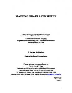
low exquisite temporal resolution of neural processing (typically on a 10 to 100 ms time scale), but suffer from relatively poor spatial resolution (between one and several centimet res). Functional MRI (fMRI) methods are in the second category, along with positron emission tomography (PET). They can be used to detect changes in regional blood perfusion, blood volume (e.g., using injected magnetic resonance contrast agents), or blood oxygenation that accompany neuronal activity. PET demands injection of radioactive tracers and highly specialised equipment, limiting the number of scans that made with any single subject and the availability of the technique. Blood Oxygenation Level Dependent (BOLD) fMRI has become the overwhelmingly most important of these methods because it has a similar spatial resolution (on the order of a few mm) to PET and a better temporal resolution (seconds, limited by the haemodynamic response itself). Moreover, the technique can be implemented on any modern high field MRI system, making it widely available at a reasonable cost. BOLD fMRI relies on detecting consequences of the locally increased blood flow (and blood volume) associated with increased neuronal activity (Fig. 1) [76– 78]. Because the increase in local blood flow is in excess of the increased metabolic demands, there is reduced oxygen extraction and a higher ratio of oxy- to deoxyhaemoglobin (“redder blood”) in the region of neuronal activation. Greater blood oxygenation leads to greater signal on an appropriately (T2 ∗ ) sensitised MRI scan. The BOLD fMRI contrast arises from the different magnetic properties of oxygenated (oxyHb) and deoxygenated haemoglobin (deoxyHb): deoxyHb is paramagnetic (and distorts an applied magnetic field for imaging), whereas oxyHb is diamagnetic (and does not perturb the applied magnetic field significantly). Thus, arterioles carrying largely oxygenated arterial blood cause little distortion to the magnetic field, while capillaries and veins containing blood that is partially deoxygenated distort the magnetic field in their vicinity. The (microscopic) magnetic field inhomogeneities associated with distortions from deoxyHb leads to destructive interference of signal within the tissue voxel, which shortens the so-called T2 ∗ “relaxation time”. A shorter T2∗ leads to lower signal on a gradient echo MRI and a darker area on the image. Alternatively, the increased oxy-/deoxyhaemoglobin ratio associated with activation leads to a longer T2 ∗ , more signal and a relatively brighter area locally on the gradient echo image. Although the increased blood flow with neuronal activity is associated with increased energy utilisation,
247
it is not the increased energy use itself that directly triggers the increase in blood flow Instead, increased blood flow appears to be a direct consequence of neurotransmitter action [3]. The increased blood flow thus reflects local neuronal signalling. Electrophysiologically, increases in the BOLD signal are correlated most strongly with the local field potential (reflecting input to neurons and local processing) rather than the neuronal firing rate [59]. The volume over which blood flow increases associated with neuronal activity are found is determined by the perfusion territory of local arterioles. In an fMRI experiment a large (typically hundreds) series of images are acquired rapidly (using a fast imaging technique, such as echo planar imaging [EPI]) while the subject performs a task in which brain activity alternates between two or more well-defined states (e.g., rest vs. movement of the hand) [66]. By correlating the signal time course in each large volume element (voxel) with the known time course of the task it is possible to identify those voxels in the brain that show task-associated signal changes corresponding to “activation”. A consequence of this approach to analysis is that, while methods such as PET provide an absolute measure of tissue metabolism, BOLD fMRI can at present be used only for determining relative signal intensity changes associated with different cognitive states during a single imaging session. The magnitude of the signal intensity changes being measured with fMRI also is small- on the order of 0.5–5.0%. As this is much smaller than the intrinsic local tissue contrast (e.g., between grey matter and CSF), one of the most significant practical confounds in fMRI is the extreme sensitivity to motion, which can mix signal from neighboring voxels. If movement occurs synchronously with the task, movement-associated signal changes will be found along with those associated specifically with brain functional changes and thus contribute to artifactual “activation”. In contrast, movement asynchronous with a task may be expected to reduce apparent signal.
4. Motor learning and a cognitive neuroscience model for neurorehabilitation 4.1. Motor control There is now a substantial literature describing brain functional changes associated with movement of both the upper and lower limbs (e.g., [89,104]). From this, some general principles of motor control can be extracted (see e.g., [20,102]). Although the specific di-
248
P.M. Matthews et al. / fMRI and neurorehabilitation
Rest
Active capillary bed
arteriole
capillary bed venules
arteriole
venule
= oxyHb = deoxyHb
Resting flow Resting level [deoxyHb] Increased T2* Resting signal
Increased flow Decreased [deoxyHb] Increased T2* Increased signal
Fig. 1. Cartoon illustrating the physiological changes allowing of Blood Oxygenation Dependent Imaging for fMRI. In the transition from a rest to an active state, there is a local increase in blood flow, which leads to a decreased ratio of oxygenated (oxyHB) to deoxygenated (deoxyHb) haemoglobin. An increasing ratio of oxyHb/deoxyHb leads to increased MRI signal.
visions of function are somewhat arbitrary (and potentially misleading, as a functional network is being described, rather than a true hierarchy), four levels can be defined: 1. Activity at the highest level mediates attention to and conception of the movement and involves prefrontal systems. These are engaged early (indeed, as demonstrated by the presence of the bereitshaftspotential, significantly before) [36]. In considering alternative approaches to neurorehabilitation, it is interesting to note that parts of this “preparation for action” system are used in similar ways in tasks involving motor imagery and even in observation of action [45,62]]. 2. A second level involves the sensorimotor transformations necessary to develop the framework for a more detailed action plan. This makes use of widely distributed mechanisms involving both the primary perceptual and association cortex which contribute to development of a detailed plan for subsequent movement [19,26,70]. 3. A third level of activity is responsible particularly for execution of the action plan. For explicit motor tasks this involves supplementary motor areas, premotor cortex and the primary motor cortex. More implicit (or automated) tasks involve circuits that are distinguished by a greater reliance on subcortical networks, e.g, with increased activity in the cerebellum and basal ganglia, in addition to primary motor cortex. Subcortical grey matter (basal ganglia) has an important role in selection of movements [46]. The potential direct contributions of secondary pathways
from premotor and supplementary motor areas may become more important under conditions of pathology (although, even then their significance is debated- see discussion below)(see e.g., [13, 55,91,93,121]). Limb movements normally are associated with highly hemispherically lateralised activity, driven primarily from the contralateral hemisphere [49]. For truncal and other midline musculature the motor control network intrinsically is more bihemispheric [64]. 4. An additional important level of action control relevant to neurorehabilitation is that of feedback to correct errors or to reinforce correctly performed actions. The cerebellum has an important role in the on-line comparison of a “forward model” of movement with afferent information coming from proprioceptive and somatosensory systems during movement [68]. Discrepancies between the forward model for movement and the perceived movement itself then are used as the basis for modulation of movement execution. Exciting recent work has begun to explore more general mechanisms by which feedback relevant to the action plan is processed. Ramnani et al used a simple visuomotor association learning task and distinguished between activity time-locked to positive, negative and neutral (control) feedback [88]. Relative to the neural feedback, meaningful (positive and negative) feedback activated areas including the ventral striatum/pallidum and the amygdala. Negative feedback was associated specifically with activation of additional regions, including the orbital and anterior prefrontal cortex and
P.M. Matthews et al. / fMRI and neurorehabilitation
the parietal cortex. Positive feedback activated distinct areas including the anterior temporal cortex and cerebellum. Two conclusions relevant to the current discussion can be drawn from this. First, specific mechanisms are responsible for responding to the feedback necessary to modify behaviour. Second, there are common pathways for reinforcement of motor responses and for reinforcement of other beh aviours that involve so-called “reward pathways” of the ventral prefrontal area (e.g., orbital frontal cortex, amygdala and ventral striatum). 4.2. Motor learning Motor learning can occur over either the short or long-term. Different patterns of behavioural and brain functional activity changes are associated with the two time frames for learning. With short-term learning, there is a rapid improvement in performance associated with a decrease in the specific attentional demands of the task [28]. This is associated with decreasing involvement of prefrontal cortical areas and a progressive “focusing” of activity in primary motor cortex (see e.g., [16,34,115]). With increasing practice, performance changes are slower, but task performance becomes more automatic. Accompanying this is an increased motor representation in the primary motor cortex [47]. Acquisition of motor skills in general are associated with altered primary motor cortex representations, e.g., qualitatively similar phenomena are found after learning an isometric tracking task (Floyer et al., unpublished) or in skilled badminton players [83]. Analogous changes have been describ ed in the nonhuman primate primary motor cortex based on invasive cortical mapping. As monkeys learn to draw peanuts from smaller wells there is reorganisation of motor representations for limb movement in the primary motor cortex [73]. However, it is important to note that both human and primate studies have emphasised that the patterns of brain activity associated with learning are task specific [28,47,50]. For example, after one a specific sequence of finger movements is overlearned, significant differences in motor cortex activity are associated with execution of a similar (e.g., same fingers and same number of finger movements), but distinct (e.g., altered order of finger movements) sequence [47]. This suggests that the substrate for general skill learning (e.g., being able to play the piano, as opposed to being able to play any specific piece on the piano) is not simply a change in motor representation in the primary motor cortex. One may speculate that this form
249
of learning- which possibly is central to understanding neurorehabilitation- is mediated (at least in part) by fronto-parietal circuits. Activity in the dorsolateral prefrontal cortex is associated with problem solving and shows performance-related increases in activity [21]. Parietal activity seems particularly involved in evaluating the potential significance of stimuli [19,70]. The posterior parietal cortex is concerned with aspects of visual and spatial stimulus selection [26]. Consistent with this, the preparation to move is associated with fronto-parietal activation. The brief discussion above has strongly emphasised neocortical mechanisms. This is partly a consequence of the observational biases in the experiments. It has been more difficult to study subcortical circuits. It is clear that substantial changes occur in these circuits with learning, as well. Subcortical circuits are involved particularly for learning implicit tasks [18,52] or as tasks become more automatic (when performance is less affected by distracters) [28]. 4.3. Is motor rehabilitation simply motor learning redux? Does neurorehabilitation involve simply a specialised form of motor learning? It is attractive to link the broad range of examples of functional reorganisation of the brain in response to altered external stimuli or internal state by common mechanisms. Studies in animal models have suggested qualitatively similar functional changes in sensorimotor cortical areas to those during learning with peripheral nerve injury [107], amputation [27], or repeated direct stimulation [72]. Peripheral deafferentation in monkeys leads to a reduction of the area of cortical responsiveness for the deafferented limb [33,67] that mirrors the enlargements of cortical responsiveness with motorskill learning [50] or repetitive intra-cortical microstimulation [72] Changes in functional organisation of the cortex related to altered afferent or efferent activity must share at least some common mechanisms, whatever the specific cause of the altered activity. However, while motor learning provides a useful a framework of understanding neurorehabilitation, it is unlikely that the problem will turn out to be so simple. As noted above, a number of special mechanisms may alter the potential for functional reorganisation in the context of brain injury [126]. Brain injury triggers complex changes in cortical excitability [127]: local to the infarct excitability is decreased [71], while more distantly excitability is increased. The latter occurs in
250
P.M. Matthews et al. / fMRI and neurorehabilitation
association with down-regulation of GABA receptor levels [98]. Studies in the healthy brain suggest that functional reorganisation in motor cortex is strongly affected by GABAergic activity [6]. Deafferentation induces increased excitability in the contralateral hemisphere [124]. Even acute loss of sensory afferents can reduce local GABA concentrations, potentially helping to drive short-term functional reorganisation [56]. These factors may even contribute to the structural and functional changes t hat occur in the brain even many months following the injury, when primary repair may be expected to have slowed or stopped [113]. A cognitive neuroscience approach suggests another distinction between simple motor learning and neurorehabilitation, as well. Neurorehabilitation is actually a special case of re-learning to perform a previously learned task in a different way- either compensatory strategies must be developed or alternative pathways adaptively recruited. In general, this must involve some systems distinct from those engaged for na¨ıve motor learning. For example, in the Ramnani et al. study exploring feedback-related responses with visuomotor learning, when the contingencies between stimuli and actions were changed and in a re-learning trial, meaningful feedback activated the supramarginal gyrus (leftlateralised for negative feedback, right-lateralised for positive feedback) to a greater degree than with learning [88]. Thus, while studies of motor learning in the healthy brain can provide a guide to understanding neurorehabilitation, rules specific to the pathological brain still need to be explored.
5. Direct observations on patients after brain injury: common brain mechanisms contribute to adaptive changes after corticospinal tract injury 5.1. Cortical plasticity Adaptive functional reorganisation is an example of the more general phenonmenon of “plasticity”, the ability of brain networks to reorganise functionally, either in response to such external factors such as altered stimulus-response associations, or internal changes arising from injury. These changes can be defined by fMRI (or related methods) as an expansion or recruitment of additional cortical areas, or as local shifts in centres of activity associated with specific behaviours. Direct electrophysiological studies (still perhaps the “gold standard”) have shown empirically that
there must be a dual focus in defining post-injury plasticity: both local and distant changes can be found. For example, reorganisation after injury to the corticospinal tract involves both a local expansion (or shift) in motor representations [74,75] and new involvement of motor cortex in the undamaged hemisphere [31,124]. Both of these changes suggest that post-injury reorganisation depends on modifications to existing pathways rather than development of entirely new circuits, e.g., recruitment of existing motor pathways in the hemisphere ipsilateral to the hand moved [11] that may be used to only a minor extent by the healthy brain [49] or only under different conditions, e.g., with more complex tasks [89]. 5.2. Evidence for cortical plasticity with white matter injury Multiple sclerosis (MS) is characterized by multifocal lesions of the white matter in the brain and spinal cord that are disseminated in time. We have chosen MS as a model disease for the study of brain plasticity as most patients show good recovery from symptomatic expression of new lesions (relapses) in the earlier stages of the illness, despite evidence for axonal injury with each attack, both locally and remote from lesions in the normal appearing white matter [22,32]. Some aspects of axonal injury may be partially reversible [15], which must contribute to clinical recovery. However, irreversible axonal injury is substantial [22,116], suggesting that other factors, such as adaptive functional reorganisation, also may operate in the disease [9]. What limits this adaptive plasticity? The fact that the brain cannot maintain normal behaviours with even isolated focal lesions, if they are large enough, suggests an intrinsic limitation based on local specialisations for processing and connectivity. In a diffuse disease such as MS, other factors also play a role. Principle among these factors must be diffuse cortical injury and neurodegeneration [8,85,128]. Several studies have showed impaired cortical function in MS. Positron emission tomography (PET) measures of resting cerebral blood flow and metabolism have shown decreases of cortical metabolism associated with clinical progression in MS [4]. Sun et al [111] reported a correlation between decreased oxidative metabolism and increasing disability. Yousry et al first provided fMRI-based evidence suggesting that MS patients with motor weakness show a potentially adaptive increased activation of ipsilateral and accessory motor areas [129]. This recalled earlier
P.M. Matthews et al. / fMRI and neurorehabilitation
findings with subcortical ischaemic stroke [7]. Later studies have related such functional changes directly with measures of injury or disability (see e.g., [24,54, 79,94–96,100]). To interpret this type of change in brain functional organisation as evidence for adaptive plasticity, two conditions should be able to be fulfilled: 1. The extent of the functional reorganisation must be related to the burden of disease and thus evolve dynamically through the course of the disease (although not necessarily in a monotonic fashion); 2. Evidence for functional reorganisation can be found even without disease-associated behavioural impairment, consistent with the notion that adaptive plasticity limits clinical expression of the disease. Further strong evidence is derived from demonstration that any abnormal patterns of functional activation in patients are independent of volitional activation. This finding helps to distinguish between changes that are “adaptive” from those that are “compensatory”. Imaging techniques provide a number of measures for quantifying disease burden that can be used in tests for relationships with functional changes. Subcortical injury can be assessed using MRI and correlated with the extents of activation as measured by fMRI. Using lesion load as assessed from T2-weighted MRI (though not a highly predictive the extent of disability), for example [103]. The extent of axonal injury can be assessed more specifically with magnetic resonance spectroscopy (MRS) by measuring the relative concentration of N-acetylaspartate (NAA), a compound found only in neurons (including dendrites and axons) in the mature brain [69]. NAA concentration is reduced in both acute and chronic lesions of MS, as well as in the normal appearing white matter in MS patients [2]. Functional reorganisation defined by fMRI in patients with MS is related to the burden of disease. Using a simple hand movement task, Lee et al found brain T2 lesion load was correlated with a decreasing motor cortical activation lateralisation index (LI, a measure of relative activation in contralateral to ipsilateral hemisphere so that a lower value corresponds with a more bilateral activation) [55]. There was a posterior shift in the geometric centre of activation of the sensorimotor area contralateral to the hand moved in patients. Reddy et al. demonstrated a similar correlation between NAA decreases in the corticospinal tract and changes in patterns of functional activity [93]. Pantano et al showed a similar correlation between changes in the pattern of
251
brain activation and lesion load pathology in the corticospinal tract of MS patients after a first episode of hemiparesis [79,101]. Aspects of this phenomenon can be generalised to other systems. Staffen et al. [110] studied MS patients and healthy controls performing a sustained attention task. Although performance was equivalent between the two groups, MS patients showed activation in right frontal and left parietal cortex not found in healthy controls, suggesting additional, adaptive activation. Werring et al examined MS patients who had recovered from optic neuritis using visual stimulation with fMRI [125]. Although the patients showed decreased activation of the visual cortex receiving projections from the affected optic nerve, there was additional extensive activation in the claustrum, the lateral temporal and posterior parietal cortices and the thalamus. All of these brain regions are involved in higher order visual processing or object recognition. The results suggest that parallel pathways in these areas may have been recruited adaptively or to compensate for impairment of the primary visual pathway. The extent to which this extra-striate circuit was recruited was related to the severity of pathology. Functional reorganisation observed by fMRI is dynamic, changing with the pathological evolution of lesions. In a report by Reddy et al. [93], a patient with a new large demyelinating lesion in the corticospinal tract was followed after the onset of hemiparesis with serial MRI, MRS and fMRI studies, as well as disability scoring. Clinical recovery preceded normalization of NAA concentrations (an index of lesion repair) and was associated with relative increases in premotor area and supplementary motor area activation in the hemisphere ipsilateral to the hand moved (an index of functional adaptation). The general correlation between changes in fMRI responses and MRS measures suggests a progressive normalisation of patterns of functional organisation in motor cortex in response to the repair of axonal injury associated with the lesion. A key point is that this behaviour is (at least to a large degree) independent of the specific cause of neuroaxonal damage. The pattern of change discussed above for MS is similar to that found with serial fMRI studies of motor activation in patients with stroke, which have shown a similar initial bilateral activation with lesions of the contralateral motor cortex that reverts toward a more lateralised activation pattern with progression of recovery [23,63,120]. Altered patterns of recruitment may help to maintain functions (e.g., via adaptive recruitment of additional descending motor pathways) despite persistent injury to the corticospinal tract.
252
P.M. Matthews et al. / fMRI and neurorehabilitation
The second criterion for adaptive functional reorganisation- that it can be found even without behavioural changes- also has been demonstrated. Changes in cortical activation occur even very early in the course of MS and in patients without symptoms in the affected functional system. MS patients without motor or sensory impairment of the upper limbs were investigated with fMRI and MRS using a hand-tapping paradigm [93]. LI was abnormally low in the patients and decreased progressively with decreases in relative brain NAA concentrations. Since these results were obtained from patients who had normal hand function, the potential confound that arises from performance differences was absent. In fact, even patients with optic neuritis as their only clinical manifestation of MS show functional changes in motor cortex compared with healthy controls [79]. Again, emphasising that the changes are not determined solely by the pathology, qualitatively similar changes were noted in the well recovered patients with capsular infarcts studied by PET a decade ago [121]. The third criterion for adaptive brain plasticity- that changes are independent of volitional activity- also can be confirmed in some patients. Because fMRI assesses predominantly input and local processing of information (rather than neuronal output spiking [59] and there is strong afferent sensory input into motor cortical regions, passive movement of a limb can define a network of brain activation also associated with the same movement performed actively [92,122]. A passive movement task therefore can be used to test for abnormal patterns of brain activity in patients relative to healthy controls free from potential confounds of differences in movement preparation and planning [90,92]. Reddy et al. tested a group of patients with MS using both an active and a passive hand movement task, verifying with surface EMG that the passive task was not associated with motor unit activation. A strong, quantitative correlation was found between indices of relative activation with the acti ve and passive tasks (Fig. 2). The recruitment particularly of ipsilateral motor cortex during movement with this pathology therefore is not dependent on factors related to volition. The integration of specific functional systems (e.g., cognitive, perceptual, action) in distributed networks controlling task performance suggests that adaptive functional changes need not be confined to the primary effector system for the task. Abnormal activation in MS patients performing a simple motor task may be found not only in primary motor control regions, but
Fig. 2. Relative activation in the contra- and ipsilateral motor cortex is highly correlated (r = 0.93, p = 0.001) between passive and active hand movement tasks and in patients with multiple sclerosis there is increased ipsilateral motor cortex activation relative to healthy controls. The graph shows the relationship between the relative lateralisation of activity (LI [see text], which varies between +1 and −1 as activation moves from being fully contralateral to fully ipsilateral) for the two types of tasks. This provides evidence that the functional changes found in the motor cortex of patients are independent of volition [97].
also in sensory and association cortex including the insula and temporal, parietal, and occipital areas [25,99]. However, some of the additional activation in patients may be a consequence of changes in patterns of use (associated with disability) that accompany injury in patients with significant impairments rather than adaptive functional responses to injury itself. Altered patterns of somatosensory feedback and use certainly lead to changes in the functional organisation of the brain in other contexts, for example [41,87]. A recent fMRI study directly tested whether the effects of injury and disability could b e distinguished [97] (Fig. 3). Patients with greater brain injury but no functional impairment showed larger ipsilateral premotor and bilateral supplementary motor area activations. A separate contrast testing effects of greater disability with a similar extent of injury showed greater activation in bilateral primary and secondary somatosensory cortex. MS has been a useful model disease with which to study functional reorganisation because of the slow progression of the pathology and the availability of wellvalidated MRI indices of pathological progression and clinical indices of disability. There is evidence that the fundamental principles are generalisable. As noted above, functional cortical changes have been demonstrated in patients after strokes (e.g., [13,121]) and also with the growth of tumours [108], for example. Reddy et al tested the notion of generalisability more directly using a vascular disease “analogue” of MS in
P.M. Matthews et al. / fMRI and neurorehabilitation
Fig. 3. Patterns of brain activation with hand movement may show distinct patterns of change with differences in the extent of injury (A) and disability (B)(right hemisphere to the viewer’s left). Three groups of multiple sclerosis patients were defined, differing in relative axonal injury in the corticospinal tract and disability. Increasing corticospinal tract injury is associated with relative changes in the premotor cortex and supplementary motor area (A, shown at the level of [Talairach coordinate] z = 66 for the left image and z = 64 for the right image). Increasing disability is associated with relative changes in the secondary (left image, z = 48) and primary somatosensory (right image, z = 64) cortex (B) [92].
a study of patients with diffuse ischaemic changes in white matter from cerebral autosomal dominant arteriopathy with subcortical infarcts and leukoencephalopathy (CADASIL) using both MRS and fMRI to study the relationship between axonal injury, disability, and cortical functional organisation [91]. The CADASIL patients chosen had a wide range of disability. Just as was found with MS patients, for a si mple motor task, increases in ipsilateral motor cortical activation correlated with severity of the disease in CADASIL patients.
6. Evidence that the ipsilateral premotor cortex (iPMC) shows specific functional adaptations in patients with corticospinal tract injury Are these cortical functional changes truly adaptive? Does recruitment of ipsilateral motor cortex, for example, clearly limit clinical manifestation of the disease? Serial functional imaging data has been interpreted as showing that increased ipsilateral activation is a marker of poor recovery, based on simple association between outcomes and activation patterns [63]. This data does not really assess the relative functional sig-
253
nificance of new patterns of activation, however. It is only a strong test of whether new patterns of activation can fully compensate for functional impairments (they cannot in general). The functional role of a specific cortical region can be tested directly by use of TMS to transiently disrupt its activity [39,119]. Applying a TMS pulse over a region introduces localised electrical “noise” that will interfere with any task-relevant processing taking place. If TMS of a region causes a disruption of behaviour (e.g., TMS of primary motor cortex slowing reaction times [14]), then it can be inferred that the stimulated region plays a functionally significant role in task performance. TMS is a powerful tool for introducing reversible “virtual lesions” in human subjects. TMS therefore provides an approach with which one can test the functional significance of potentially adaptive functional changes in specific cortical areas [44, 123]. For example, does increased fMRI activity in the primary motor cortex in the undamaged hemisphere reflect functional changes contributing to recovery [7, 13]? Werhahn et al. tested this by transiently disrupting this region with TMS and observing effects on hand movement [123]. While stimulation of the primary motor cortex in the undamaged hemisphere disrupted movements of the contralateral, unaffected hand, it had no effect on movement of the ipsilateral, paretic hand, suggesting that it does not play a crucial role in movement of the paretic limb after recovery. FMRI studies also show increased activity in the premotor cortex of the hemisphere ipsilateral to the hand moved with injury to the corticospinal tract, as described above. Johansen-Berg et al used TMS to compare the effects of disruption to the premotor and primary motor cortex of the undamaged hemisphere [44]. As found by Werhahn et al., effects of primary motor cortical stimulation did not differ between patients and healthy controls. However, stimulation of the premotor cortex had significant and distinct effects in the patient group. TMS over the dorsal premotor cortex in the hemisphere ipsilateral to the (paretic) hand slowed response times for a simple movement by the patients (and not for healthy controls). The magnitude of response slowing with TMS over the undamaged premotor cortex ipsilateral to the paretic hand correlated with the magnitude of the ipsilateral premotor fMRI activation during the same motor task. It also increased with the extent of the original brain inju ry. Is ipsilateral dorsal premotor cortex (iPMd) recruited in the injured brain simply using mechanisms also used by the healthy brain under other conditions? FMRI
254
P.M. Matthews et al. / fMRI and neurorehabilitation
studies show that the iPMd is involved in complex motor responses in healthy controls [89,106]. The time course for interference with activity using TMS over the iPMd in patients performing simple movements is similar to that in healthy controls performing complex movements [44]. Increased iPMd involvement in patients could simply reflect the increased difficulty of even simple motor tasks for this group of subjects. However, while ipsilateral PMd involvement is predominantly a phenomenon of the left hemisphere in healthy controls, it can be found in the right hemisphere in patients with corticospinal tract injury [44]. This suggests that there is a qualitative change in mechanisms as well as an enhanced recruitment. Animal studies support the hypothesis that injury to motor pathways is associated with structural changes in motor cortex ipsilateral to the paretic hand. Areas of the uninjured hemisphere corresponding to lesion in the injured hemisphere show structural and functional changes as a consequence of the injury, e.g., new dendritic growth occurs with the injury [74]. These are changes that are not expected to accompany simple motor learning and may be triggered by specific factors, e.g., release of growth factors [48]. The increased activity in the iPMd may lead to increased activation of uncrossed corticospinal projections [51]. The notion that ipsilateral projections might be “unmasked” is supported by direct stimulation studies with TMS. In patients after strokes, ipsilateral motor unit activity can be induced by iPMd stimulation with latencies that are shorter than with stimulation of the premotor cortex contralateral to the hand moved. This activity is associated with a better motor outcome [1]. However, it is difficult to understand how these changes alone could mediate recovery. Ipsilateral PMd projections are predominantly concerned with more proximal movements and show differences in distribution and functional properties relative to primary motor cortex [65]. This suggests that they work in conjunction with additional adaptive changes elsewhere in the CNS, e.g., in the brainstem or spinal cord.
7. Adaptation as a dynamic phenomenon: applications of functional imaging methods to the study of neurorehabilitation A basic tenet of brain functional plasticity in the context of injury is that multiple representations can be recruited to control a desired behaviour. As well as occurring spontaneously with brain injury, dynamic
Fig. 4. Changes in brain activity with hand movement occur in specific brain regions with improvements in performance following an intensive 2 week period of rehabilitation in patients after stroke. Panels (A) show areas (cerebellum, with z = − 28 [extrapolated from Talairach coordinates], and premotor cortex with z = 58) in which increased activity is associated with improved performance. Panel (B) shows a single area in the primary motor cortex (z = 64) ipsilateral to the hand moved that shows a decrease in activity (right= left hemisphere, left= right hemisphere) [43].
changes in motor representations also can be induced by rehabilitation training. Significant behavioural improvements can be achieved by patients even long after a stroke with an intervention such as constraint-induced therapy [112]. What are the functional brain changes that are related to a behavioural improvements after therapy? Johansen-Berg et al. used a rehabilitation protocol based on the principles of constraint-induced therapy to define neural correlates of this therapy-induced functional change [44]. Chronic stroke patients varied in the degree to which they benefited from a two-week therapy regime. The range of recovery outcomes enabled direct correlations between outcome and fMRI activation changes. FMRI scans taken before and after therapy identified specific regions of the motor system (premotor cortex, cerebellum and secondary somatosensory cortex) where increases in activation during movement of the paretic hand after therapy correlated with functional improvements (Fig. 4). The premotor region is complex and multi-functional. Studies in healthy subjects had suggested that the lateral dorsal premotor cortex codes responses to perceptual (visual and somatosensory) cues, while a more medial stream is involved in selection of movements and initiation [37]. Changes associated with rehabilitation in the stroke patients with injury to the corti-
P.M. Matthews et al. / fMRI and neurorehabilitation
cospinal tract appeared more localised to the latter (although this was not tested directly). The importance of the premotor cortex in recovery is supported by animal studies. Recovery of dexterity after unilateral primary motor cortex lesions in the macaque appears to involve activity of the premotor cortex of the damaged hemisphere, as (reversible) inhibition of the premotor cortex abolishes recovered movement abilities [58], for example. The specific regions of cerebellar cortex that were implicated in rehabilitation-mediated recovery can show increased resting metabolism after brain injury generally [117] and activation in these regions is associated with improved prognosis in patients after stroke [109]. Cerebellar activation may be critical for sensory processing [46], specifically in monitoring and optimising movements based on proprioceptive feedback. This role could link these changes to those in secondary somatosensory areas, recalling the specifically disability associated activations reported by Reddy et al. [97]. Involvement of the premotor cortex and the superior posterior cerebellar hemispheres in rehabilitation-mediated recovery also supports the hypothesis that processes underlying a successful rehabilitation response make use of mechanisms responsible for motor learning in the healthy brain. Therapy-related changes also are seen during movement of the unaffected limb: decreased activation in the contralesional primary motor cortex during movements of the unaffected hand after neurorehabilitation also correlates with improved functional outcome [44] (Fig. 4). A similar phenomenon was reported in a study that used TMS to map motor output regions before and after constraint-induced movement therapy [57]. The findings are consistent with the notion that reduced use of the unaffected limb during the therapy period results in a decreased representation of this limb in the primary motor cortex. Other recent examples provide consistent, additional illustrations of changes in the functional organisation of the brain with rehabilitation. Jang et al have shown that clinical recovery after intensive rehabilitation poststroke correlates increased contralateral and decreased ipsilateral sensorimotor activation during hand movement [40,43]. Papathanassiou et al have shown with TMS that in patients with hemiparesis and agraphia post-stroke, rehabilitation involving a writing task with the paretic hand resulted in greater ipsilateral motor excitability by TMS, suggesting a reduction in GABAergic inhibition could mediate the functional recruitment of this area [80].
255
8. A reversible lesion model for the study of motor system plasticity in the human brain The consequences of inactivation of specific regions on brain activity and for behaviour can be probed with animal models in a variety of ways, e.g., selective freezing, pharmacological inhibition or direct lesioning. The effects of focal brain injury and its consequences can be mimicked safely in the healthy human brain using slower (e.g., 1 Hz) repetitive trains of TMS to induce a brief, reversible disruption in specific regions of the motor system. Lee et al. recently used this approach to interfere with activity in the primary motor cortex while monitoring the metabolic response of the entire motor system using PET [53]. Although the TMS parameters used (30 minutes of 1 Hz stimulation) should have decreased excitability of the left motor cortex, no behavioural consequences of the disruption were observed with a simple motor task, suggesting that functionally intact regions of the motor network were able to compensate for the effects of disruption in the primary motor cortex. Immediately acquired PET scans showed that, following repetitive TMS interference to the left primary motor cortex, increased activation of the ipsilateral (right) premotor cortex was seen during right hand movement. This recalls the changes that occur in the premotor cortex of the undamaged hemisphere after corticospinal tract damage (see above). A novel element in this study was the demonstration of an approach to characterisation of functional changes using measures of functional coupling between regions of the brain. Covariance analyses of PET signals demonstrated increased coupling between activity in a deep region of sensorimotor cortex unlikely to have been stimulated directly and activity in non-primary motor cortices after TMS. The changes in functional coupling were argued to reflect adaptively increased connectivity. While there are still many uncertainties in the interpretation of this type of analysis, it suggests that new opportunities may be found by modelling dynamic behaviours in terms of interactions, as well as localised activities [30].
9. Consequences of studying brain recovery and rehabilitation using the approaches from cognitive neuroscience: the importance of “context” Functional imaging has allowed an increasingly informative study of motor control and related cognition
256
P.M. Matthews et al. / fMRI and neurorehabilitation
in the healthy brain. This has provided a cognitive neuroscience framework for understanding neurorehabilitation. There are several potential benefits to using this approach to the analysis of brain recovery and rehabilitation. These can be summarised as emphasising the importance of “context”. One benefit is a more precise appreciation for the role of the context of learning in neurorehabilitation. Ramnani et al. highlighted that there were distinct differences in the way in which the brain processes meaningful feedback regarding behaviour for initial learning and relearning [88]. Thus, while studies of motor learning offer useful insights into potential mechanisms of neurorehabilitation, it also must be appreciated that neurorehabilitation- a recovery of movement- is more accurately a specialised form of relearning. The importance of context has been emphasised in a different way by a recent study showing that the way in which the motor cortex is activated also is important in determining outcome: Lotze and Cohen showed that motor learning depends on more than just movement even with control for alertness and kinematic aspects of training [60]. Although both active and passive movements led to activation of the same general area within the primary motor cortex, active training led to more prominent increases in the extent of activation in the primary motor cortex over time, as well as changes in TMS recruitment curves reflecting the relative degree of intracortical facilitation. The pathological context of the injury also must be important. It long has been recognised that the heterogeneity of localisation, size and severity of strokes, for example, is associated with heterogeneity in outcome. With appreciation of the interacting cognitive systems for perception, movement planning and sensorimotor transformation and execution of the movement and its control by feedback, new approaches to characterisation of motor impairment in terms of deficits in specific functional systems becomes possible. This promises a potential for better targeted therapies- moving away from the notion that one therapeutic approach will be optimally suited for all patients.
ses. In the context of motor recovery, current understanding of movement control offers guidance for new ways of characterising deficits in terms of neurobiologically distinct elements involved in motor control. This strategy promises direct applications to clinical trials. It can be used to establish markers of response that may allow more sensitive monitoring, leading to smaller and shorter trials. It offers the opportunity to explore the potential for tailoring rehabilitation to specific deficits or patient groups, potentially improving the benefits. Finally, in the longer term, it may help in better establishing prognosis, if only to help identify those patients who could benefit most from a given therapy. With an understanding of the brain activity of the systems level also can come an opportunity for making inferences of the neurotransmitter changes underlying these activities. This can be used to develop strategies by which pharmacological neuromodulators might be used to enhance recovery. Measures of outcome can be more specific. One of the challenges in such work is to most appropriately integrate the range of tools that are available. While it is clear that brain imaging modalities have a considerable potential independently, it is even more exciting to consider the way in which they can be used together. For example, fMRI provides good correlates of brain activity changes, but cannot be used to test the degree to which a given area of the brain is necessary for performing a task. In addition, it suffers from the need for the task to be performed in an entirely constrained or artificial environment, making it difficult in applications for long-term studies. TMS, which can be used to transiently interfere with activity in a region tests directly the importance of its functional significance. In the future, use of optical imaging techniques may allow brain activity to be studied over time in an even more dynamic (and even ambulatory) fashion, although with a lower spatial resolution.
Acknowledgements 10. Conclusions and practical implications FMRI and related methods provide powerful new ways of exploring systems level responses of the brain after injury and with neurorehabilitation. Cognitive neuroscience provides a useful framework for interpreting these observations and for generating new hypothe-
PMM is grateful to the MRC (UK) for personal support and for core support of the FMRIB centre. We thank the Wellcome Trust for award of a postdoctoral fellowship to HJ-B and the Rhodes Trust for support of HR during work in Oxford.
P.M. Matthews et al. / fMRI and neurorehabilitation
References [18] [1]
[2]
[3] [4]
[5]
[6]
[7]
[8]
[9]
[10] [11] [12]
[13]
[14]
[15]
[16]
[17]
G. Alagona, V. Delvaux, P. Gerard, V. De, Pasqua, G. Pennisi, P.J. Delwaide, F. Nicoletti and Maertens, de Noordhout. Ipsilateral motor responses to focal transcranial magnetic stimulation in healthy subjects and acute-stroke patients, Stroke 32 (2001), 1304–1309. D.L. Arnold, J.S. Wolinsky, P.M. Matthews and A. Falini, The use of magnetic resonance spectroscopy in the evaluation of the natural history of multiple sclerosis, J Neurol Neurosurg Psychiatry 64 (Suppl 1) (1998), S94–101. D. Attwell and C. Iadecola, The neural basis of functional brain imaging signals, Trends Neurosci 25 (2002), 621–625. M. Blinkenberg, C.V. Jensen, S. Holm, O.B. Paulson and P.S. Sorensen, A longitudinal study of cerebral glucose metabolism, MRI, and disability in patients with MS, Neurology 53 (1999), 149–153. S. Boniface and U. Ziemann, Plasticity in the human nervous system: investigations with transcranial magnetic stimulation, 2003. C.M. Butefisch, B.C. Davis, S.P. Wise, L. Sawaki, L. Kopylev, J. Classen and L.G. Cohen, Mechanisms of usedependent plasticity in the human motor cortex. Proc. Natl. Acad. Sci. USA 97 (2000), 3661–3665. F. Chollet, V. DiPiero, R.J. Wise, D.J. Brooks, R.J. Dolan and R.S. Frackowiak, The functional anatomy of motor recovery after stroke in humans: a study with positron emission tomography, Ann Neurol 29 (1991) 63–71. A. Cifelli, M. Arridge, P. Jezzard, M.M. Esiri, J. Palace and P.M. Matthews, Thalamic neurodegeneration in multiple sclerosis, Ann Neurol 52 (2002), 650–653. A. Cifelli and P.M. Matthews, Cerebral plasticity in multiple sclerosis: insights from fMRI, Mult. Scler. 8 (2002), 193– 199. M.C. Cirstea and M.F. Levin, Compensatory strategies for reaching in stroke, Brain 123(Pt 5) (2000), 940–953. J. Cole and P. Glees, Ipsilateral impairment following area 4 lesions in monkeys, J Physiol 117 (1952), 54P. M.C. Comelli, D. Guidolin, M.S. Seren, R. Zanoni, R. Canella, R. Rubini and H. Manev, Time course, localization and pharmacological modulation of immediate early inducible genes, brain-derived neurotrophic factor and trkB messenger RNAs in the rat brain following photochemical stroke, Neuroscience 55 (1993), 473–490. S.C. Cramer, G. Nelles, R.R. Benson, J.D. Kaplan, R.A. Parker, K.K. Kwong, D.N. Kennedy, S.P. Finklestein and B.R. Rosen, A functional MRI study of subjects recovered from hemiparetic stroke, Stroke 28 (1997), 2518–2527. B.L. Day, J.C. Rothwell, P.D. Thompson, Maertens, de Noordhout, K. Nakashima, K. Shannon and C.D. Marsden, Delay in the execution of voluntary movement by electrical or magnetic brain stimulation in intact man. Evidence for the storage of motor programs in the brain, Brain 112(Pt 3) (1989), 649–663. N. De Stefano, P.M. Matthews and D.L. Arnold, Reversible decreases in N-acetylaspartate after acute brain injury, Magn Reson Med 34 (1995), 721–727. P. De Weerd, K. Reinke, L. Ryan, T. McIsaac, P. Perschler, D. Schnyer, T. Trouard and A. Gmitro, Cortical mechanisms for acquisition and performance of bimanual motor sequences, Neuroimage 19 (2003), 1405–1416. J.P. Donoghue, S. Suner and J.N. Sanes, Dynamic organization of primary motor cortex output to target muscles in adult
[19] [20] [21]
[22]
[23]
[24]
[25]
[26]
[27]
[28]
[29]
[30] [31]
[32]
[33]
[34]
[35]
257
rats. II. Rapid reorganization following motor nerve lesions, Exp. Brain Res. 79 (1990), 492–503. J. Doyon, A.W. Song, A. Karni, F. Lalonde, M.M. Adams, and L.G. Ungerleider, Experience-dependent changes in cerebellar contributions to motor sequence learning, Proc. Natl. Acad. Sci. USA 99 (22-1-2002), 1017–1022. J. Driver and J.B. Mattingley, Parietal neglect and visual awareness, Nat. Neurosci. 1 (1998), 17–22. R.P. Dum and P.L. Strick, Motor areas in the frontal lobe of the primate, Physiol Behav 77 (2002), 677–682. J. Duncan, R.J. Seitz, J. Kolodny, D. Bor, H. Herzog, A. Ahmed, F.N. Newell and H. Emslie, A neural basis for general intelligence, Science 289 (2000), 457–460. N. Evangelou, M.M. Esiri, S. Smith, J. Palace and P.M. Matthews, Quantitative pathological evidence for axonal loss in normal appearing white matter in multiple sclerosis, Ann Neurol 47 (2000), 391–395. A. Feydy, R. Carlier, A. Roby-Brami, B. Bussel, F. Cazalis, L. Pierot, Y. Burnod and M.A. Maier, Longitudinal study of motor recovery after stroke: recruitment and focusing of brain activation, Stroke 33 (2002), 1610–1617. M. Filippi, M. Bozzali, M. Rovaris, O. Gonen, C. Kesavadas, A. Ghezzi, V. Martinelli, R.I. Grossman, G. Scotti, G. Comi and A. Falini, Evidence for widespread axonal damage at the earliest clinical stage of multiple sclerosis, Brain 126 (2003), 433–437. M. Filippi, M.A. Rocca, B. Colombo, A. Falini, M. Codella, G. Scotti and G. Comi, Functional magnetic resonance imaging correlates of fatigue in multiple sclerosis, Neuroimage 15 (2002), 559–567. G.R. Fink, P.W. Halligan, J.C. Marshall, C.D. Frith, R.S. Frackowiak and R.J. Dolan, Where in the brain does visual attention select the forest and the trees? Nature 382 (1996), 626–628. S.L. Florence, H.B. Taub and J.H. Kaas, Large-scale sprouting of cortical connections after peripheral injury in adult macaque monkeys, Science 282 (1998), 1117–1121. A. Floyer-Lea and P.M. Matthews, Transition of activity from prefrontal cortex to pre-SMA during early motor learning, Neuroimage 19(Part 2) (2003), 156. K.M. Friel and R.J. Nudo, Recovery of motor function after focal cortical injury in primates: compensatory movement patterns used during rehabilitative training, Somatosens. Mot. Res. 15 (1998), 173–189. K.J. Friston, L. Harrison and W. Penny, Dynamic causal modelling, Neuroimage 19 (2003), 1273–1302. S.B. Frost, S. Barbay, K.M. Friel, E.J. Plautz and R.J. Nudo, Reorganization of remote cortical regions after ischemic brain injury: a potential substrate for stroke recovery, J Neurophysiol 89 (2003), 3205–3214. L. Fu, P.M. Matthews, N. De Stefano, K.J. Worsley, S. Narayanan, G.S. Francis, J.P. Antel, C. Wolfson and D.L. Arnold, Imaging axonal damage of normal-appearing white matter in multiple sclerosis. Brain 121 (1998), 103–113. P.E. Garraghty and J.H. Kaas, Large-scale functional reorganization in adult monkey cortex after peripheral nerve injury, Proc. Natl. Acad. Sci. USA 88 (1991), 6976–6980. S.T. Grafton, J.C. Mazziotta, S. Presty, K.J. Friston, R.S. Frackowiak and M.E. Phelps, Functional anatomy of human procedural learning determined with regional cerebral blood flow and PET, J Neurosci 12 (1992), 2542–2548. C. Halfpenny, T. Benn and N. Scolding, Cell transplantation, myelin repair, and multiple sclerosis, Lancet Neurol 1 (2002), 31–40.
258 [36] [37]
[38]
[39]
[40]
[41]
[42] [43]
[44]
[45]
[46]
[47]
[48]
[49]
[50]
[51]
[52]
[53]
P.M. Matthews et al. / fMRI and neurorehabilitation M. Hallett, Movement-related cortical potentials. Electromyogr, Clin. Neurophysiol. 34 (1994), 5–13. E. Hoshi and J. Tanji, Integration of target and body-part information in the premotor cortex when planning action, Nature 408 (2000), 466–470. K.M. Jacobs and J.P. Donoghue, Reshaping the cortical motor map by unmasking latent intracortical connections, Science 251 (1991), 944–947. M. Jahanshahi and J. Rothwell, Transcranial magnetic stimulation studies of cognition: an emerging field, Exp Brain Res 131 (2000), 1–9. S.H. Jang, Y.H. Kim, S.H. Cho, J.H. Lee, J.W. Park and Y.H. Kwon, Cortical reorganization induced by task-oriented training in chronic hemiplegic stroke patients, Neuroreport 14(1) (2003), 137–141. W.M. Jenkins, M.M. Merzenich, M.T. Ochs, T. Allard and E. Guic-Robles, Functional reorganization of primary somatosensory cortex in adult owl monkeys after behaviorally controlled tactile stimulation, J Neurophysiol 63 (1990), 82– 104. P. Jezzard, P.M. Matthews and S.M. Smith, Functional MRI: an introduction to methods 2001. H. Johansen-Berg, H. Dawes, C. Guy, S.M. Smith, D.T. Wade and P.M. Matthews, Correlation between motor improvements and altered fMRI activity after rehabilitative therapy, Brain 125(Pt. 12) (Dec. 2002), 2731–2742. H. Johansen-Berg, M.F. Rushworth, M.D. Bogdanovic, U. Kischka, S. Wimalaratna and P.M. Matthews, The role of ipsilateral premotor cortex in hand movement after stroke, Proc. Natl. Acad. Sci. USA 99 (2002), 14518–14523. S.H. Johnson-Frey, F.R. Maloof, R. Newman-Norlund, C. Farrer, S. Inati and S.T. Grafton, Actions or hand-object interactions? Human inferior frontal cortex and action observation, Neuron 39 (2003), 1053–1058. M. Jueptner and C. Weiller, A review of differences between basal ganglia and cerebellar control of movements as revealed by functional imaging studies, Brain 121(Pt 8) (1998), 1437– 1449. A. Karni, G. Meyer, P. Jezzard, M.M. Adams, R. Turner and L.G. Ungerleider, Functional MRI evidence for adult motor cortex plasticity during motor skill learning, Nature 377 (1995), 155–158. T. Kawamata, W.D. Dietrich, T. Schallert, J.E. Gotts, R.R. Cocke, L.I. Benowitz and S.P. Finklestein, Intracisternal basic fibroblast growth factor enhances functional recovery and up-regulates the expression of a molecular marker of neuronal sprouting following focal cerebral infarction, Proc. Natl. Acad. Sci. USA 94 (1997), 8179–8184. S.G. Kim, J. Ashe, K. Hendrich, J.M. Ellermann, H. Merkle, K. Ugurbil and A.P. Georgopoulos, Functional magnetic resonance imaging of motor cortex: hemispheric asymmetry and handedness, Science 261 (1993), 615–617. J.A. Kleim, S. Barbay and R.J. Nudo, Functional reorganization of the rat motor cortex following motor skill learning, J Neurophysiol 80 (1998), 3321–3325. H.G. Kuypers and J. Brinkman, Precentral projections to different parts of the spinal intermediate zone in therhesus monkey, Brain Res 24 (1970), 29–48. R. Laforce Jr. and J. Doyon, Distinct contribution of the striatum and cerebellum to motor learning, Brain Cogn 45 (2001), 189–211. L. Lee, H.R. Siebner, J.B. Rowe, V. Rizzo, J.C. Rothwell, R.S. Frackowiak and K.J. Friston, Acute remapping within the motor system induced by low-frequency repetitive tran-
[54]
[55]
[56]
[57]
[58]
[59]
[60]
[61]
[62]
[63]
[64]
[65]
[66]
[67]
[68]
[69]
[70]
[71]
scranial magnetic stimulation, J Neurosci 23 (2003), 5308– 5318. M. Lee, H. Reddy, H. Johansen-Berg, S. Pendlebury, M. Jenkinson, S. Smith, J. Palace and P.M. Matthews, The motor cortex shows adaptive functional changes to brain injury from multiple sclerosis, Ann Neurol 47 (2000), 606–613. M. Lee, H. Reddy, H. Johansen-Berg, S. Pendlebury, M. Jenkinson, S. Smith, J. Palace and P.M. Matthews, The motor cortex shows adaptive functional changes to brain injury from multiple sclerosis, Ann Neurol 47 (2000), 606–613. L.M. Levy, U. Ziemann, R. Chen and L.G. Cohen, Rapid modulation of GABA in sensorimotor cortex induced by acute deafferentation, Ann Neurol 52 (2002), 755–761. J. Liepert, W.H. Miltner, H. Bauder, M. Sommer, C. Dettmers, E. Taub and C. Weiller, Motor cortex plasticity during constraint-induced movement therapy in stroke patients, Neurosci. Lett. 250 (1998), 5–8. Y. Liu and E.M. Rouiller, Mechanisms of recovery of dexterity following unilateral lesion of the sensorimotor cortex in adult monkeys, Exp. Brain Res. 128 (1999), 149–159. N.K. Logothetis, J. Pauls, M. Augath, T. Trinath and A. Oeltermann, Neurophysiological investigation of the basis of the fMRI signal, Nature 412 (2001), 150–157. M. Lotze, C. Braun, N. Birbaumer, S. Anders and L.G. Cohen, Motor learning elicited by voluntary drive, Brain 126 (2003), 866–872. I. Loubinoux, J. Pariente, O. Rascol, P. Celsis and F. Chollet, Selective serotonin reuptake inhibitor paroxetine modulates motor behavior through practice. A double-blind, placebocontrolled, multi-dose study in healthy subjects, Neuropsychologia 40 (2002), 1815–1821. F. Malouin, C.L. Richards, P.L. Jackson, F. Dumas and J. Doyon, Brain activations during motor imagery of locomotor-related tasks: a PET study, Hum. Brain Mapp 19 (2003), 47–62. R.S. Marshall, G.M. Perera, R.M. Lazar, J.W. Krakauer, R.C. Constantine and R.L. DeLaPaz, Evolution of cortical activation during recovery from corticospinal tract infarction, Stroke 31 (2000), 656–661. R.E. Martin, B.G. Goodyear, J.S. Gati and R.S. Menon, Cerebral cortical representation of automatic and volitional swallowing in humans, J Neurophysiol 85 (2001), 938–950. A.M. Martino and P.L. Strick, Corticospinal projections originate from the arcuate premotor area, Brain Res 404 (1987), 307–312. P.M. Matthews and P. Jezzard, Functional magnetic resonance imaging, J Neurol Neurosurg. Psychiatry, In press, 2003. M.M. Merzenich, J.H. Kaas, J. Wall, R.J. Nelson, M. Sur and D. Felleman, Topographic reorganization of somatosensory cortical areas 3b and 1 in adult monkeys following restricted deafferentation, Neuroscience 8 (1983), 33–55. R.C. Miall, G.Z. Reckess and H. Imamizu, The cerebellum coordinates eye and hand tracking movements, Nat. Neurosci 4 (2001), 638–644. J.R. Moffett, M.A. Namboodiri, C.B. Cangro and J.H. Neale, Immunohistochemical localization of N-acetylaspartate in rat brain, Neuroreport 2 (1991), 131–134. D.J. Mort, P. Malhotra, S.K. Mannan, C. Rorden, A. Pambakian, C. Kennard and M. Husain, The anatomy of visual neglect, Brain 126 (2003), 1986–1997. T. Neumann-Haefelin and O.W. Witte, Periinfarct and remote excitability changes after transient middle cerebral artery occlusion, J Cereb. Blood Flow Metab 20 (2000), 45–52.
P.M. Matthews et al. / fMRI and neurorehabilitation [72]
[73]
[74]
[75]
[76]
[77]
[78]
[79]
[80]
[81]
[82]
[83]
[84]
[85]
[86]
[87]
R.J. Nudo, W.M. Jenkins and M.M. Merzenich, Repetitive microstimulation alters the cortical representation of movements in adult rats, Somatosens. Mot. Res. 7 (1990), 463– 483. R.J. Nudo, G.W. Milliken, W.M. Jenkins and M.M. Merzenich, Use-dependent alterations of movement representations in primary motor cortex of adult squirrel monkeys, J Neurosci 16 (1996), 785–807. R.J. Nudo, E.J. Plautz, and S.B. Frost, Role of adaptive plasticity in recovery of function after damage to motor cortex, Muscle Nerve 24 (2001), 1000–1019. R.J. Nudo, B.M. Wise, F. SiFuentes and G.W. Milliken, Neural substrates for the effects of rehabilitative training on motor recovery after ischemic infarct, Science 272 (1996), 1791– 1794. S. Ogawa, T.M. Lee, A.S. Nayak and P. Glynn, Oxygenationsensitive contrast in magnetic resonance image of rodent brain at high magnetic fields, Magn Reson Med 14 (1990), 68–78. S. Ogawa, R.S. Menon, D.W. Tank, S.G. Kim, H. Merkle, J.M. Ellermann and K. Ugurbil, Functional brain mapping by blood oxygenation level-dependent contrast magnetic resonance imaging. A comparison of signal characteristics with a biophysical model, Biophys. J. 64 (1993), 803–812. S. Ogawa, D.W. Tank, R. Menon, J.M. Ellermann, S.G. Kim, H. Merkle and K. Ugurbil, Intrinsic signal changes accompanying sensory stimulation: functional brain mapping with magnetic resonance imaging, Proc. Natl. Acad. Sci. USA 89 (1992), 5951–5955. P. Pantano, G.D. Iannetti, F. Caramia, C. Mainero, S. Di Legge, L. Bozzao, C. Pozzilli and G.L. Lenzi, Cortical motor reorganization after a single clinical attack of multiple sclerosis, Brain 125 (2002), 1607–1615. I. Papathanassiou, S.R. Filipovic, R. Whurr and M. Jahanshahi, Plasticity of motor cortex excitability induced by rehabilitation therapy for writing, Neurology 61 (2003), 977–980. J. Pariente, I. Loubinoux, C. Carel, J.F. Albucher, A. Leger, C. Manelfe, O. Rascol and F. Chollet, Fluoxetine modulates motor performance and cerebral activation of patients recovering from stroke, Ann Neurol 50 (2001), 718–729. A.M. Parry, R.B. Scott, J. Palace, S. Smith and P.M. Matthews, Potentially adaptive functional changes in cognitive processing for patients with multiple sclerosis and their acute modulation by rivastigmine, Brain 126 (2003), 1–11. A.J. Pearce, G.W. Thickbroom, M.L. Byrnes and F.L. Mastaglia, Functional reorganisation of the corticomotor projection to the hand in skilled racquet players, Exp. Brain Res. 130 (2000), 238–243. I.K. Penner, M. Rausch, L. Kappos, K. Opwis and E.W. Radu, Analysis of impairment related functional architecture in MS patients during performance of different attention tasks, J Neurol 250 (2003), 461–472. J.W. Peterson, L. Bo, S. Mork, A. Chang and B.D. Trapp, Transected neurites, apoptotic neurons, and reduced inflammation in cortical multiple sclerosis lesions, Ann Neurol 50 (2001), 389–400. C. Pittenger and K.R. Kandel, In search of general mechanisms for long-lasting plasticity: Aplysia and the hippocampus, Philos. Trans. R. Soc. Lond B Biol. Sci. 358 (2003), 757–763. E.J. Plautz, G.W. Milliken and R.J. Nudo, Effects of repetitive motor training on movement representations in adult squirrel monkeys: role of use versus learning, Neurobiol. Learn. Mem. 74 (2000), 27–55.
[88]
[89]
[90]
[91]
[92]
[93]
[94]
[95]
[96]
[97]
[98]
[99]
[100]
[101]
259
N. Ramnani, T.E.J. Behrens, H. Johansen-Berg, O. Ciccarelli, M. Woolrich, S.M. Smith, C.A.M. WheelerKingshott, P.A. Boulby, G.J. Barker, A.J. Thompson and P.M. Matthews, Mapping corticocerebellar projections in the human brain: an ‘in – vivo’ diffusion imaging study, AbstractViewer/Itinerary Planner 2003; Washington, DC: Society for Neuroscience: 882.8 S.M. Rao, J.R. Binder, P.A. Bandettini, T.A. Hammeke, F.Z. Yetkin, A. Jesmanowicz, L.M. Lisk, G.L. Morris, W.M. Mueller and L.D. Estkowski, Functional magnetic resonance imaging of complex human movements, Neurology 43 (1993), 2311–2318. H. Reddy, D. Bendahan, M.A. Lee, H. Johansen-Berg, M. Donaghy, D. Hilton-Jones and P.M. Matthews, An expanded cortical representation for hand movement after peripheral motor denervation, J Neurol Neurosurg Psychiatry 72 (2002), 203–210. H. Reddy, N. De Stefano, M. Mortilla, A. Federico and P.M. Matthews, Functional reorganization of motor cortex increases with greater axonal injury from CADASIL, Stroke 33 (2002), 502–508. H. Reddy, A. Floyer, M. Donaghy and P.M. Matthews, Altered cortical activation with finger movement after peripheral denervation: comparison of active and passive tasks, Exp. Brain Res. 138 (2001), 484–491. H. Reddy, S. Narayanan, B. Arnoutelis, M. Jenkinson, J. Antel, P.M. Matthews and D.L. Arnold, Evidence for adaptive functional changes in the cerebral cortex with axonal injury from multiple sclerosis, Brain 123(Pt 11) (2000), 2314–2320. H. Reddy, S. Narayanan, R. Arnoutelis, M. Jenkinson, J. Antel, P.M. Matthews and D.L. Arnold, Evidence for adaptive functional changes in the cerebral cortex with axonal injury from multiple sclerosis, Brain 123 (2000), 2314–2320. H. Reddy, S. Narayanan, P.M. Matthews, R.D. Hoge, G.B. Pike, P. Duquette, J. Antel and D.L. Arnold, Relating axonal injury to functional recovery in MS, Neurology 54 (2000), 236–239. H. Reddy, S. Narayanan, M. Woolrich, T. Mitsumori, Y. Lapierre, D.L. Arnold and P.M. Matthews, Functional brain reorganization for hand movement in patients with multiple sclerosis: defining distinct effects of injury and disability, Brain 125 (2002), 2646–2657. H. Reddy, S. Narayanan, M. Woolrich, T. Mitsumori, Y. Lapierre, D.L. Arnold and P.M. Matthews, Functional brain reorganization for hand movement in patients with multiple sclerosis: defining distinct effects of injury and disability, Brain 125 (2002), 2646–2657. C. Redecker, W. Wang, J.M. Fritschy and O.W. Witte, Widespread and long-lasting alterations in GABA(A)receptor subtypes after focal cortical infarcts in rats: mediation by NMDA-dependent processes, J Cereb. Blood Flow Metab 22 (2002), 1463–1475. M.A. Rocca, P.M. Matthews, D. Caputo, A. Ghezzi, A. Falini, G. Scotti, G. Comi and M. Filippi, Evidence for widespread movement-associated functional MRI changes in patients with PPMS, Neurology 58 (2002), 866–872. M.A. Rocca, D.M. Mezzapesa, A. Falini, A. Ghezzi, V. Martinelli, G. Scotti, G. Comi and M. Filippi, Evidence for axonal pathology and adaptive cortical reorganization in patients at presentation with clinically isolated syndromes suggestive of multiple sclerosis, Neuroimage 18 (2003), 847–855. M.A. Rocca, D.M. Mezzapesa, A. Falini, A. Ghezzi, V. Martinelli, G. Scotti, G. Comi and M. Filippi, Evidence for axonal pathology and adaptive cortical reorganization in patients at
260
[102]
[103]
[104]
[105]
[106]
[107] [108]
[109]
[110]
[111]
[112]
[113]
[114]
[115]
P.M. Matthews et al. / fMRI and neurorehabilitation presentation with clinically isolated syndromes suggestive of multiple sclerosis, Neuroimage 18 (2003), 847–855. P.E. Roland and K. Zilles, Functions and structures of the motor cortices in humans, Curr. Opin. Neurobiol. 6 (1996), 773–781. M. Rovaris, G. Comi, D. Ladkani, J.S. Wolinsky and M. Filippi, Short-term correlations between clinical and MR imaging findings in relapsing-remitting multiple sclerosis, AJNR Am. J Neuroradiol 24 (2003), 75–81. C. Sahyoun, A. Floyer, H. Johansen-Berg and P.M. Matthews, Towards an understanding of gait control: brain activation during the anticipation, preparation and execution of foot movements, Neuroimage, in press, 2003. W.R. Schabitz, S. Schwab, M. Spranger and W. Hacke, Intraventricular brain-derived neurotrophic factor reduces infarct size after focal cerebral ischemia in rats, J Cereb. Blood Flow Metab 17 (1997), 500–506. R.I. Schubotz and D.Y. von Cramon, A blueprint for target motion: fMRI reveals perceived sequential complexity to modulate premotor cortex, Neuroimage 16 (2002), 920–935. F.J. Seil, Recovery and repair issues after stroke from the scientific perspective, Curr. Opin. Neurol 10 (1997), 49–51. R.J. Seitz, Y. Huang, U. Knorr, L. Tellmann, H. Herzog, and H.J. Freund, Large-scale plasticity of the human motor cortex, Neuroreport 6 (1995), 742–744. S.L. Small, P. Hlustik, D.C. Noll, C. Genovese and A. Solodkin, Cerebellar hemispheric activation ipsilateral to the paretic hand correlates with functional recovery after stroke, Brain 125 (2002), 1544–1557. W. Staffen, A. Mair, H. Zauner, J. Unterrainer, H. Niederhofer, A. Kutzelnigg, S. Ritter, S. Golaszewski, B. Iglseder and G. Ladurner, Cognitive function and fMRI in patients with multiple sclerosis: evidence for compensatory cortical activation during an attention task, Brain 125 (2002), 1275– 1282. X. Sun, M. Tanaka, S. Kondo, K. Okamoto and S. Hirai, Clinical significance of reduced cerebral metabolism in multiple sclerosis: a combined PET and MRI study, Ann Nucl. Med. 12 (1998), 89–94. E. Taub, N.E. Miller, T.A. Novack, E.W. Cook, W.C. III, Fleming, C.S. Nepomuceno, J.S. Connell and J.E. Crago, Technique to improve chronic motor deficit after stroke, Arch. Phys. Med. Rehabil. 74 (1993), 347–354. E. Taub, G. Uswatte and T. Elbert, New treatments in neurorehabilitation founded on basic research, Nat. Rev. Neurosci. 3 (2002), 228–236. S. Tonegawa, K. Nakazawa and M.A. Wilson, Genetic neuroscience of mammalian learning and memory, Philos. Trans. R. Soc. Lond B Biol. Sci. 358 (2003), 787–795. I. Toni, N. Ramnani, O. Josephs, J. Ashburner and R.E. Passingham, Learning arbitrary visuomotor associations: temporal dynamic of brain activity, Neuroimage 14 (2001), 1048– 1057.
[116]
[117]
[118] [119]
[120]
[121]
[122]
[123]
[124]
[125]
[126]
[127]
[128]
[129]
B.D. Trapp, J. Peterson, R.M. Ransohoff, R. Rudick, S. Mork and L. Bo, Axonal transection in the lesions of multiple sclerosis, N. Engl. J Med. 338 (1998), 278–285. Y. Vandermeeren, E. Olivier, G. Sebire, G. Cosnard, A. Bol, M. Sibomana, C. Michel and A.G. De Volder, Increased FDG uptake in the ipsilesional sensorimotor cortex in congenital hemiplegia, Neuroimage 15 (2002), 949–960. D.T. Wade, Measurement in neurologic rehabilitation, Curr. Opin. Neurol 6 (1993), 778–784. V. Walsh and M.A. Rushworth, primer of magnetic stimulation as a tool for neuropsychology, Neuropsychologia 37 (1999), 125–135. N.S. Ward, M.M. Brown, A.J. Thompson and R.S. Frackowiak, Neural correlates of outcome after stroke: a crosssectional fMRI study, Brain 126 (2003), 1430–1448. C. Weiller, F. Chollet, K.J. Friston, R.J. Wise and R.S. Frackowiak, Functional reorganization of the brain in recovery from striatocapsular infarction in man, Ann Neurol 31 (1992), 463–472. C. Weiller, M. Juptner, S. Fellows, M. Rijntjes, G. Leonhardt, S. Kiebel, S. Muller, H.C. Diener and A.F. Thilmann, Brain representation of active and passive movements, Neuroimage 4 (1996), 105–110. K.J. Werhahn, A.B. Conforto, N. Kadom, M. Hallett and L.G. Cohen, Contribution of the ipsilateral motor cortex to recovery after chronic stroke, Ann Neurol 54 (2003), 464– 472. K.J. Werhahn, J. Mortensen, A. Kaelin-Lang, B. Boroojerdi and L.G. Cohen, Cortical excitability changes induced by deafferentation of the contralateral hemisphere, Brain 125 (2002), 1402–1413. D.J. Werring, E.T. Bullmore, A.T. Toosy, D.H. Miller, G.J. Barker, D.G. MacManus, M.J. Brammer, V.P. Giampietro, A. Brusa, P.A. Brex, I.F. Moseley, G.T. Plant, W.I. McDonald and A.J. Thompson, Recovery from optic neuritis is associated with a change in the distribution of cerebral response to visual stimulation: a functional magnetic resonance imaging study, J Neurol Neurosurg. Psychiatry 68 (2000), 441–449. O.W. Witte, Lesion-induced plasticity as a potential mechanism for recovery and rehabilitative training, Curr. Opin. Neurol 11 (1998), 655–662. O.W. Witte, H.J. Bidmon, K. Schiene, C. Redecker, and G. Hagemann, Functional differentiation of multiple perilesional zones after focal cerebral ischemia, J Cereb. Blood Flow Metab 20 (2000), 1149–1165. M. Wylezinska, A. Cifelli, P. Jezzard, J. Palace, M. Alecci and P.M. Matthews, Thalamic neurodegeneration in relapsingremitting multiple sclerosis, Neurology 60 (2003), 1949– 1954. T.A. Yousry, I. Berry and M. Filippi, Functional magnetic resonance imaging in multiple sclerosis, J Neurol Neurosurg. Psychiatry 64(Suppl 1) (1998), S85–S87.


![[PDF] Download Mapping the Brain and Its Functions - Google Sites](https://m.moam.info/img/260x300/pdf-download-mapping-the-brain-and-its-functions-g_6477dc9f097c474d228c82f0.jpg)