Apidologie 35 (2004) 311–318 © INRA/DIB-AGIB/ EDP Sciences, 2004 DOI: 10.1051/apido:2004015
311
Original article
Non-lethal sampling of honey bee, Apis mellifera, DNA using wing tips Nicolas CHÂLINEa*, Francis L.W. RATNIEKSa, Nigel E. RAINEa, Nichola S. BADCOCKa, Terry BURKEb a Laboratory
of Apiculture & Social Insects, Department of Animal and Plant Sciences, University of Sheffield, Western Bank, Sheffield, S10 2TN, UK Ecology Laboratory, Department of Animal and Plant Sciences, University of Sheffield, Western Bank, Sheffield, S10 2TN, UK
b Molecular
(Received 6 February 2003; revised 28 July 2003; accepted 25 August 2003)
Abstract – DNA sampling of insects frequently relies upon lethal or invasive methods. Because insect colonies contain numerous workers it is often possible to destructively sample workers for genetic analysis. However, this is not possible if queens or workers must remain alive after sampling. Neither is it possible to remove an entire leg, wing or other appendage as this will often hinder normal behaviour. This study investigates the possibility of genotyping queen honey bees Apis mellifera using DNA extracted from wing tips so that flight and other activities are unaffected. Our results show that wing tip samples (c. 1.3 mm2) provide good quality DNA which gives reliable genotypes when PCR amplified (94.3% success rate). Wing tip DNA sampling will permit a variety of novel research approaches, including genotyping of queens at emergence in breeding programs where certain patrilines or genotypes are preferred, and genotyping workers and queens which must behave normally following sampling. Apis mellifera / DNA microsatellite / non-destructive sampling / selection program / DNA extraction
1. INTRODUCTION Non-lethal sampling for DNA fingerprinting is becoming increasingly important for conservation, behavioural and population studies (Gerken et al., 1998; Lushai et al., 2000; Starks and Peters, 2002). It is also important in selection and breeding programs. For small animals such as insects, one methodological challenge is to develop tissue sampling methods that do not affect individual survivorship while still providing adequate quality DNA for genetic analysis (Gerken et al., 1998). A study on damselflies (Fincke and Hadrys, 2001) has shown that removal of one tibia provides sufficient tissue for DNA extraction but does not kill the insect. Haemolymph from larval and adult scorpionflies is another non-lethal tissue source * Corresponding author:
[email protected]
(Gerken et al., 1998; Kurtz and Sauer, 1999), although the subsequent effect on adult survivorship was not recorded. Studies on butterflies successfully used 2 mm2 of wing edge (Rose et al., 1994) and 3 mm2 of wing tip (Lushai et al., 2000) to extract usable DNA without killing the insects. In social insects, the survival of sampled individuals is not always important. In species with large numbers of workers, individuals can be sacrificed to provide the samples needed for many types of genetic analyses, such as for determining kinship and relatedness among progeny (e.g. honey bees: Châline et al., 2002; wasps: Foster et al., 2001; ants: Bourke et al., 1997). However, lethal sampling can be problematic when small colonies are studied, and is unsuitable for genotyping queens destined to
312
N. Châline et al.
head colonies or workers whose subsequent behaviour must be studied (Starks and Peters, 2002). In addition, extensive sampling from a population can alter the subsequent population structure (Starks and Peters, 2002). Non-lethal sampling of an entire leg has been used in Polistes wasps (Starks and Peters, 2002) but it had significant effects on the behaviour and survivorship of sampled workers. The removal of one to three tarsi for marking purposes has also been used in Leptothorax acervorum (Bourke, 1991, 1993) without apparently harming the queens or hindering their behaviour. In honey bees, Apis mellifera L., non-lethal sampling would be valuable in several types of studies such as behavioural studies of workers in relation to genotype or patriline, and studies of queens. Non-lethal sampling of queens would also permit novel breeding programs, such as selecting among newly-emerged queens reared from a single mother colony according to patriline or genotype. Any potential tissue sampling method should not interfere with the queen’s ability to mate (unless instrumental insemination is used) or to carry out colony duties. The purpose of this study was to determine whether small pieces of wing tip could be used for the extraction of DNA suitable for genotyping queen bees with polymerase chain reaction (PCR) amplification. Our results show that small areas of wing tip (c. 1.3 mm2) taken from newly-emerged queens provided good quality DNA in 95% of cases. The genotypes scored from wing tips were the same as those from large tissue samples (whole wings or antennae).
2. MATERIALS AND METHODS Two DNA extraction experiments were carried out using worker and queen honey bees (Apis mellifera) taken from colonies kept at the Laboratory of Apiculture and Social Insects, University of Sheffield. The first experiment used workers to determine the suitability of two methods (freezing at –20 °C and ethanol at room temperature) for storing two tissue samples (wings, tarsi) for later DNA analysis. The second experiment determined whether wing tips of newly-emerged queens could provide sufficient DNA for PCR amplification and analysis at 4 commonly used nuclear microsatellite loci. These wing tip genotypes were compared with those obtained from extractions of whole forewing and antenna samples.
2.1. Worker samples A frame of capped worker brood was incubated overnight at 34 °C. The following day 30 newlyemerged workers were marked using numbered tags (Opalithplättchen). The tarsi from one middle and one hind leg were removed with fine forceps, and one forewing was clipped 3 mm from the thorax. The forewing was then cut in half across the length, yielding a proximal and a distal sample. Each half (proximal or distal) was randomly allocated to one of the two storage methods: (1) in 1 ml of 95% ethanol at room temperature and (2) dry (without buffer) at –20 °C. Tarsi were similarly allocated to these two storage methods. Subsequently, workers were kept in a cage at 34 °C with syrup, pollen and water ad libitum. After ten days, ten workers were killed by freezing and used to collect a second set of the same appendages which were then stored in the same ways. In addition, the heads were collected and frozen at –20 °C as a control. DNA extractions and genetic analyses were made one month after the final samples were taken.
2.2. Queen samples The queens used were reared during the spring and summer of 2002 following standard queen rearing methods (larval grafting into artificial queen cells in a two-storey queenright starter-finisher colony which was fed sucrose syrup; Laidlaw and Page, 1997). Larvae from five different mother colonies were used during the season. Eight days after grafting larvae into queen cups the sealed queen cells containing pupae were removed from the hive and incubated at 34 °C until emergence. Newlyemerged queens were marked with numbered tags (Opalithplättchen) and kept in individual cages in a “queen bank” colony unless they were introduced into mating nucleus colonies (see below). A first set of 12 queens were introduced into small queenless colonies (queen mating nucleus colonies) 2–14 days after emergence. The purpose of this first set of queens was to see if it was possible to genotype mated queens from their clipped wings, because clipping wings of mated queens is a common beekeeping practise to reduce swarming (Laidlaw and Page, 1997). These colonies were regularly inspected for egg-laying. Three days after extensive egg-laying was observed, the queen was removed from her colony, and her right fore and hindwing were clipped 3 mm from the thorax and frozen at –20 °C. These queens were subsequently caged and returned to the queen bank colony for later use and stored at –20 °C after death. In September, any queens still alive were killed and stored at –20 °C. Seven of the queens were confirmed to have mated successfully. Two did not mate. Three laid eggs but we were unable to determine
Non-lethal DNA sampling of honey bees
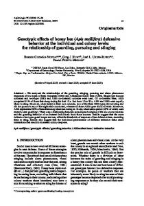
Figure 1. Forewings of two queens, one complete and the other clipped for DNA extraction from the wing tip (scale bar = 3.4 mm). if they were mated or not because their spermathecae were damaged when they were collected. A second set of 35 queens were reared and at emergence a small piece of the tip of each forewing was cut off using fine scissors and stored at –20 °C (Fig. 1). We estimated the area removed from each wing at 1.3 mm2 by approximating the wing tip as a triangle with dimensions given by graticule measurements made under a binocular microscope. The removed area was around 7.5% of each forewing surface. Seven of these queens were introduced to mating nuclei, five of them mated successfully, whilst two became drone layers. These seven queens were collected from the mating nuclei and kept in the queen bank colony until they died. Following emergence, the remaining 28 queens were kept in individual cages in the queen bank until death, after which they were stored at –20 °C. The purpose of keeping the queens in the bank colony was to determine if it was possible to obtain DNA extractions from wings of older bees. Because wings are mainly dried cuticle through which a few veins circulate haemolymph (Snodgrass, 1956) and the epidermis cells degenerate after emergence (Richards and Davies, 1977), it is possible that wings will become unsuitable with time for DNA extractions. For all genetic analyses, both antennae of each dead queen were collected and stored at –20 °C as control samples. The remaining part of one forewing of the queens in the second set of queens was also clipped 3 mm from the thorax at death and stored at –20 °C.
2.3. DNA extractions DNA was extracted from worker heads using high-salt extractions (Bruford et al., 1998; Miller et al., 1988). Heads were added to 250 µl of proteinasing solution (0.2 mg/mL proteinase K, 50 mM Tris, 120 mM NaCl, 1% SDS, 20 mM EDTA, pH 8.0) and crushed thoroughly. The remaining cuticle of the
313
head was then removed and the solution digested (with constant agitation) at 55 °C for 3 h. An equal volume of 4M ammonium acetate was then added and the solution was vortexed and left at room temperature for 15 min. The sample was centrifuged at 8000 g for 10 min and the supernatant decanted into an autoclaved labelled eppendorf tube. To precipitate the DNA from the supernatant, two volumes of 100% ethanol were added and the sample was centrifuged at 8000 g for 10 min. The supernatant was decanted and the pellet was rinsed in 1 ml of 70% ethanol and air-dried for 30 min. DNA samples were dissolved overnight in 250 µl of 10 mM Tris, 0.1 mM EDTA. All the other tissue samples (tarsus, antenna, full forewing and wing tip) were extracted using chelex®100 extraction (Walsh et al., 1991). The samples were placed in liquid nitrogen for 5 min and then crushed thoroughly with a disposable pestle. Different amounts of 5% chelex®100 solution were added according to the nature of the sample: 200 µl was added to antenna and tarsus samples, 100 µl was added to full forewing samples and 50 µl was added to wing tip samples. The samples were then incubated at 56 °C for 2 hours with constant agitation, vortexed for 10 s, boiled at 100 °C for 15 min and vortexed for another 10 s. Following 3 min of centrifugation at 8000 g, 20 µl of the supernatant was pipetted into 200 µl microtitre plates. All the extractions were used neat for PCRs reactions. All the steps following the incubation period were repeated if more DNA samples were needed.
2.4. Microsatellite analysis Polymerase chain reaction (PCR) amplifications were used to amplify 4 microsatellite markers: A76, A107, A113 and B124 (Tab. I). A76, A107 and A113 were previously isolated from Apis mellifera (Estoup et al., 1994, 1995) and B124 was isolated from Bombus terrestris L. (Estoup et al., 1994). PCRs were performed with a Hybaid thermal cycler in a 10.5 µl volume containing 1.5 µl of DNA sample, 1.0 µM of each primer, 0.2 mM of each dNTP, 1.5 or 2.0 mM MgCl2, and 0.05 units of Taq DNA polymerase (Thermoprime plus, Advanced Biotechnologies), in the manufacturer’s buffer at a final concentration of 20 mM (NH4)2SO4, 75 mM Tris-HCl pH 9.0 and 0.01% (w/v) Tween. The reaction profile for each locus was 94 °C for 1 min, followed by 39 cycles of 94 °C for 30 s, annealing temperature (Tab. I) for 30 s, and 72 °C for 30 s, followed by a last elongation stage of 5 min at 72 °C. The forward primer of each marker was 5’ end-labelled with a fluorescent phosphoramidite (NED, 6-FAM or HEX). The PCR products were visualised on an Applied Biosystems (ABI) 377 DNA sequencer using an internal size-standard (ROX). Because of the size and dye differences
314
N. Châline et al.
Table I. The 4 DNA microsatellite markers used. Locus
Fluorescent label
Ta (°C)
Dilution (µl)
MgCl2
Number of alleles
Size-range (bp)
Heterozygosity
A107
Hex
58
0
1.0
15
160-186
0.946
A113
6-Fam
58
30
1.2
6
202-234
0.875
A76
Ned
58
10
1.2
18
208-315
0.875
B124
Hex
54
10
1.5
13
207-251
0.786
Ta annealing temperature. MgCl2 published concentrations of MgCl2 for PCR reactions. Numbers of alleles and heterozygosities, calculated with CERVUS (Marshall et al., 1998) and based only on the 6 colonies of honey bees used in the experiments (n = 46 queens and 10 workers).
between the PCR products for the 4 loci we were able to multiplex them in a single set of markers after diluting them with different amounts of ddH2O (Tab. I). The gels were analysed using ABI Genescan software (version 3.1) and Genotyper DNA fragment analysis software (version 2.5). The annealing temperatures used and the 1.5 mM concentration of MgCl2 were obtained previously by optimisation on phenol extracted samples and were used in the first reactions. The worker samples were only amplified once at these conditions. However, the presence of chelating agents in the DNA samples might cause amplification problems and for the queen samples, any individuals with failed reactions after the first PCRs were redone at two MgCl2 concentrations, 1.5 and 2.0 mM. All PCR reactions were performed using both negative (water) and positive controls (DNA extracted from worker heads using classic phenol technique and of known genotypes). Because we amplified several samples from the same individuals and performed more than one successful PCR amplification on some samples we could check the reliability of the genotypes obtained from the wing tip samples and that using very little tissue for the extraction did not cause allelic dropout during the amplifications, as sometimes occurs (Taberlet et al., 1999).
2.5. Statistical analyses A generalised linear model with binomial error structure was used to test whether the amplification efficiencies of the 4 microsatellite loci were significantly different for the various sampling and storage regimes. For this purpose the individual samples were scored as 1 if they successfully amplified at the 4 loci and as 0 if at least one of them did not. When multiple pair-wise comparisons were done, we used the Bonferroni correction to adjust the level of significance.
2.6. Behavioural analysis Twenty forager bees were collected at the entrance of an observation hive and anaesthetised by chilling at 4 °C for ten minutes. They were then marked with a dot of white paint and their wing tips clipped in the same way as the newly-emerged queens (i.e., removal of 1.3 mm2). They were released 20 m from their original colonies to determine if they could fly back to their colony.
3. RESULTS 3.1. Worker samples There was no significant effect of type of tissue, storage method or age of bee on the mean amplification success of the 4 loci for the worker DNA samples (Tab. II, n = 86, P > 0.2) although the B124 and A113 loci amplified for fewer individuals than the other two loci, A76 and A107. It was therefore decided to use the simpler method, freezing, as the storage method for the subsequent queen samples. 3.2. Queen samples We analysed 44 antenna samples, 46 full forewing samples and 35 wing tip samples. The 46 queens were aged between 2 and 142 days (mean ± s.e.: 34.45 ± 5.31) before the final sampling (antennae for the first set, full forewing and antennae for the second). After the first PCRs, the tissue type had a significant effect on the amplification success (Tab. III, n = 125, P < 0.001). The pair-wise comparisons of the 3 sampling methods showed that only the wing tip and the antennae samples were significantly different from each other (n = 79, P < 0.001). Locus
Non-lethal DNA sampling of honey bees
315
Table II. Experiment 1. Number of unsuccessful PCR amplifications for each sampling method for the worker samples at the 4 loci that were tested. Locus Tissue sample (sample size)
A107
A113
A76
B124
Total
Mean %
Head (n = 10)
0
1
0
4
5
12.50
Tarsus ethanol (n = 9)
1
1
2
1
5
13.89
Tarsus ethanol ten days (n = 9)
0
0
0
1
1
2.78
Tarsus frozen (n = 10)
0
0
0
1
1
2.50
Tarsus frozen ten days (n = 10)
0
1
1
0
2
5.00
Half wing ethanol (n = 8)
1
2
0
2
5
15.62
Half wing ethanol ten days (n = 10)
0
1
0
1
2
5.00
Half wing frozen (n = 10)
1
2
1
1
5
12.50
Half wing frozen ten days (n = 10)
0
0
1
0
1
2.50
Total Mean %
3
8
5
11
27
3.48
9.30
5.81
12.79
7.85
Table III. Experiment 2. Number of unsuccessful PCR amplifications in the first/second PCR amplifications performed on the queen samples. Locus Tissue sample
A107
A113
A76
B124
Mean %
Antennae (n = 44)
2/0
2/0
0/0
2/0
3.4/0
Wing tips (n = 35)
11/1
13/2
4/1
14/2
30.0/4.3 8.7/0
Full wing (n = 46) Mean %
3/0
1/0
0/0
12/0
12.8/0.8
12.8/1.6
3.2/0.8
22.4/1.6
A76 amplified for more samples than all the others (3.2% of failures vs. 12.8% for A107 and A113 and 22.4% for B124). We amplified all individuals with missing genotypes again at all loci with two different magnesium concentrations: 1.5 mM and 2.0 mM. Locus B124 amplified better at 2.0 mM MgCl2 which is 0.5 above the recommended concentration with the published sequence (Tab. I), and could be necessary because of the presence of a chelating agent in the DNA samples. 1.5 mM MgCl2 was already above the recommended concentration for the other markers (Tab. I). Following the second round of amplifications, all samples could be scored at all loci except for two of the wing tip samples, giving an overall amplification failure of 4.3% for wing tips (n = 140 genotypes), 0% for full wings (n = 184) and 0% for antennae (n = 176). One of the wing tip samples did not give any product with any of the markers used. The other
unsuccessful wing tip sample gave a product only for two loci (A107 and A76; Tab. III). For all other amplifications, the genotypes were identical and consistent for the different samples of the same individual and different PCR amplifications of the same samples. In addition, the five different colony origins of the queens could be identified using their genotypes. Sufficient DNA was extracted from the wing tip samples to perform at least 20 PCR amplification reactions using wing tips and at least 50 with DNA extracted from whole wings.
3.3. Behavioural analysis All the wing tip clipped workers released 20 m from their observation hives were seen flying back home and on subsequent days some of them were seen leaving the hive on foraging trips and on the combs inside. It was
316
N. Châline et al.
not possible to record all the bees as only two combs of the nine frame observation hive were observable. The mating success of the wing tip clipped queens introduced to mating nuclei (5/7) and of unclipped queens (7/9) was not significantly different (Fisher’s exact test, P = 0.3) suggesting no adverse effect of wing clipping. Poor weather conditions and robbing of the mating nucleus colonies by other colonies may have caused the failure of some of the queens to mate.
4. DISCUSSION Our results clearly show that it is possible to extract DNA from wings and wing tips using standard and simple techniques and that the quality of the DNA is good enough to perform PCR amplifications. The wing tip samples proved to be harder to amplify but the success rate of 33/35 (94.3%) at the four loci was still very good. If minimal impact on survival or behaviour is desired then this is clearly the preferred method. The removal of legs or tarsi in honey bees for DNA sampling, even if suitable for genetic analyses (Starks and Peters, 2002), is probably not the best option as they are essential for conducting many colony activities. In addition, queen and worker tarsi produce important pheromones (Lensky and Slabezki, 1981; Winston, 1987) and queens with missing tarsi are superseded more frequently (Woyke, 1988). Queens with missing legs, which sometimes occur naturally, appear less able to move around in the colony and lay eggs more slowly (F.L.W. Ratnieks, personal observation). Although queens need their wings for making mating flights (and workers obviously use their wings for foraging, defence, removal of corpses from the nest, etc.) both queens and workers frequently have worn wing tips, showing that they can fly despite losing part of their wings. A study on bumblebee wing wear (Hedenström et al., 2001) showed that a 10% reduction of the wing surface did not significantly affect forager survivorship. Clipping full wings of mated queens in colonies is a common beekeeping practice which does not affect the queen’s ability to carry out her in-nest duties, but prevents them from swarming (honey bee queens never remate once egg-laying has begun; Laidlaw and Page, 1997).
Because it is more difficult to amplify DNA from wing tip samples, it is recommended that PCR conditions be optimised for all the markers using control samples. Although each PCR product that was obtained could be scored reliably, we still recommend performing two amplifications of each locus to ensure maximal scoring accuracy (Taberlet et al., 1999). For the whole wing extractions, no amplification problems occurred and even wings of older queens (up to 142 days) gave good quality DNA. Whole wing and wing tip sampling of queens can be a useful method in honey bee breeding and conservation programs. Honey bees are economically important for their honey production and as major pollinators of crops and wild plants (e.g. Roubik, 2002). Typically, breeding programs select for desirable traits such as low defensiveness and high disease resistance (Spivak, 1996; Spivak and Reuter, 1998) or can attempt to conserve local races (Cooper, 1986). Being able to select queens before allowing them to mate naturally or before instrumental insemination has the potential to speed up the selection process and reduce the amount of work involved. Sometimes the presence of only one or a few patrilines with the desired trait is sufficient to make the whole colony express a desirable phenotype such as hygienic behaviour (e.g. Trump et al., 1967), a phenomenon known as behavioural dominance (Craig, 1980). The standard breeding approach of randomly selecting queens from colonies with a desirable phenotype can then be a relatively inefficient way of artificial selection. By using the wing tip sampling methods, however, a breeder could specifically select queens from the preferred patrilines if these have been determined by behavioural studies of workers. Such within colony selection can increase the response to selection in breeding programs (Wenseleers and Ratnieks, unpublished). In addition, bee genetics and behavioural studies are now well developed and quantitative trait loci responsible for certain behaviour like foraging (Hunt et al., 1995), defence (Hunt et al., 1998) and hygienic behaviour (Lapidge et al., 2002) are starting to be identified so that newly-emerged queens could also be selected based on specific genes or markers. Non-destructive tissue sampling could also be used for other purposes. For example, in behavioural studies, the patrilines
Non-lethal DNA sampling of honey bees
of particular workers could be determined before they are studied, which could give better designed experiments (e.g., equal sample sizes per patriline). Or in parentage studies, the queens could be non-destructively sampled to increase confidence in parentage assignments.
ACKNOWLEDGEMENTS NC was funded by the EC network ‘Beekeeping and Apis Biodiversity in Europe’ (BABE). NER was funded by NERC (GR3/12816). We thank Tom Wenseleers and Deborah Dawson for comments on the manuscript. Résumé – Échantillonnage non létal d’ADN de l’Abeille domestique (Apis mellifera) à partir des extrémités des ailes. L’échantillonnage de l’ADN des insectes repose souvent sur des méthodes d’échantillonnage des tissus, qui sont létales ou qui ôtent des parties importantes du corps. Parce que les sociétés d’insectes comportent de nombreuses ouvrières, il est souvent possible de prélever des échantillons d’ouvrières pour analyse génétique en les tuant. Ce n’est par contre pas possible s’il s’agit d’analyser des reines ou si les individus doivent rester vivants après l’échantillonnage. Il n’est pas non plus possible de prélever une patte entière, une aile ou tout autre appendice, car cela empêche généralement les individus de poursuivre normalement leurs activités. Nous avons étudié la possibilité de déterminer le génotype de reines d’abeilles à l’aide de l’ADN extrait des extrémités de leurs ailes (environ 1,3 mm2), de sorte que le vol et les autres activités restent non affectées. Nos résultats montrent que les échantillons des extrémités des ailes peuvent fournir de l’ADN de bonne qualité. 94,3 % des 35 échantillons d’extrémités d’ailes ont pu être amplifiés par PCR en utilisant 4 marqueurs microsatellites. Lorsque l’amplification a réussi, les génotypes étaient fiables et cohérents dans toutes les amplifications par PCR et toujours identiques à ceux obtenus avec des échantillons témoins plus grands. L’échantillonnage d’ADN dans les extrémités des ailes va permettre toute une série de nouvelles approches comme (i) déterminer le génotype de reines fraîchement écloses dans le cadre de programmes de sélection où seules les reines de certaines lignées paternelles ou de certains génotypes doivent être utilisées ou (ii) déterminer le génotype d’individus qui doivent rester intacts afin de se comporter normalement après l’échantillonnage. Apis mellifera / ADN / microsatellite / extraction / échantillonnage non destructif / programme de sélection
317
Zusammenfassung – Nichdestruktive DNS – Beprobung von Honigbienen (Apis mellifera) durch Nutzung der Flügelspitzen. DNS-Proben bei Insekten verwenden zumeist Gewebsproben, deren Entnahme tödlich ist oder die wichtige Körperteile entfernten. Da soziale Insekten in Gemeinschaften mit sehr vielen Arbeiterinnen leben, kann man meist genügend Arbeiterinnen für genetische Analysen entnehmen. Dies ist jedoch nicht möglich, wenn die Königin analysiert werden soll oder Arbeiterinnen nach der Beprobung aktiv bleiben müssen. Hier können weder ganze Beine, Flügel oder andere Körperanhänge verwendet werden, da dies oft die Individuen an der Ausführung ihrer normalen Beschäftigungen hindert. Diese Studie untersucht ob es möglich ist, Königinnen von Honigbienen anhand der aus Flügelspitzen (ca. 1,3 mm2) extrahierten DNA zu genotypisieren, ohne ihren Flug und andere Aktivitäten zu beeinflussen, wie dies bei Entnahme anderer Teile wie ganzer Flügel, Antennen, Beinen oder Füssen der Fall wäre. Unsere Resultate zeigen, dass man bei Entnahme der Flügelspitzen DNA in guter Qualität erhält. Von 35 Flügelspitzenproben konnten 94,3 % erfolgreich an vier Mikrosatellitenloci amplifiziert werden. Bei erfolgreicher Amplifikation konnten die Genotypen zuverlässig und bei mehreren Amplifikaten übereinstimmend bestimmt werden und waren identisch zu größeren Kontrollproben erhaltenen. Die Beprobung von Flügelspitzen-DNA ermöglicht eine Reihe neuer Forschungsansätze. Dies schließt die Genotypisierung von frischgeschlüpften Königinnen als Teil von Zuchtprogrammen ein, in denen aus den Völkern nur Königinnen bestimmter Vaterlinien oder Genotypen genutzt werden sollen, oder von Individuen, die nach der Beprobung unbeeinträchtigt bleiben müssen um eine normales Verhalten zu zeigen. Apis mellifera / DNS Mikrosatelliten / Nichtdestruktive Probennahme / Selektionsprogramme / DNS Extraktion
REFERENCES Bourke A.F.G. (1991) Queen behaviour, reproduction and egg cannibalism in multiple-queen colonies of the ant Leptothorax acervorum, Anim. Behav. 42, 295–310. Bourke A.F.G. (1993) Lack of experimental evidence for pheromonal inhibition of reproduction among queens in the ant Leptothorax acervorum, Anim. Behav. 45, 501–509. Bourke A.F.G., Green H.A.A., Bruford M.W. (1997) Parentage, reproductive skew and queen turnover in a multiple-queen ant analysed with microsatellites, Proc. R. Soc. London B 264, 277–283. Bruford M.W., Hanotte O., Brookfield J.F.Y, Burke T. (1998) Multilocus and single-locus DNA fingerprinting, in: Hoelzel A.R. (Ed.), Molecular
318
N. Châline et al.
Genetic Analysis of Populations: A Practical Approach, IRL Press, Oxford, pp. 287–336. Châline N., Ratnieks F.L.W., Burke T. (2002) Anarchy in the UK: Detailed genetic analysis of worker reproduction in a naturally occurring British anarchistic honeybee, Apis mellifera, colony using DNA microsatellites, Mol. Ecol. 11, 1795–1803. Cooper B.A. (1986) The honeybees of the British Isles, British Isles Bee Breeders’ Association, Derby, UK. Craig R. (1980) Sex investment ratios in social Hymenoptera, Am. Nat. 116, 311–323. Estoup A., Solignac M., Cornuet J.M. (1994) Precise assessment of the number of patrilines and of genetic relatedness in honeybee colonies. Proc. R. Soc. London B 258, 1–7. Estoup A., Garnery L., Solignac M., Cornuet J.M. (1995) Microsatellite variation in honey bee (Apis mellifera L) populations – hierarchical genetic structure and test of the infinite allele and stepwise mutation models, Genetics 140, 679–695. Fincke O.M., Hadrys H. (2001) Unpredictable offspring survivorship in the damselfly, Megaloprepus coerulatus, shapes parental behavior, constrains sexual selection, and challenges traditional fitness estimates, Evolution 55, 762–772. Foster K.R., Ratnieks F.L.W., Gyllenstrand N., Thoren P.A. (2001) Colony kin structure and male production in Dolichovespula wasps, Mol. Ecol. 10, 1003–1010. Gerken T., Kurtz J., Sauer K.P., Lubjuhn T. (1998) DNA preparation and efficient microsatellite analysis from insect haemolymph, Electrophoresis 19, 3069–3070. Hedenstrom A., Ellington C.P., Wolf T.J. (2001) Wing wear, aerodynamics and flight energetics in bumblebees (Bombus terrestris): an experimental study, Funct. Ecol. 15, 417–422. Hunt G.J., Page R.E., Fondrk M.K., Dullum C.J. (1995) Major quantitative trait loci affecting honey bee foraging behaviour, Genetics 141, 1537–1545. Hunt G.J., Guzman-Novoa E., Fondrk M.K., Page R.E. (1998) Quantitative trait loci for honey bee stinging behavior and body size, Genetics 148, 1203–1213. Kurtz J., Sauer K.P. (1999) The immunocompetence handicap hypothesis: testing the genetic predictions, Proc. R. Soc. London B 266, 2515–2522. Laidlaw H.H., Page R.E. (1997) Queen rearing and bee breeding, Wicwas Press, Cheshire, Connecticut. Lapidge K.L., Oldroyd B.P., Spivak M. (2002) Seven suggestive quantitative trait loci influence hygienic behavior of honey bees, Naturwissenschaften 89, 565–568.
Lensky Y., Slabezki Y. (1981) The inhibiting effect of the queen bee (Apis mellifera L.) foot-print pheromone on the construction of swarming queen cups, J. Insect Physiol. 27, 313–323. Lushai G., Fjellsted W., Marcovitch O., Aagaard K., Sherratt T.N., Allen J.A., Maclean N. (2000) Application of molecular techniques to non-lethal tissue samples of endangered butterfly populations (Parnassiuss apollo L.) in Norway for conservation management, Biol. Conserv. 94, 43–50. Marshall T.C., Slate J., Kruuk L.E.B., Pemberton J.M. (1998) Statistical confidence for likelihood-based paternity inference in natural populations, Mol. Ecol. 7, 639–655. Miller S.A., Dykes D.D., Polesky H.F. (1988) A simple salting out procedure for extracting DNA from human nucleated cells, Nucl. Acids Res. 16, 1215. Richards O.W., Davies R.G. (1977) Imms' general textbook of entomology, Vol. 1, Chapman and Hall, London. Rose O.C., Brookes M.I., Mallet J.L.B. (1994) A quick and simple nonlethal method for extracting DNA from butterfly wings, Mol. Ecol. 3, 275– 275. Roubik D.W. (2002) The value of bees to the coffee harvest, Nature 417, 708. Snodgrass R.E. (1956) Anatomy of the honey bee, Vail-Ballou Press, Inc., Binghamton, N.Y. Spivak M. (1996) Honey bee hygienic behavior and defense against Varroa jacobsoni, Apidologie 27, 245–260. Spivak M., Reuter G.S. (1998) Performance of hygienic bee colonies in a commercial apiary, Apidologie 29, 291–302. Starks P.T., Peters J.M. (2002) Semi-nondestructive genetic sampling from live eusocial wasps, Polistes dominulus and Polistes fuscatus, Insectes Soc. 49, 20–22. Taberlet P., Waits L.P., Luikart G. (1999) Noninvasive genetic sampling: look before you leap, Trends Ecol. Evol. 14, 323–327. Trump R.F., Thompson V.C., Rothenbuhler W.C. (1967) Behaviour genetics of nest cleaning in honeybees. V. Effect of previous experience and composition of mixed colonies on response to disease-killed brood, J. Apic. Res. 6, 127–131. Walsh P.S., Metzger D.A., Higuchi R. (1991) Chelex100 as a medium for simple extraction of DNA for PCR-based typing from forensic material, BioTechniques 10, 506–513. Winston M.L. (1987) The Biology of the Honeybee, Harvard University Press, Cambridge, MA. Woyke J. (1988) Problems with Queen Banks, Am. Bee J. 128, 276–278.