Alessandra De Riva,* Christine Bourgeois,* George Kassiotis,â and Brigitta .... 2 Address correspondence and reprint requests to Dr. Brigitta Stockinger, Division ...
The Journal of Immunology
Noncognate Interaction with MHC Class II Molecules Is Essential for Maintenance of T Cell Metabolism to Establish Optimal Memory CD4 T Cell Function1 Alessandra De Riva,* Christine Bourgeois,* George Kassiotis,† and Brigitta Stockinger2* CD4 memory T cells surviving in the absence of MHC class II contact lose their characteristic memory function. To investigate the mechanisms underlying the impaired function of memory T cells in the absence of MHC class II molecules, we analyzed gene expression profiles of resting memory T cells isolated from MHC class II-competent or -deficient hosts. The analysis focused on five transcripts related to T cell activation, metabolism, and survival that are underexpressed in resting memory T cells from MHC class II-deficient hosts compared with MHC class II-competent hosts. CD4 memory cells isolated from MHC class II-deficient hosts display alterations in their degree of differentiation as well as metabolic activity, and these changes are already manifest in the effector phase despite the presence of Ag-expressing dendritic cells. Our data suggest that the absence of interactions with noncognate MHC class II molecules compromises the progressive accumulation of signals that ensure optimal survival and fitness to sustain the metabolic activity of activated T cells and shape the functional capacity of the future memory compartment. Signals via AKT coordinate survival and metabolic pathways and may be one of the crucial events linking interaction with MHC class II molecules to the successful generation of a long-lived functional memory CD4 T cell population. The Journal of Immunology, 2007, 178: 5488 –5495.
T
cell activation requires both the engagement of the TCR with cognate peptide presented in the context of MHC molecules on APCs (1) and costimulatory signals that are necessary to enforce T cell commitment to activation (2). The exact nature and the kinetics of the interactions occurring during the effector phase remain unclear despite the increasing amount of information now available. Overall, variations in the duration and strength of TCR stimulation, the abundance of specific epitopes, the nature of the costimulatory molecules, and the cytokine environment shape the differentiation of effector and memory T cells (3– 6), especially within the CD4 subset. In addition IL-7 is involved in the transition of effector cells to memory CD4 T cells (7, 8). Although MHC molecules are generally (9 –16), albeit not universally (17, 18), thought to be essential for maintaining survival of naive T cells, memory T cell survival can be sustained by cytokines in the absence of MHC molecules (19, 20). However, we have previously shown that absence of MHC interactions compromises the typical function of memory CD4 T cells (19). To investigate the mechanisms underlying the impaired function of memory T cells in the absence of MHC class II molecules, we analyzed gene expression profiles of resting memory T cells isolated from MHC class II-competent or -deficient hosts using an Affymetrix microarray approach. We focused on five transcripts related to T cell activation, metabolism, and survival that are underexpressed
†
*Division of Molecular Immunology and Division of Immunoregulation, Medical Research Council Institute for Medical Research, London, United Kingdom Received for publication October 27, 2006. Accepted for publication February 14, 2007. The costs of publication of this article were defrayed in part by the payment of page charges. This article must therefore be hereby marked advertisement in accordance with 18 U.S.C. Section 1734 solely to indicate this fact. 1
in resting memory T cells from MHC class II-deficient hosts compared with MHC class II-competent hosts. We provide evidence that CD4 memory cells isolated from MHC class II-deficient hosts display alterations in their degree of differentiation as well as metabolic activity. These changes are already manifest in the effector phase despite the presence of Ag-expressing dendritic cells (DC),3 suggesting that interactions with noncognate MHC molecules of the host environment are essential to sustain the metabolic activity of activated T cells and shape the functional capacity of the future memory compartment.
Materials and Methods Mice Female A1 TCR-transgenic Rag1⫺/⫺ (H2k) mice (harboring T cells specific for the male H-Y Ag) (21) were used between 6 and 10 wk of age. Recipient mice were either allogeneic H2bRag2⫺/⫺Il2rg⫺/⫺ (referred to as MHC class II-competent mice) or allogeneic H2bRag2⫺/⫺Il2rg⫺/⫺H2A⫺/⫺ (referred to as MHC class II-deficient mice). CBA and C57BL/6 mice were used to provide bone marrow-derived DCs. All animals were bred under specified pathogen-free conditions, and all experiments were done in conventional but pathogen free-facilities in accordance with institutional guidelines and Home Office regulations.
Generation of effector and memory T cells Lymph node T cells (2– 4 ⫻ 106) from female A1 TCR-transgenic H2kRag1⫺/⫺ mice were cotransferred with syngeneic bone marrow-derived DCs pulsed with 1 M H-Y peptide into allogeneic adoptive hosts which were either H2bRag2⫺/⫺Il2rg⫺/⫺ (MHC class II competent) or H2bRag2⫺/⫺Il2rg⫺/⫺H2-A⫺/⫺ (MHC class II deficient). DCs were generated from the bone marrow of syngeneic (CBA) or allogeneic (C57BL/6) mice by culture with GM-CSF as previously described (22).
This work was supported by the Medical Research Council U.K.
2
Address correspondence and reprint requests to Dr. Brigitta Stockinger, Division of Molecular Immunology, The Medical Research Council National Institute for Medical Research, The Ridgeway, Mill Hill, London, U.K. E-mail address: bstocki@nimr. mrc.ac.uk www.jimmunol.org
3 Abbreviations used in this paper: DC, dendritic cell; ␥c, common ␥-chain; PdBu, phorbol dibutyrate.
Copyright © 2007 by The American Association of Immunologists, Inc. 0022-1767/07/$2.00
The Journal of Immunology Cell suspension, flow cytometry, and cell sorting Lymph nodes and spleen cell suspensions were prepared in IMDM (SigmaAldrich). DC isolation was performed using Liberase CI Purified Enzyme Blend (Roche). All Abs were purchased from e-Bioscience with the exception of PeTexas Red anti-CD4 from Caltag Laboratories; biotin antimouse H-2Kk, PE anti-IL-2, PE anti-DO.11.10, FITC-conjugated Armenian hamster anti-mouse Bcl-2 mAb, and isotype control from BD Pharmingen; phospho-S6 ribosomal protein (Ser235/236) and phospho-Stat5 (Tyr694) primary Abs from Cell Signaling Technology; and FITC-labeled goat anti-rabbit Ig from BD Pharmingen as secondary Ab. For determination of intracellular proteins, cells were fixed on ice in 100 l of 2% paraformaldehyde in PBS for 15 min and permeabilized with 0.1% Nonidet-P40, PBS for 3 min, followed by staining with specific Abs. Analytical flow cytometry was conducted using a FACSCalibur (BD Biosciences), and the data were processed using FlowJo software (Tree Star). Cell sorting was done on a MoFlo cell sorter (Cytomation).
Analysis of gene expression Resting memory CD4⫹ T cells recovered from pools of 8 –10 mice per group were purified by FACS sorting on a MoFlo cell sorter to ⬎98% purity. Total RNA was isolated (Qiagen) and assessed for quality and quantity on an Agilent Bioanalyser 2100 (Agilent Technologies) using a RNA 6000 Nano LabChip Kit (Agilent). Using a GeneChip Two-Cycle cDNA Synthesis Kit (manufactured by Invitrogen for Affymetrix), 600 ng of total RNA were amplified and hybridized on Mouse 430 A Plus chips (Affymetrix). The results were analyzed using GeneSpring version 7.0 software (Silicon Genetics), and genes differentially expressed with a cutoff point of 1.5 were considered for further investigation. Reverse transcription from total RNA was performed using a GeneAmp RNA PCR Core Kit (PerkinElmer). Il-2 and Glut1 gene expression was assessed using Assays-on-Demand Gene Expression Products (Applied Biosystems) on the ABI PRISM 7000 Sequence detection system (Applied Biosystems). Target gene expression was calculated using the comparative method for relative quantitation upon normalization to Hprt1 gene expression. For graph representation, data were normalized to the expression levels on naive CD4 T cells. The raw data are deposited at www.ebi.ac.uk/ arrayexpress/, accession number E-MEXP-890.
In vitro activation and cytokine production assays A1 CD4 T cells were stimulated in 96-well plates with serial dilutions of plate-bound anti-CD3 (2C11) in the absence or presence of 10 g/ml platebound anti-CD28. IL-2 production was assessed on day 2 in culture supernatant with an Alamar blue-based (23) CTLL assay. For intracellular detection, cells were stimulated with 500 ng/ml phorbol dibutyrate (PdBu; Sigma-Aldrich), 500 ng/ml ionomycin (Sigma-Aldrich), and 10 g/ml brefeldin (Sigma-Aldrich) for 6 h at 37°C before fixation.
Statistical analysis p values were obtained using the Mann-Whitney two-tailed t test.
Results Altered gene expression in memory cells from MHC class II-deficient hosts CD4 memory T cells were generated by transferring naive T cells from A1 TCR-transgenic Rag⫺/⫺ hosts specific for H-Y peptide in the context of H-2Ek class II molecules together with Ag-pulsed syngeneic DCs into adoptive hosts that were either expressing allogeneic MHC (Rag2 and common ␥-chain (␥c) deficient) or lacking conventional MHC class II (Rag2, ␥c, and H-2A deficient) (19). The allogeneic H-2b host MHC haplotype is neutral for the A1 TCR (24, 25); it cannot present cognate Ag to A1 T cells and neither positively nor negatively selects during thymic development. Therefore, Ag presentation is limited by the availability of syngeneic DCs, which disappear ⬃3 wk after transfer, thus ensuring the complete absence of antigenic stimulation (24). As a result, a pure population of resting memory CD4 T cells persists in these hosts. As previously described, A1 memory T cells recovered from MHC class II-deficient hosts show distinct functional defects upon reencountering their Ag, such as reduced capacity to produce IL-2, lack of providing help to B cells, and failure to reject H-Y expressing skin grafts (19).
5489 Table I. Microarray data of gene expression in memory CD4 T cells recovered from MHC-competent and -deficient hostsa
Gene Name
Signal transduction and intracellular signaling 1418110_a_at 1421469_a_at 1460700_at 1422103_a_at 1460280_at 1424527_at 1449393_at 1448713_at 1417936_at 1418497_at 1418990_at 1420572_at 1449984_at Apoptosis and antiapoptosis 1416657_at 1422938_at 1449193_at 1448784_at 1427843_at 1418901_at Cell cycle and cell cycle arrest 1453307_a_at 1417326_a_at 1416868_at 1417457_at 1449519_at 1428570_at Metabolism 1427604_a_at 1432372_a_at 1449237_at 1416838_at 1448261_at 1426260_a_at Cytokinesis and cytokines 1426205_at 1448713_at 1417457_at 1428570_at Inflammatory responses 1449990_at 1449984_at
Fold Change
Common Name
GenBank Reference
2.288 2.076 1.968 1.922 1.878 1.763 0.492 0.475 0.422 0.308 0.295 0.27 0.188
Inpp5d Stat5a Stat3 Stat5b Mona Ppp2r2d Sh2d1a Stat4 Ccl9 Fgf13 Ms4a4d Ms4a3 Cxcl2
U39203 U36502 AK004083 BC024319 NM_010815 AF366393 NM_011364 NM_011487 AF128196 AF020737 NM_025658 NM_133246 NM_009140
2.354 1.877 1.873 0.503 0.391 0.298
Akt1 Bcl2 Cd5l Taf10 Kua Cebpb
NM_009652 NM_009741 NM_009690 NM_020024 AB012278 NM_009883
2.019 2.009 0.501 0.427 0.408 0.406
Anapc5 Anapc11 Cdkn2c Cks2 Gadd45a Ccnc
AK003821 NM_025389 BC027026 NM_025415 NM_007836 AK009615
1.969 1.805 1.77 0.432 0.343 0.342
Atp9a Spr Aloxe3 Mut Cdh1 Ugt1a2
AF011336 AK004941 NM_011786 NM_008650 NM_009864 D87867
0.524 0.475 0.427 0.406
Ppp1cb Stat4 Cks2 Ccnc
M27073 NM_011487 NM_025415 AK009615
3.271 0.188
Il2 Cxcl2
AF065914 NM_009140
a Gene name, fold change of expression (MHC competent : MHC deficient), common name, and GenBank accession number of differentially expressed transcripts. The classification of genes is according to gene ontology biological processes of the GeneSpring software. The five transcripts chosen for further analysis are shown in bold.
To further investigate the mechanisms underlying the impaired function of memory T cells in the absence of MHC class II, we analyzed the gene expression profiles of resting memory T cells isolated from MHC class II-competent or -deficient hosts ⬎70 days after immunization using the Affymetrix Mouse Genome 430 version 2.0 GeneChip. Approximately 400 genes were found differentially expressed in memory cells isolated from MHC-competent or -deficient hosts and a selection of transcripts is shown in Table I. The full set of raw data is available from www.ebi.ac.uk/ arrayexpress/, accession number E-MEXP-890. Here we focused on five transcripts related to T cell activation and survival that are underexpressed in resting memory T cells from MHC class IIdeficient hosts compared with MHC class II-competent hosts (Table I bold transcripts). Memory T cells from MHC class II-deficient hosts expressed reduced amounts of prestored transcripts for
5490
MHC INTERACTION MAINTAINS MEMORY FUNCTION
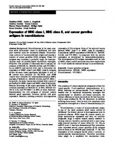
FIGURE 1. FACS analysis of resting A1 memory T cells. A1 memory T cells isolated from either MHC class II-competent hosts (full line) or MHC class II-deficient hosts (shaded histograms) 6 –7 wk after adoptive transfer/activation were analyzed for their expression of Bcl-2 (A), IL-7R␣ (B), p437-Akt (C), and pStat5 (D) in comparison with naive A1 T cells (stippled lines). Isotype control staining in A and B is shown in black histograms. Mean fluorescence intensities (MFI), SD and p values are shown for all markers.
IL-2, compared with memory T cells from MHC-competent hosts. The transcripts for the two isoforms of Stat5, Stat5a and Stat5b, were down-regulated in CD4 memory T cells from MHC class II-deficient hosts. STAT5 phosphorylation after signaling through IL-2 and IL-7 receptors results in dimerization and translocation into the nucleus where it binds to target genes, such as the antiapoptotic molecule Bcl-2 (26). The transcript for Bcl-2 was also underexpressed in A1 memory T cells from MHC class II-deficient hosts. The transcript for an important molecule involved in cell metabolism, survival, and signaling, Akt1 was underexpressed in memory T cells from MHC class II-deficient hosts, suggesting a potential impairment of survival. FACS analysis verified that resting memory T cells recovered from MHC class II-deficient hosts expressed lower amounts of Bcl-2 than memory T cells recovered from MHC class II-competent hosts (Fig. 1A). Levels of Bcl-2 expression are correlated with expression of IL-7R (27) and indeed A1 memory T cells from MHC class II-deficient hosts display lower levels of IL-7R than those from MHC class II-competent hosts (Fig. 1B). Furthermore, the levels of phosphorylated Akt (Fig. 1C) and Stat5 (Fig. 1D) were reduced in memory T cells from MHC class II-deficient hosts. The expression levels for all markers were intermediate between naive T cells and memory T cells isolated from MHC class II-competent hosts. Reduced metabolic activity in memory T cells from MHC class II-deficient hosts The PI3K/AKT pathway has effects on multiple aspects of T cell survival and activation. Signal transduction through Akt can be assessed by measuring the level of phosphorylation of the small ribosomal S6 subunit (28), which is required for the assembly of the ribosomal complex for protein synthesis (29). In T cells, S6 phosphorylation is mainly due to S6K1 activation, which is induced by TCR stimulation in a PI3K/AKT-dependent manner. As
FIGURE 2. Metabolic impairment of memory CD4 T cells in the absence of noncognate MHC class II. Memory CD4 T cells were recovered as described in the legend to Fig. 1. Intracellular staining for pS6 after 30 min in vitro stimulation with PdBU and ionomycin (A) or soluble anti-CD3 (B) is shown for memory A1 T cells from MHC class II-competent hosts (open histograms); gray histograms, MHC class II-deficient hosts; black histogram, resting unstimulated memory T cells. There is no difference in pS6 levels in resting memory A1 T cells whether they are isolated from MHC class II-competent or -deficient hosts. Mean fluorescence intensities (MFI), SD, and p values are shown for all markers. C, Glut1 mRNA expression after in vitro stimulation with anti-CD3 and anti-CD28 over a 3-day culture period in FACS-sorted memory A1 T cells from MHC class II-competent hosts (open dots) and MHC class II-deficient hosts (gray squares). D, IL-2 expression after stimulation for 4 h with PdBU, ionomycin, and brefeldin A in A1 memory T cells generated in MHC class IIcompetent hosts (open histogram) or MHC class II-deficient hosts (gray histogram). Naive A1 T are shown as reference (stippled lines). E, Percentage of cycling and apoptotic cells in memory cells isolated from MHC class II-competent hosts (open bars) and MHC class II-deficient hosts (gray bars). Data are derived from pooled FACS-sorted cells of 10 mice per group.
a result, small and metabolically inactive T cells increase protein synthesis and acquire the size required for proliferation and optimal effector functions (30). Levels of pS6 on resting memory cells are similar whether they are isolated from MHC class II-competent or -deficient hosts (Fig. 2, A and B, black histograms), but upon in vitro reactivation, memory cells from MHC class II-deficient hosts showed lower levels of S6 phosphorylation than those from MHC class II-competent hosts (Fig. 2, A and B). Akt also has a major role in glucose metabolism and regulates not only transcription of
The Journal of Immunology
5491
the main glucose transporter, Glut1 (31, 32), but also its cellular localization (33). Analysis of Glut1 mRNA expression from purified memory A1 T cells recovered from MHC class II-deficient and -competent hosts during in vitro restimulation with antiCD3 and anti-CD28 showed that memory A1 T cells from MHC class II-deficient hosts did not reach the same levels of expression of Glut1 mRNA as memory A1 T cells from MHC class II-competent hosts (Fig. 2C). In accordance with our previously described defect in IL-2 production, memory T cells recovered from MHC class II-deficient hosts show reduced IL-2 production assessed by intracellular staining after a short in vitro restimulation (Fig. 2D). Thus, functional impairment of the IL-2 response as well as a reduction in metabolic activity suggests that the absence of MHC class II molecules compromises CD4 memory T cells on several levels. Although similar numbers of memory T cells can be recovered from both types of hosts, memory CD4 T cells surviving in the absence of MHC class II molecules divide more rapidly, assessed by BrdU labeling (19). This suggests that on a per cell basis the survival of memory CD4 T cells lacking MHC contact may be compromised in line with the observed alterations in metabolic activity. Indeed, propidium iodide staining of FACSsorted memory T cells established that a higher proportion of memory T cells recovered from MHC class II-deficient hosts is in cell cycle and that there are more proapoptotic cells in this population (Fig. 2E).
Functional defects in memory A1 T cells lacking noncognate MHC class II interactions manifest themselves early in memory generation To assess whether contact with noncognate MHC class II molecules was solely required during the maintenance of established memory cells or was needed also in the early phase of memory generation, we analyzed A1 T cells 14 days after adoptive transfer of naive A1 T cells with Ag-pulsed DCs into MHCcompetent or -deficient hosts for the expression of Bcl-2 and IL-7R as well as functional activity. A time point of 42 days after transfer resembling the established memory phase was analyzed in parallel. The number of T cells recovered from MHC class II-deficient hosts was not significantly different from those in MHC class IIcompetent hosts (data not shown). Already at day 14 after transfer/ activation, A1 T cells transferred into MHC class II-deficient hosts showed intermediate expression levels of Bcl-2 between naive A1 T cells and memory A1 T cells generated in MHC class II-competent hosts (Fig. 3A). IL-7R expression (Fig. 3B) was consistently lower in A1 T cells isolated from MHC class II-competent hosts than in A1 T cells from MHC class II-competent hosts and like Bcl-2 slightly higher than in naive T cells. Functionally, as early as day 14 after activation/transfer, A1 T cells isolated from MHC class II-deficient hosts were less efficient than A1 T cells from MHC class II-competent hosts in IL-2 production, as assessed by in vitro reactivation with anti-CD3 with or without anti-CD28 (Fig. 3, C and D). In addition, lower levels of phosphorylated S6 seen in established memory T cells also were already evident in A1 T cells isolated 14 days after transfer/activation in MHC class II-deficient hosts (Fig. 3E). These results suggest that differences in the functional and metabolic activity of A1 memory T cells in MHC class II-deficient hosts are already imprinted in the early stages of memory generation.
FIGURE 3. Kinetics of T cell defect in memory A1 T cells generated in the absence of MHC class II. FACS analysis of Bcl-2 (A) and IL-7R␣ (B) ex vivo expression of memory A1 T cells from MHC class II-competent hosts (open histograms) and MHC class II-deficient hosts (gray histograms) recovered 14 or 42 days (d) after adoptive transfer/activation. Naive A1 T cells (stippled histograms) and isotype control (black histograms) are shown as reference. Mean fluorescence intensities (MFI), SD, and p values are shown for all markers. C and D, IL-2 production by memory A1 T cells recovered 14 days after adoptive transfer/activation into MHC class IIcompetent hosts (open dots) or MHC class II-deficient hosts (gray squares). IL-2 production after was assessed after 2 days of in vitro stimulation with either plate-bound anti-CD3 (C) or anti-CD3 and anti-CD28 (D). Mean values and SD for four mice per group are shown. Where SDs are not visible the bars are smaller than the symbol used. E, Intracellular staining for pS6 after 30 min in vitro stimulation with PdBU and ionomycin showing memory cells from MHC class II-competent hosts (open histogram), MHC class II-deficient hosts (gray histogram), as well as resting unstimulated memory T cells (black histogram).
5492
MHC INTERACTION MAINTAINS MEMORY FUNCTION
FIGURE 4. Impairment in A1 T cells activation in the absence of noncognate MHC class II molecules. A1 T cells were isolated from MHC class II-competent (open histograms) or -deficient (gray histograms) hosts and analyzed at day 4 (left panels), 8 (middle panels), and 14 (right panels) for expression of CD44 (A), CD71 (B), CD98 (C), and intracellular staining for pStat5 (D). Intracellular expression of IL-2 after a 4-h restimulation with PdBu-ionomycin and brefeldin A (E). Naive A1 T cells are shown as stippled histograms and isotype controls as black histograms. Histograms represent values of cells from four mice per group.
Altered expression of activation markers and reduced functional activity in the absence of noncognate MHC class II during the effector phase Evidence of functional impairment as early as 14 days after transfer/activation into MHC class II-deficient hosts suggests a role for noncognate MHC contact during the effector phase of the immune response. We therefore studied the expression of activation markers 4, 8, and 14 days after transfer of naive A1 T cells with Ag-pulsed DCs into MHC class II-deficient or -competent hosts. Similar numbers of A1 T cells were recovered from both hosts at day 4, but at day 8 A1 T cells seemed to have expanded to a lesser extent in MHC class II-deficient hosts than in MHC class II-competent hosts. On day 14 after transfer/activation, the numbers of A1 T cells recovered reflected the numbers usually recovered in the memory phase, and there was no significant difference in recovery of T cells from the two host types (data not shown). We next determined the status of activation achieved by transferred T cells by measuring the expression of CD44, IL7R-␣, CD71, CD98 and pStat5 at each time point (Fig. 4). In both hosts, A1 T cells up-regulated the expression of CD44 throughout the effector phase, although A1 T cells from MHC class II-deficient hosts followed a slower kinetics than those from MHC class II-
competent hosts. There was no difference in the expression of the early activation markers CD69 and CD25 at any of the time points tested (data not shown). IL7R␣ expression was down-regulated to a similar degree in A1 T cells from both hosts during the acute effector stage in accordance with reduced expression of this marker after activation. CD71, the transferrin receptor, and CD98 (4F2 Ag or Ly-10), the common H chain subunit component of amino acid transporters, (34) were expressed at lower levels in A1 T cells from MHC class II-deficient hosts. This indicates a reduction in metabolic activity, which is necessary to sustain activation (and possibly proliferation). However, CD71 and CD98 expression were transient because the expression of both molecules was downregulated in A1 T cells from both types of hosts at days 8 and 14. Stat5 phosphorylation on day 4 after transfer/activation was evident only in a subset of A1 T cells from MHC class IIdeficient hosts, whereas virtually all A1 T cells transferred into MHC class II-competent hosts displayed Stat5 phosphorylation at this stage. We also tested A1 T cells isolated from MHC class II-deficient or -competent hosts for their capacity to produce IL-2 upon a short restimulation in vitro (Fig. 4F). Effector cells recovered 4 days after transfer/activation were refractory to restimulation, and no intracellular IL-2 was detected. By days 8 and 14 after transfer, A1
The Journal of Immunology
5493 cient hosts activated in the presence of allogeneic B cells showed a recovery of Stat5 phosphorylation to levels seen in memory T cells from MHC class II-competent hosts (Fig. 5A). Fourteen days after transfer, memory cells from the residual mice were isolated and tested for their capacity to produce IL-2 by intracellular staining as well as in vitro stimulation with anti-CD3. Although the presence of B cells in MHC class II-competent hosts did not influence the IL-2 response of the A1 memory population, the IL-2 response in adoptive hosts lacking MHC class II molecules was significantly improved by the presence of allogeneic B cell to levels comparable with those in MHC class II-competent hosts (Fig. 5B). Thus, the availability of noncognate MHC class II interaction in addition to that of Ag-presenting MHC class II is an essential component in the development of functionally competent CD4 memory T cells.
Discussion FIGURE 5. Restoration of A1 T cell function by cotransfer of allogeneic B cells. A, Histograms show the level of phosphorylated Stat5 in A1 memory T cells from MHC class II-competent hosts (open histograms) or MHC class II-deficient hosts (gray histograms). Left, pStat5 levels in memory cells from hosts that did not receive any B cells; right, pStat5 in memory cells isolated from hosts that received cotransfer of allogeneic B cells expressing noncognate MHC class II molecules. Histograms represent values of cells from four mice per group. B, Percentage of A1 T cells expressing IL-2 detected by intracellular staining following a 4-h restimulation with PdBU-ionomycin and brefeldin A. Open bar and striped bar on the left represent A1 T cells isolated from MHC class II-competent hosts without (open) or with (striped) B cells; gray bar and striped bar on the right represent A1 T cells isolated from MHC class II-deficient hosts without (gray) or with (striped) B cells. Values are the means and SDs from four mice. p values were obtained by Mann-Whitney U test.
T cells from MHC class II-competent hosts showed a high proportion of IL-2 producers comparable with that seen in established memory A1 T cells. In contrast, A1 T cells from MHC class IIdeficient hosts showed impaired IL-2 production on day 8 after activation. As expected, the reduced capability to synthesize IL-2 was sustained at day 14 and similar to that observed at later time points in the memory phase. The manifestation of similar defects as seen in the resting memory stage as early as 4 days after activation was surprising, because at that stage cotransferred APCs should be still present. Indeed, we found similar numbers of donor type DCs in MHC class II-deficient and -competent hosts at all three time points (data not shown), indicating that the defect cannot be attributed to a lack of Ag-presenting DCs but instead may reflect the lack of signal from noncognate MHC class II molecules present in the MHC-competent host, but lacking in MHC class II-deficient hosts. Restoration of T cell functions by transfer of B cells expressing noncognate MHC class II To ascertain whether noncognate MHC class II molecules could rescue the functional and metabolic defects observed in A1 memory cells developing in MHC class II-deficient hosts, we injected MHC class II-competent or -deficient hosts at the time of adoptive transfer of naive A1 T cells and Ag-pulsed syngeneic DCs with allogeneic H-2b B cells. Four days after transfer, A1 T cells were isolated from both types of hosts and analyzed for expression of Stat5 phosphorylation. Memory T cells from MHC class II-defi-
Self-peptide recognition by mature T cells in the periphery is an important phenomenon that affects many aspects of T cell behavior, such as survival (35), homeostatic expansion, and antigenic reactivity (19, 35, 36). It was demonstrated that naive CD4 T cells require continuous MHC class II contact to maintain their responsiveness to subsequent Ag stimulation. Even a short loss of this contact (as short as 20 min) resulted in a reduction in the ability to respond to Ag as measured by proliferation, IL-2 production, and determination of cell size (18, 36). Although MHC contact seemed to be of less importance for memory T cells as far as survival and overall functionality are concerned, we had previously shown that more physiological tests of memory function based on either in vivo readouts such as skin graft rejection or on more subtle in vitro analysis clearly indicate a substantial loss of functional ability in memory cells that do not have continuous exposure to MHC class II molecules (19). These studies relied on an adoptive transfer of naive transgenic T cells together with Ag-pulsed syngeneic DCs into allogeneic hosts that were either MHC competent or MHC class II deficient. Formally, the H-2A knockout is not completely devoid of MHC class II molecules given that these mice can form heterodimers between H-2A␣ and H-2E molecules which can interact with some CD4 T cells (37). However, the presence of this hybrid form was not sufficient to ensure survival of naive A1 Tg CD4 T cells (data not shown) and to preserve survival and functionality of the A1 memory cells. Using an allogeneic adoptive transfer system allowed us to control the quality and quantity of specific Ag/MHC class II presentation, because Ag presentation was restricted to the donor bone marrow-derived DCs injected without any cross-presentation by allogeneic host DC whether or not they were expressing MHC class II molecules. The absence of the cytokine ␥c and Rag in these hosts furthermore guaranteed that there would be no NK-mediated rejection of the injected allogeneic T cells and DC. A criticism that is sometimes applied to adoptive transfer models into lymphopenic hosts concerns the potential for lymphopenia-driven expansion of the transferred cells due to unlimited IL-7 in the lymphopenic host. However, we have previously shown that lymphopenia-driven expansion of A1 T cells is minor in comparison with Ag-driven expansion which occurs in cotransfers of T cells and Ag-pulsed DCs (38), so that it seems unlikely that lymphopenia constitutes a major disturbance in this experimental system. To define the basis for the functional impairment of memory T cells that lost MHC contact, gene expression profiles of resting
5494 memory CD4 T cells generated in MHC class II-deficient or -competent hosts were compared. Among the transcripts differentially expressed, our attention was drawn to a group of genes that suggested a reduced survival capacity (Bcl-2 and Akt1) and a decreased potential to efficiently respond to Ag restimulation (Stat5 and Il-2) in resting memory CD4 T cells recovered from MHC class II-deficient hosts. Although similar cell recovery from both types of hosts superficially suggested similar survival capacity, we had initially observed that CD4 memory T cells maintained in MHC class II-deficient hosts have higher rates of division as measured by BrdU incorporation (19). Here we show by propidium iodide staining of freshly isolated and sorted cells that memory A1 cells from MHC class II-deficient hosts not only proliferate at a higher rate than memory cells from MHC class II-competent hosts but also exhibit a higher proportion cells undergoing apoptosis. Restoration of a functional memory population was achieved by cotransfer of allogeneic MHC-expressing B cells during initiation of the T cell response. Although B cells were clearly sufficient to provide the necessary signals, it is quite likely that other MHC class II-expressing cells, notably DC, are involved in this process. Reconstitution of MHC class II expression after initiation of the effector phase (14 days after T cell transfer) did not restore a functional memory response (data not shown). Thus, the data shown here suggest that the functional defects apparent in the memory phase are already imprinted during the effector phase despite the fact that syngeneic DCs presenting cognate Ag to the transferred T cells are maintained throughout the effector phase. The absence of allogeneic noncognate MHC class II contact in this early phase of activation weakened the metabolic activity of effector cells, impaired their survival capacity, and probably as a consequence of these events compromised their functional capacity, an effect that was imprinted in the persisting memory population. Because T cells do not all receive identical signals from APCs, heterogeneity of the effector pool is expected, and indeed it was shown that T cells accumulate signals and acquire functional fitness progressively (39). Although only agonist peptide-MHC class II complexes can initiate T cell activation, noncognate MHC class II complexes expressing self peptides also accumulate in the immunological synapse, suggesting such noncognate ligands contribute to synapse formation and T cell signaling (40). The involvement of neutral, nonselecting MHC molecules in interaction with memory T cells is not an unusual finding and has been reported previously (19, 41, 42). However, our data indicate that the requirement for noncognate MHC interaction for shaping the functional capacity of memory cells is already imprinted in the effector phase. Whether recognition of allogeneic MHC molecules involves the TCR itself or whether interaction of nonpolymorphic regions with CD4 suffices as signal is currently not clear. Our data suggest that the absence of interactions with noncognate MHC class II molecules compromises the progressive accumulation of signals that ensure optimal survival and fitness and that AKT-mediated coordination of survival and metabolic pathways may be one of the crucial events linking signals via MHC class II molecules to the successful generation of a long-lived functional memory CD4 T cell population.
Acknowledgments We thank Trisha Norton and Hannah Boyes for expert animal husbandry and maintenance and Chris Atkins and Graham Preece for FACS.
Disclosures The authors have no financial conflict of interest.
MHC INTERACTION MAINTAINS MEMORY FUNCTION
References 1. Viret, C., and C. A. Janeway, Jr. 1999. MHC and T cell development. Rev. Immunogenet. 1: 91–104. 2. Guerder, S., and R. A. Flavell. 1995. T-cell activation: two for T. Curr. Biol. 5: 866 – 868. 3. Liwski, R. S., J. C. Chase, W. H. Baldridge, I. Sadek, G. Rowden, and K. A. West. 2006. Prolonged costimulation is required for naive T cell activation. Immunol. Lett. 106: 135–143. 4. Kaech, S. M., E. J. Wherry, and R. Ahmed. 2002. Effector and memory T-cell differentiation: implications for vaccine development. Nat. Rev. Immunol. 2: 251–262. 5. Lee, W. T., G. Pasos, L. Cecchini, and J. N. Mittler. 2002. Continued antigen stimulation is not required during CD4⫹ T cell clonal expansion. J. Immunol. 168: 1682–1689. 6. Obst, R., H. M. van Santen, D. Mathis, and C. Benoist. 2005. Antigen persistence is required throughout the expansion phase of a CD4⫹ T cell response. J. Exp. Med. 201: 1555–1565. 7. Kondrack, R. M., J. Harbertson, J. T. Tan, M. E. McBreen, C. D. Surh, and L. M. Bradley. 2003. Interleukin 7 regulates the survival and generation of memory CD4 cells. J. Exp. Med. 198: 1797–1806. 8. Li, J., G. Huston, and S. L. Swain. 2003. IL-7 promotes the transition of CD4 effectors to persistent memory cells. J. Exp. Med. 198: 1807–1815. 9. Takeda, S., H. R. Rodewald, H. Arakawa, H. Bluethmann, and T. Shimizu. 1996. MHC class II molecules are not required for survival of newly generated CD4⫹ T cells, but affect their long-term life span. Immunity 5: 217–228. 10. Kirberg, J., A. Berns, and H. von Boehmer. 1997. Peripheral T cell survival requires continual ligation of the T cell receptor to major histocompatibility complex-encoded molecules. J. Exp. Med. 186: 1269 –1275. 11. Brocker, T. 1997. Survival of mature CD4 T lymphocytes is dependent on major histocompatibility complex class II-expressing dendritic cells. J. Exp. Med. 186: 1223–1232. 12. Ernst, B., D. S. Lee, J. M. Chang, J. Sprent, and C. D. Surh. 1999. The peptide ligands mediating positive selection in the thymus control T cell survival and homeostatic proliferation in the periphery. Immunity 11: 173–181. 13. Witherden, D., N. van Oers, C. Waltzinger, A. Weiss, C. Benoist, and D. Mathis. 2000. Tetracycline-controllable selection of CD4⫹ T cells: half-life and survival signals in the absence of major histocompatibility complex class II molecules. J. Exp. Med. 191: 355–364. 14. Labrecque, N., L. S. Whitfield, R. Obst, C. Waltzinger, C. Benoist, and D. Mathis. 2001. How much TCR does a T cell need? Immunity 15: 71– 82. 15. Polic, B., D. Kunkel, A. Scheffold, and K. Rajewsky. 2001. How ␣ T cells deal with induced TCR␣ ablation. Proc. Natl. Acad. Sci. USA 98: 8744 – 8749. 16. Martin, B., C. Becourt, B. Bienvenu, and B. Lucas. 2006. Self-recognition is crucial for maintaining the peripheral CD4⫹ T-cell pool in a nonlymphopenic environment. Blood 108: 270 –277. 17. Clarke, S. R., and A. Y. Rudensky. 2000. Survival and homeostatic proliferation of naive peripheral CD4⫹ T cells in the absence of self peptide:MHC complexes. J. Immunol. 165: 2458 –2464. 18. Dorfman, J. R., I. Stefanova, K. Yasutomo, and R. N. Germain. 2000. CD4⫹ T cell survival is not directly linked to self-MHC-induced TCR signaling. Nat. Immunol. 1: 329 –335. 19. Kassiotis, G., S. Garcia, E. Simpson, and B. Stockinger. 2002. Impairment of immunological memory in the absence of MHC despite survival of memory T cells. Nat. Immunol. 3: 244 –250. 20. Swain, S. L., H. Hu, and G. Huston. 1999. Class II-independent generation of CD4 memory T cells from effectors. Science 286: 1381–1383. 21. Zelenika, D., E. Adams, A. Mellor, E. Simpson, P. Chandler, B. Stockinger, H. Waldmann, and S. P. Cobbold. 1998. Rejection of H-Y disparate skin grafts by monospecific CD4⫹ Th1 and Th2 cells: no requirement for CD8⫹ T cells or B cells. J. Immunol. 161: 1868 –1874. 22. Stockinger, B., and B. Hausmann. 1994. Functional recognition of in vivo processed self antigen. Int. Immunol. 6: 247–254. 23. Ahmed, S. A., R. M. Gogal, Jr., and J. E. Walsh. 1994. A new rapid and simple non-radioactive assay to monitor and determine the proliferation of lymphocytes: an alternative to [3H]thymidine incorporation assay. J. Immunol. Methods 170: 211–224. 24. Garcia, S., J. DiSanto, and B. Stockinger. 1999. Following the development of a CD4 T cell response in vivo: from activation to memory formation. Immunity 11: 163–171. 25. Barthlott, T., and B. Stockinger. 2001. Lineage fate alteration of thymocytes developing in an MHC environment containing MHC/peptide ligands with antagonist properties. Eur. J. Immunol. 31: 3595–3601. 26. Buitenhuis, M., P. J. Coffer, and L. Koenderman. 2004. Signal transducer and activator of transcription 5 (STAT5). Int. J. Biochem. Cell Biol. 36: 2120 –2124. 27. Bradley, L. M., L. Haynes, and S. L. Swain. 2005. IL-7: maintaining T-cell memory and achieving homeostasis. Trends Immunol. 26: 172–176. 28. Hinton, H. J., D. R. Alessi, and D. A. Cantrell. 2004. The serine kinase phosphoinositide-dependent kinase 1 (PDK1) regulates T cell development. Nat. Immunol. 5: 539 –545. 29. Holz, M. K., B. A. Ballif, S. P. Gygi, and J. Blenis. 2005. mTOR and S6K1 mediate assembly of the translation preinitiation complex through dynamic protein interchange and ordered phosphorylation events. Cell 123: 569 –580. 30. Lafont, V., E. Astoul, A. Laurence, J. Liautard, and D. Cantrell. 2000. The T cell antigen receptor activates phosphatidylinositol 3-kinase-regulated serine kinases protein kinase B and ribosomal S6 kinase 1. FEBS Lett. 486: 38 – 42.
The Journal of Immunology 31. Chakrabarti, R., C. Y. Jung, T. P. Lee, H. Liu, and B. K. Mookerjee. 1994. Changes in glucose transport and transporter isoforms during the activation of human peripheral blood lymphocytes by phytohemagglutinin. J. Immunol. 152: 2660 –2668. 32. Barthel, A., S. T. Okino, J. Liao, K. Nakatani, J. Li, J. P. Whitlock, Jr., and R. A. Roth. 1999. Regulation of GLUT1 gene transcription by the serine/threonine kinase Akt1. J. Biol. Chem. 274: 20281–20286. 33. Rathmell, J. C., C. J. Fox, D. R. Plas, P. S. Hammerman, R. M. Cinalli, and C. B. Thompson. 2003. Akt-directed glucose metabolism can prevent Bax conformation change and promote growth factor-independent survival. Mol. Cell. Biol. 23: 7315–7328. 34. Edinger, A. L., and C. B. Thompson. 2002. Akt maintains cell size and survival by increasing mTOR-dependent nutrient uptake. Mol. Biol. Cell 13: 2276 –2288. 35. Jameson, S. C. 2002. Maintaining the norm: T-cell homeostasis. Nat. Rev. Immunol. 2: 547–556. 36. Stefanova, I., J. R. Dorfman, and R. N. Germain. 2002. Self-recognition promotes the foreign antigen sensitivity of naive T lymphocytes. Nature 420: 429 – 434.
5495 37. Martin, B., C. Bourgeois, N. Dautigny, and B. Lucas. 2003. On the role of MHC class II molecules in the survival and lymphopenia-induced proliferation of peripheral CD4⫹ T cells. Proc. Natl. Acad. Sci. USA 100: 6021– 6026. 38. Bourgeois, C., G. Kassiotis, and B. Stockinger. 2005. A major role for memory CD4 T cells in the control of lymphopenia-induced proliferation of naive CD4 T cells. J. Immunol. 174: 5316 –5323. 39. Lanzavecchia, A., and F. Sallusto. 2002. Progressive differentiation and selection of the fittest in the immune response. Nat. Rev. Immunol. 2: 982–987. 40. Wulfing, C., C. Sumen, M. D. Sjaastad, L. C. Wu, M. L. Dustin, and M. M. Davis. 2002. Costimulation and endogenous MHC ligands contribute to T cell recognition. Nat. Immunol. 3: 42– 47. 41. Tanchot, C., F. A. Lemonnier, B. Perarnau, A. A. Freitas, and B. Rocha. 1997. Differential requirements for survival and proliferation of CD8 naive or memory T cells. Science 276: 2057–2062. 42. Kassiotis, G., R. Zamoyska, and B. Stockinger. 2003. Involvement of avidity for major histocompatibility complex in homeostasis of naive and memory T cells. J. Exp. Med. 197: 1007–1016.