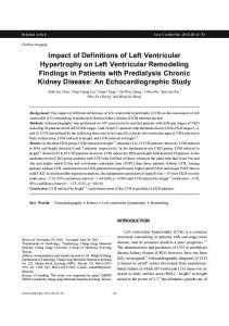
the. 12 patients with normal left ventricular ejection fraction, mean right atrial pressure ..... 'stiff heartâ syndrome. Am. J. Cardiol. 1975;. 36:438-44. 4 Swanton. RH,.
with a low (
Jun 10, 2014 - Victor Utomi,1 David Oxborough,1 Euan Ashley,2 Rachel Lord,1 Sarah ... Mike Stembridge,4 Rob Shave,4 Martin D Hoffman,5 Greg Whyte,1 ...
Twenty children (10.2 ± 0.4 years of age) classified as overweight, on the basis of ... ficantly lower in the overweight than in the normal-weight groups, whereas ...
Oct 22, 2008 - Dipti Gupta*, Sunil T Mathew, Deepu Alexander, William Schapiro,. Jing Han, Michael Passick, Mark J Goldman and Nathaniel Reichek.
Jul 30, 2016 - tionships of vascular ageing to changes in left ventricular systolic and diastolic function have .... house Matlab based program and determine pulse wave velocity PWV = ÎX/ÎT [17]. Ad- ..... This last point ap- pears to agree ...
end-diastolic left ventricular posterior wall thickness (LVPWd) and end-diastolic interventricular septum ... evaluation of a heart murmur which was found to.
Oct 26, 2009 - Step 5: spironolactone (25â100 mg) (APS, Cleckheaton,. UK). The study was single blinded with regard to the doctor administering the study.
1. St George's Hospital Medical School, University of London, London, United Kingdom. 2. Brompton Centre for Fetal Cardiology, Royal Brompton Hospital, ...
Left ventricular non-compaction (LVNC) is a rare congenital cardiomyopathy characterized by the presence of an extremely thickened endocardial layer with ...
2) Attenhofer J CH, Connolly HM, Warnes CA, O'leary P, Tajik AJ, Pellikka. PA, et al. ... 7) Van Engelen K, Postma AV, van de Meerakker JB, Roos-Hesselink JW,.
A46 year old previously fit and healthy male was admitted with a right femoral artery embolus, which was immediately removed by percutanous thrombectomy.
LOUIS E. TEICHHOLZ, AND MICHAEL V. HERMAN. From the Division of Cardiology, Department of Medicine of The Mount Sinai Hospital and The Mount .... ventricular contractility without asynergic seg- ments. .... C. V. Mosby, St. Louis.
over, evidence for a good inter-vendor agreement of 2-D strain data from the ... With the 2-D speckle tracking software from each vendor, radial, circum- ferential and ...... Medicine; Yoshihiro Seo, MD, Faculty of Medicine, University of Tsukuba;.
volume (LVEDV) and left ventricularend-systolic volume. (LVESV) and left ventricularejection fraction (LVEF). Validation studies indicate that measurements ...
can then be used to assess left ventricular end-diastolic volume (LVEDV) and ... determine normal measurements for both gated SPECT LVV and gated SPECT ...
In patients with STEMI treated by PCI, the recovery of LV subendocardial shortening strain seen in the longitudinal direction underlies the improvement in LV ...
Mar 22, 2014 - autopsy cases of sudden death with suspicion of this disease. ... Key words: Left ventricular non-compaction of the myocardium, autopsy, ...
Jan 27, 2016 - Presence Of Left Ventricular Non Compaction In. Hypertrophic Cardiomyopathy Is Associated With. Arrhythmia. Ganesh Kumar Gnanappa1,2*, ...
our recent study11 had shown that septal E/Em is better than IVRT to .... 0.076. Late diastolic velocity (Am), cm/s. 9.4 ± 1.6. 9.4 ± 1.8. 0.995. Em/Am. 0.66 ± 0.19.
ventricular images showing the effectiveness of the parametric features for normal/abnormal discrimination. Index Termsâ echocardiography, 4-chamber views,.
Left ventricular noncompaction (LVNC) is a myocardial dis- ... The most common cardiac abnormality was dilated cardiomyopathy (n = 15,. 24%). There was no ...
Left ventricular hypertrophy in athletes. John Rawlins, Amit Bhan, and Sanjay Sharma*. King's College Hospital, Denmark Hill, London SE5 9RS, UK. Received ...
Normal Data of Left Ventricular Parameters in Non ...
Juergens KU, Grude M, Fallenberg EM, Opitz C,. Wichter T, Heindel W, et al. Using ECG-gated multidetector CT to evaluate global left ventricular. Cardiac.
Normal Data of Left Ventricular Parameters in Non-Hypertensive Patients Using 320-Slice Cardiac CT: A Study on Intra- and Interobserver Variability Sutipong Jongjirasiri MD*, Chanika Sritara MD*, Phanloet Waeosak MD*, Jiraporn Laothamatas MD* * Ramathibodi Hospital, Mahidol University, Bangkok, Thailand
Objective: Delineate normal data in a Thai population for global left ventricular function, left ventricular mass (LVM), and LV mass index (LVMI) by gated volumetric CT angiography (CTA). Material and Method: Two hundred twenty seven patients with intermediate risk factors for CAD were referred for CT coronary angiography. The non-hypertensive and non-diabetic asymptomatic group was selected with negative coronary CTA. Pre-condition of mild stenosis or less than 50% area stenosis was consistently met for inclusion in the present study. One hundred fifteen patients were included (31 males, 84 females, age range 38-76 years, mean 54.6 + 7.2 for males and 54.0 + 6.8 for females respectively). Results: Mean EDV, ESV, SV, CO, and EF in male and females were EDV 122.6 + 17.3 vs. 94.9 + 16.6 ml, ESV 48.2 + 9.9 vs. 34.1 + 9.1 ml, SV 74.5 + 14.2 vs. 60.8 + 10.6 ml, CO 4.2 + 0.8 vs. 3.6 + 0.7 L and EF 60.6 + 6.6 vs. 64.3 + 5.6% respectively. The mean LVM and LVMI were higher in males than females (LVM 134.6 + 21.4 vs. 96.9 + 20.3 g, and LVMI 76.8 + 12.4 vs. 61.1 + 11.56 g/m2). Altman and Bland plot for each of them showed that the mean and standard deviation of the differences was constant throughout the range of measurements. Each histogram of differences showed that these differences correspond to approximately normal distribution, indicating that the required assumptions held for the use of 95% limits of agreement are valid. Conclusion: Data from this group represents healthy volunteers and this may serve as an important source of information representing normal reference values to be used for Thai patients. Keywords: 320-slice cardiac CT, Left ventricular parameters, Normal data
J Med Assoc Thai 2011; 94 (9): 1053-60 Full text. e-Journal: http://www.mat.or.th/journal Left ventricular (LV) function is an independent predictor of morbidity and mortality in patients with coronary artery disease (CAD)(1,2). Patients with both coronary artery disease and depressed LV function are at high risk for sudden death. LV hypertrophy (LVH) is also associated with a significant increase in cardiovascular mortality and morbidity independent of the presence of coronary artery disease or arterial hypertension(3-6). Prevailing noninvasive imaging modalities include echocardiography, radionuclide ventriculography, gated perfusion single-photon emission CT, and multislice CT are being used to evaluate left ventricular function (LVF). Volume mediated CT scan using 320-slice CT Correspondence to: Jongjirasiri S, Ramathibodi Hospital, Mahidol University, Rama VI Rd, Bangkok 10400, Thailand. Phone: 0-2201-2465, 0-2201-1212, Fax: 0-2220-1297 E-mail: [email protected]
J Med Assoc Thai Vol. 94 No. 9 2011
technology has been recently released for general use. One heart beat scan time is very short and will better enhance the contour of intraventricular cavities. The measurement of the LV function and mass should be ideally depicted when only single volume data is used. The error from interpolation data between slices from spiral mode cardiac such as respiratory registration artifacts causing overlapping or missing of interslice data information can be eliminated with this one volume scan obtained from this technology. The present study outlines non-clinical data of normal LV function and mass acquired from single volume, single heart beat data information. Material and Method Between August and October 2009, a cohort of 227 consecutive asymptomatic patients with intermediate risk factors for CAD (NYHA I) referred to undergo coronary CT angiogram (CCTA) using
1053
320-slice volume cardiac CT (Aquilion one Toshiba, Japan) at Ramathibodi Hospital, Mahidol University. The results were retrospectively reviewed for normative data LV parameters. Prior institutional review board approval and the informed consent were dutifully obtained. Exclusion criteria included hypertension (Blood pressure > 140/90 mmHg), receiving antihypertensive therapy, history of angina pectoris, diabetes mellitus with peripheral vascular disease, iodinated contrast hypersensitivity, and creatinine blood level > 1.5 mg/dl. Of these patients, a group of 115 patients who had non-significant coronary artery stenosis were included. All patients who had initial heart rate more than 70 BPM were administered 50-100 mg metoprolol one hour prior to scanning. CCTA was performed using the 320-slice CT scanner. Patients were examined with retrospective ECG-triggered, ECG modulated tube current dose reduction methodology scanning a range between 30% and 80% of the cardiac cycle. The tube voltage varied with patient size (120-135 kVP, 380-500 mA). The rotation time was 0.35 second at 0.5 mm. collimation. An 18 gauge intravenous catheter was placed in the right antecubital vein. An enhanced scan was obtained following injection of 70-90 ml non-ionic contrast (Ultravist, 370 mg%) given at 5 ml/second followed by 20 ml of saline at 5 ml/second. Automated bolus tracking was used. A scan was performed only after a pre- condition of 170 HU density was reached in the descending aorta. After this threshold was met at 170 HU, the scan started after a four seconds delay. Ten phases of the cardiac cycle were reconstructed at 10% intervals from 0% R-R interval to 90% R-R interval and sent to an independent workstation (Vitrea FX version 1.1, Vital Images, Inc., Minnesota, USA) for LV function analysis and LV mass calculation. Optimal phase, being the best phase, usually locates at 75% level was reconstructed to give a better picture. Interpretation for coronary artery stenosis included analysis of axial source images, 3 mm maximum intensity projection (MIP) in axial, right anterior oblique (RAO), left anterior oblique (LAO), post processed 3-D reconstruction and multiplanar reformats, and curve reformats for each coronary arteries. LV function and LV mass analysis The analysis and calculation of LV function and LV mass were done by two independent observers to obtain a measure of inter-observer variability.
1054
Observer 1 and 2 were senior technologists with more than five years of experience in cardiac MDCT. Two observers delineated the outer and inner contours of the LV independently and in a random order of patients. Furthermore, the data was also reanalyzed and recalculated by one of the observers several weeks after the first analysis of the images. The intraobserver study was repeated separately, 1 month apart. In this way, a measure of intra-observer variability was also clearly delineated. The papillary muscle was not included in the LV myocardial mass (Fig. 1). The observers were asked to analyze the software in a similar manner starting with automatic phase selection of systolic and diastolic phases followed by manually phase correction. The systolic and diastolic volume was automatically calculated by delineation of endocardial border of the left ventricle inside the inner layer of the left ventricular muscle. The myocardial mass was calculated from volume of the area between inner and outer contours of the left ventricle (Fig. 2). The four-layout screen was an automatic setup allowing the operator to set apical and basal myocardial limits. Correction along axis
Fig. 1
This demonstrates diastolic phase of the vertical long axis, horizontal four chamber and short axis (a, b and d), the outlined endocardial border in d represents end-diastolic volume. The papillary muscle was not included. Automatic contour detection is excellent due to isophasic contrast and no stair step artifact. The systolic and diastolic phases are chosen to calculate for global left ventricular function. 3-D image of the heart with normal left coronary artery is also demonstrated (c)
J Med Assoc Thai Vol. 94 No. 9 2011
Results
Fig. 2
This demonstrates automatic contour detection along the epicardial (outer dots line) and endocardial (inner dots line) borders. The area between these lines represents volume of left ventricular mass. No artifact demonstration is shown
angulations in three directions, vertical two chambers, four chambers, and short axis views were done in the same manner (Fig. 1). Statistical analysis Descriptive statistics were used to summarize all data in term of frequency (percentage), mean, median, range standard deviation (SD) and 95% confidence interval where appropriate. Regression and correlation analysis were applied between the functional parameters and mass correlate with intraobserver and inter-observer variables.
Only 115 from 227 patients ( 50.66%) were included for analysis. There were 31 male and 84 females in the present study. The patients’ demographic data are shown in Table 1. The mean + SD calcium score was 6.3 + 11.1 in males and 6.6 + 18.5 in females (Table 1). The body surface area (BSA) is calculated using DuBois & DuBois formula(7). The results of the left ventricular parameters are shown in Table 2. The commercially available LVM formula is used converting calculating the mass in grams using the specific gravity of myocardial tissue (1.05 g/ml). The left ventricular mass index (LVMI) calculated as the ventricular mass in grams divided by the body surface area in square meters. Mean EDV, ESV, SV, CO, and EF in male and female are as follows: EDV 122.6 + 17.3 vs. 94.9 + 16.6 ml, ESV 48.2 + 9.9 vs. 34.1 + 9.1 ml, SV 74.5 + 14.2 vs. 60.8 + 10.6 ml, CO 4.2 + 0.8 vs. 3.6 + 0.7 L, and EF 60.6 + 6.6 vs. 64.3 + 5.6%, respectively. The mean LVM (Left ventricular mass) and LVMI (left ventricular mass index) are higher in males than females (LVM 134.6 + 21.4 vs. 96.9 + 20.3 g, LVMI 76.8 + 12.4 vs. 61.1 + 11.5 g/m2). Each Altman and Bland plot showed that the mean and SD of the differences were constant throughout the range of measurements and each histogram of differences showed that these differences have approximately normal distribution, indicating that the required assumption are valid and in agreement with use of 95% limits (Fig. 3-6). The Pearson’s
Table 1. Patient characteristics Characteristics Number of patients (total = 115), n Age, (year) Hyperlipidemia, n (%) Current cigarette smoking, n (%) Body mass index, kg/m2 + Weight, kg Height, cm Waist, inch Body surface area (BSA), m2 ++ Systolic blood pressure (SBP), mmHg Diastolic blood pressure (DBP), mmHg Heart rate at CT, bpm Total calcium score (mean + SD) (median, min-max)
Values are mean + SD unless indicated otherwise. + Calculated as the weight in kilograms divided by the square of the height in meters ++ Calculated with DuBois & DuBois formula(7)
J Med Assoc Thai Vol. 94 No. 9 2011
1055
Table 2. Results of the left ventricular parameters Left ventricular parameters
Ejection fraction (EF), % End diastolic volume (EDV), ml End systolic volume (ESV), ml Stroke volume (SV), ml Cardiac output (CO), L Left ventricular mass (LVM), g Left ventricular mass index (LVMI), g/m2 + +
Calculated as the left ventricular mass in grams divided by the body surface area in square meters
correlations analysis showed the functional parameters and mass correlated closely between intraobserver and interobserver values. The intraobserver variability for ejection fraction (EF), end diastolic volume (EDV), end systolic volume (ESV), stroke volume (SV), cardiac output (CO), and myocardial mass(M) is R = 0.92, 0.98, 0.97, 0.96, 0.96, and 0.97 respectively. The interobserver variability of EF, EDV,
Fig. 3
1056
Plot of difference against mean (Altman and Bland plot) for measurements of (a) LVEF, (b) EDV, (c) ESV (image left) and histogram of differences of the 2 measurements by same observer of the same measurement (image right) using 320-slice Cardiac CT
ESV, and SV is R = 0.69, 0.90, 0.87, 0.80, 0.80, 0.87, and 0.94 respectively (Table 3). Discussion Several noninvasive studies used to evaluate cardiac function, such as echocardiography, scintigraphy, and single-photon emission computed tomography show diagnostic reliability but low spatial
Fig. 4
Plot of difference against means (Altman and Bland plot) for measurements (a) SV, (b) CO and (c) mass (image left) and histogram of differences of the 2 measurements by same observer (image right) of the same measurement using 320-slice Cardiac CT
J Med Assoc Thai Vol. 94 No. 9 2011
Fig. 5
Plot of difference against mean (Altman and Bland plot) for measurements (image left) of (a) LVEF, (b) EDV, (c) ESV and histogram of differences of the 2 measurements by two different observers (image right) of the same measurements using 320-slice Cardiac CT
resolution and poor anatomic details(8). The parallel increase in the number of detectors and acquisition angle and the change from pencil to fan beam contribute to improved image quality and spatial resolution(8). Left ventricular function parameters in cardiac CT was initially introduced by electron beam CT but the access to this modality is restricted to inadequate spatial resolution, contrast, and z-resolution(2,9). In 1985, Gunthaner et al demonstrated the ability of CT for volume measurements (10). The accuracy for determination of ejection using electron beam computed tomography was also validated against Tc-99m sestamibi first-pass angiography(11,12). Wiese et al compared multislice CT (MSCT) using four detectors CT with electron beam CT to evaluate left ventricular (LV) volumes and function in an animal model and found that MSCT had a higher contrast-to noise ratio and delineated the myocardial contours more sharply than electron beam CT (EBCT). They concluded that image quality in MSCT is superior to that of EBCT but the accuracy MSCT is limited by its lower temporal resolution(13).
J Med Assoc Thai Vol. 94 No. 9 2011
Fig. 6
Plot of difference against mean (Altman and Bland plot) for measurements of (a) SV, (b) CO and (c) mass (images left) and histogram of differences of the 2 measurements by two different observers (images right) of the same measurement using 320-slice Cardiac CT
MSCT has evolved to include a multirow detector from dual to 64 detectors and current 320 detectors, progressively increasing both the number of simultaneously acquired images and temporal resolution. Makhen et al. showed that MSCT has been acceptable agreement with MR imaging(14). Automated analysis of left ventricular parameters was significantly quicker than manual drawing, indicating a potential improvement in workflow and data analysis. Schlosser et al showed that improvement of software with automatic contour detection allows a reliable assessment of left ventricular ejection fraction and a rough estimation of left ventricular volumes and mass of a CT angiography using 16-MDCT scanner(5). They concluded that automatic contour detection is feasible for CT data sets and results in fast and reliable measurements without significant differences compared with manual contour tracing and improvement in workflow and data analysis. There is still a problem with visual quality assessment, because the quality assessment for LV functional analysis focuses more on opacification of the LV cavity and the presence of stair step artifacts
1057
Table 3. Intra- and inter-observer variability of the technique of each cardiac parameter in all subjects (n = 115) Cardiac parameters EF
compared it with MRI. He categorized image quality from dual source CT under four headings. These four categories were good quality image, minor artifacts, major artifacts, and poor image quality respectively. The good image quality and minor artifacts were called when the image has no artifacts, and minor artifacts that made LV delineation sufficiently possible. When there were major artifacts, the LV delineation is not reliable. Poor image quality was called when there is no full coverage. The era of single volume CT data using 320-slice computed tomography (CT) for LV function evaluation has begun. It has high performance apparatus to collect data information not only for coronary arteries but also left ventricular volume and left ventricular (LV) mass within one heartbeat. One volume data means that there is no interpolation of the image data set. In this technique, stair step artifacts, and no full coverage defects are eliminated. There is one limitation for dose modulation technique, which is important in reducing radiation dose, in which it decreases the image quality during ventricular systole. This may affect accuracy of the automatic contour detection. However, the single volume data acquisition with isophasic contrast alleviates this limitation and improves post processing reproducibility. In conclusion, normative data from this group could represent a healthy volunteer Thai population. This will prove to be an important data set for normal reference. This data acquired from an ideal measurement of single volume image data acquisition. It should provide an accurate data set for both functional parameters and LV mass compared to a previous version of multislice CT. Acknowledgment The authors wish to thank Inder Singh Mayall, MD for his assistance with manuscript review.
EF = ejection fraction; EDV = end diastolic volume; ESV = end systolic volume; SV = stroke volume; CO = cardiac output; LVM = left ventricular mass
Potential conflicts of interest None.
caused by ventricular extra-systoles breathing artifacts or segmentation error(15). These data information are acquired from multiple rotation acquisition resulting into interpolated data acquisition and limited for automatic software contour detection due to nonisophasic of contrast in the inner LV contour. Van der Vleuten et al(16) assessed the global left ventricular function by dual-source computed tomography and
References 1. White HD, Norris RM, Brown MA, Brandt PW, Whitlock RM, Wild CJ. Left ventricular endsystolic volume as the major determinant of survival after recovery from myocardial infarction. Circulation 1987; 76: 44-51. 2. Juergens KU, Grude M, Fallenberg EM, Opitz C, Wichter T, Heindel W, et al. Using ECG-gated multidetector CT to evaluate global left ventricular
1058
J Med Assoc Thai Vol. 94 No. 9 2011
3.
4.
5.
6.
7.
8.
9.
10.
11.
12.
myocardial function in patients with coronary artery disease. AJR Am J Roentgenol 2002; 179: 1545-50. Levy D, Garrison RJ, Savage DD, Kannel WB, Castelli WP. Prognostic implications of echocardiographically determined left ventricular mass in the Framingham Heart Study. N Engl J Med 1990; 322: 1561-6. Ghali JK, Liao Y, Simmons B, Castaner A, Cao G, Cooper RS. The prognostic role of left ventricular hypertrophy in patients with or without coronary artery disease. Ann Intern Med 1992; 117: 831-6. Schlosser T, Pagonidis K, Herborn CU, Hunold P, Waltering KU, Lauenstein TC, et al. Assessment of left ventricular parameters using 16-MDCT and new software for endocardial and epicardial border delineation. AJR Am J Roentgenol 2005; 184: 765-73. Budoff MJ, Ahmadi N, Sarraf G, Gao Y, Chow D, Flores F, et al. Determination of left ventricular mass on cardiac computed tomographic angiography. Acad Radiol 2009; 16: 726-32. Du Bois D, Du Bois EF. A formula to estimate the approximate surface area if height and weight be known. 1916. Nutrition 1989; 5: 303-11. Daghini E, Ritman EL, Lerman LO. Examine thy heart with all diligence: evaluation of cardiac function using fast computed tomography. Hypertension 2007; 49: 249-56. Woo P, Mao S, Wang S, Detrano RC. Left ventricular size determined by electron beam computed tomography predicts significant coronary artery disease and events. Am J Cardiol 1997; 79: 1236-8. Guthaner DF, Nassi M, Bradley B, Gould EB, Mai CH, Schmidt KE. Quantitative evaluation of left ventricular function using computed tomography. Med Phys 1985; 12: 333-8. Gerber TC, Behrenbeck T, Allison T, Mullan BP, Rumberger JA, Gibbons RJ. Comparison of measurement of left ventricular ejection fraction by Tc-99m sestamibi first-pass angiography with electron beam computed tomography in patients with anterior wall acute myocardial infarction. Am J Cardiol 1999; 83: 1022-6. Baik HK, Budoff MJ, Lane KL, Bakhsheshi H,
J Med Assoc Thai Vol. 94 No. 9 2011
13.
14.
15.
16.
17.
18.
19.
20.
Brundage BH. Accurate measures of left ventricular ejection fraction using electron beam tomography: a comparison with radionuclide angiography, and cine angiography. Int J Card Imaging 2000; 16: 391-8. Wiese TH, Rogalla P, Taupitz M, Wagner S, Schnorr J, Mews J, et al. Assessment of left ventricular volumes and function: intraindividual comparison of multi-slice spiral CT and electron beam CT in an animal model. Acta Radiol 2004; 45: 819-27. Mahnken AH, Spuentrup E, Niethammer M, Buecker A, Boese J, Wildberger JE, et al. Quantitative and qualitative assessment of left ventricular volume with ECG-gated multislice spiral CT: value of different image reconstruction algorithms in comparison to MRI. Acta Radiol 2003; 44: 604-11. Kroft LJ, de Roos A, Geleijns J. Artifacts in ECGsynchronized MDCT coronary angiography. AJR Am J Roentgenol 2007; 189: 581-91. van der Vleuten PA, de Jonge GJ, Lubbers DD, Tio RA, Willems TP, Oudkerk M, et al. Evaluation of global left ventricular function assessment by dual-source computed tomography compared with MRI. Eur Radiol 2009; 19: 271-7. Stollberger C, Hollander I, Dimitrov L, Slany J. Influence of measurement inaccuracies on determination of left ventricular mass by M mode echocardiography. Heart 1996; 75: 312-3. Bastarrika G, Arraiza M, De Cecco CN, Mastrobuoni S, Ubilla M, Rabago G. Quantification of left ventricular function and mass in heart transplant recipients using dual-source CT and MRI: initial clinical experience. Eur Radiol 2008; 18: 1784-90. Yamamuro M, Tadamura E, Kubo S, Toyoda H, Nishina T, Ohba M, et al. Cardiac functional analysis with multi-detector row CT and segmental reconstruction algorithm: comparison with echocardiography, SPECT, and MR imaging. Radiology 2005; 234: 381-90. Okwuosa TM, Hampole CV, Ali J, Williams KA. Left ventricular mass from gated SPECT myocardial perfusion imaging: comparison with cardiac computed tomography. J Nucl Cardiol 2009; 16: 775-83.