Nuclear factor-B-like activity increases in murine cerebral cortex after sleep deprivation ZUTANG CHEN, JANOS GARDI, TETSUYA KUSHIKATA, JIDONG FANG, AND JAMES M. KRUEGER Department of Veterinary and Comparative Anatomy, Pharmacology, and Physiology, College of Veterinary Medicine, Washington State University, Pullman, Washington 99164 Chen, Zutang, Janos Gardi, Tetsuya Kushikata, Jidong Fang, and James M. Krueger. Nuclear factor-B-like activity increases in murine cerebral cortex after sleep deprivation. Am. J. Physiol. 276 (Regulatory Integrative Comp. Physiol. 45): R1812–R1818, 1999.—Several well-defined sleep regulatory substances, e.g., interleukin-1, activate the heterodimeric transcription factor nuclear factor-B (NF-B). Several substances that inhibit sleep, e.g., interleukin-4, inhibit NF-B activation. NF-B activation promotes production of several additional substances thought to be involved in sleep regulation, e.g., nitric oxide. We investigated, therefore, whether there are diurnal rhythms of NF-B activation in brain and changes in the activation after sleep deprivation. Mice were kept on a 12:12-h light-dark cycle. In one experiment, groups of mice were killed every 3 h across the 24-h cycle. In another experiment, mice were killed at 1500 after 6 h of sleep deprivation, and a group of control mice were killed at the same time. Nuclear proteins were extracted from each brain tissue sample, and NF-B-like activity was determined with an electrophoretic mobility shift assay. In cerebral cortex, but not other areas of brain, there was a diurnal rhythm in NF-B-like activation; highest levels were found during the light period. NF-B-like activation was higher in cerebral cortex after sleep deprivation compared with values obtained from control mice. The results are consistent with the hypothesis that sleep regulation involves multiple gene events, some of which include enhanced production of sleep regulatory substances, the actions of which involve NF-B activation. cytokine; slow-wave sleep; transcription factor; circadian rhythm
NUCLEAR FACTOR-B (NF-B) is a heterodimeric transcription factor activated by many substances that also promote sleep. In cytoplasm, NF-B is complexed to inhibitory proteins called I-B; the complex is inactive. Phosphorylation of I-B results in its dissociation from NF-B; this activation step allows NF-B to translocate to the nucleus, where it binds to a B consensus DNA sequence, most often inducing enhanced gene expression (reviewed in Refs. 1, 29). Sleep regulation is dependent, in part, on changes in gene expression and production of sleep regulatory substances (SRS) (reviewed in Refs. 19 and 21). There is extensive evidence indicating that interleukin-1 (IL-1), tumor necrosis factor-␣ (TNF-␣), and PGs are involved in sleep regulation (reviewed in Refs. 19 and 21). Each of these SRS is
The costs of publication of this article were defrayed in part by the payment of page charges. The article must therefore be hereby marked ‘‘advertisement’’ in accordance with 18 U.S.C. Section 1734 solely to indicate this fact. R1812
involved in NF-B regulation (32, 43; reviewed in Ref. 1). Furthermore, several additional somnogenic substances activate NF-B; the list includes nerve growth factor (NGF) (5, 40), epidermal growth factor (27), interferon-␣ (6, 33), acidic fibroblast growth factor (2), insulin, and insulin-like growth factor (6) (see Refs. 19 and 21 for review of effects of each of these substances on sleep). In contrast, interleukin-10 (IL-10) (39), interleukin-4 (IL-4) (9), and glucocorticoids (31, 37, 38) directly or indirectly inhibit NF-B activation and inhibit sleep (sleep inhibitory effects reviewed in Refs. 19 and 21). Finally, NF-B activation results in increased interleukin 2 (14), cycloxygenase-2 (COX-2) (25, 26, 43), nitric oxide synthase-2 (NOS-2) (28, 30, 36, 41), NGF (11), IL-1, and TNF-␣ production (reviewed in Ref. 1). NF-B enhancement of NGF, IL-1, and TNF-␣ expression results in a positive feedback loop that is likely involved in the amplification of somnogenic signals. NF-B enhancement of NOS-2 and COX-2 results in increased production of PGs and nitric oxide (NO). PGs and NO promote sleep (reviewed in Refs. 19 and 21); feedback from PGE2 and NO can also inhibit NF-B, thereby providing a downstream mechanism to dampen the NGF-TNF␣-IL-1-NF-B system. Upstream damping mechanisms include the actions of IL-4 and IL-10 on these substances. Because NF-B is also a well-characterized brain product implicated in several pathological states associated with sleep disorders, e.g., traumatic brain injury (44), ischemia (10), and Alzheimer’s disease (3), we hypothesized that NF-B could provide a common mechanism involved in SRS-altered sleep. We report herein that sleep deprivation increases NF-B activation in the cerebral cortex and that there is a diurnal rhythm of NF-B in the cortex. METHODS AND MATERIALS
Animals. Sixty-day-old B6X129F-2 adult male mice were purchased from The Jackson Laboratory (Bar Harbor, ME). Animals were individually housed in a sound-attenuated environmental chamber (Hotpack 352600) with 29 ⫾ 1°C ambient temperature and a 12:12-h light-dark cycle (lights on at 0800 and off at 2000). Food and water were available continuously throughout the experimental period. The animals were kept in that environment for at least 10 days to acclimate to the housing conditions before the experiments and were between 72 and 76 days of age at the time of death. Experimental protocol. In experiment I, mice were killed every 3 h starting at 0900 (1 h after light onset) by cervical dislocation and decapitation; 8–10 mice were used per time point. Thus four time points at 0900, 1200, 1500, and 1800 were during the light period, and the remaining four time points, 2100, 2400, 0300, and 0600, were during the dark
0363-6119/99 $5.00 Copyright r 1999 the American Physiological Society
Downloaded from www.physiology.org/journal/ajpregu by ${individualUser.givenNames} ${individualUser.surname} (139.081.195.094) on March 22, 2018. Copyright © 1999 American Physiological Society. All rights reserved.
SLEEP DEPRIVATION ACTIVATES NF-B
hours. When animals were killed during dark hours, it was necessary to turn on the lights to perform the experiment. The amount time from when the lights were turned on until when the animals were cervically dislocated was about 1 min. In experiment II, 6 h of sleep deprivation was performed from 0900 to 1500 by gently handling the mice (n ⫽ 9). Sleepdeprived animals were killed by cervical dislocation immediately after sleep deprivation. Control mice (n ⫽ 8) remained undisturbed and were killed at the same time as the sleepdeprived mice. Brains were quickly removed, and individual brain areas were dissected within 2 min after death with a sterile set of instruments for each mouse. The landmarks for the hypothalamus were optic chiasma, lateral sulci, mammillary bodies, and a depth of 1 mm. Cerebral cortex was sampled from the dorsal surface of the parietal cortex. Whole brain stem, cerebellum, and hippocampus were also separated and saved. The samples were placed in a preweighed tube and then were snap frozen in liquid nitrogen and kept at ⫺80°C until nuclear protein extraction. Nuclear protein extraction. On the day of nuclear protein extraction, the tubes containing brain tissue samples were taken out of the freezer and kept on ice, and the weight of the tissues was obtained. Brain tissues were washed with PBS and homogenized in four tissue volumes of buffer A [0.5 M sucrose, 10 mM HEPES pH 7.9, 1.5 mM MgCl2, 10 mM KCl, 10% glycerol, 1 mM EDTA, 1 mM 1,4-dithiothreitol (DTT), 1 mM phenylmethylsulfonyl fluoride (PMSF), and 1 mg/ml each of aprotinin, leupeptin, and pepstatin A], receiving 10 strokes with an Eppendoff sample pestle. The homogenate was incubated on ice for 5 min. After centrifugation at 4,000 g for 5 min at 4°C, the supernatant was discarded. The nuclear pellet was resuspended in one tissue volume of buffer B (20 mM HEPES, 1.5 mM MgCl2, 0.2 mM EDTA, 0.3 M NaCl, 0.5 mM DTT, 0.5 mM PMSF, 20% glycerol) with sonication (2 times, 5 s each; Vibra-Cell, Sonics and Materials, Danbury, CT) while the tube was kept on ice. The tubes were incubated on ice for another 15 min. The soluble nuclear proteins were recovered by centrifugation at 13,000 g for 10 min at 4°C. Nuclear protein was quantified by the Bradford method (Bio-Rad, Hercules, CA). The standards (BSA in 5 µl buffer B and 95 µl water), and 5 µl of samples in 95 µl water mixed with 900 µl dye were read at an optical density of 595 nm with the use of a Shimadzu UV-1601 spectrophotometer. The protein concentrations from each experimental sample were calculated with the standard curve. Probe labeling and electrophoresis. Oligonucleotides derived from murine IgK gene promoter enhancer B with the sequence 58-CAGAGGGGACTTTCCGAC-38 and its antisense (6 nucleotides shorter) 58-GTCGGAAAGTCC-38 were synthesized (GIBCO BRL, Grand Island, NY). Equal molarities (100 µM each) of these two oligonucleotides were annealed in Tris-EDTA (TE) buffer (0.1 mM EDTA pH 8.0, 10 mM Tris pH 7.4) by boiling on a hot block for 5 min and cooling down at room temperature. The double-stranded (both sense and antisense oligonucleotides) B sequences from mouse IgK gene promoter enhancer 58-AGCTTCAGAGGGACTTTCCTCTGA-38 (24) and mouse NOS-2 gene promoter 58-TGCTAGGGGG ATTTTCCCTCTC-38 (42) were made by the same method. Underlined are the NF-B-binding consensus sequences. These two double-stranded oligonucleotides were used in a competitive binding assay to demonstrate DNA protein binding specificity. The double-stranded oligonucleotide (1 µl of 50 µM) was labeled with 1 µl of 10 mM dA/C/GTP and 5 µl 32P-labeled dCTP (10 mCi/ml; NEN, Du Pont, Boston, MA) and 2 µl of Klenow fragment in 50 µl of 1X buffer (Promega, Madison, WI). The free uncoupled nucleotides were removed by filtration through a G-50 column as follows: the reaction
R1813
mixture (50 µl) was loaded onto a prespun TE midi select-D micro G-50 column (5–3 Prime, Boulder, CO) and spun at 6,000 g for 5 min (same speed and time for spinning out buffer before loading sample). The labeled probe was collected into a fresh tube, and the dpm of the probe was determined with a scintillation counter. Nuclear protein extracted from each brain tissue sample was individually examined with an electrophoretic mobility shift assay. The electrophoretic mobility shift assay has been used for the study of sequence-specific DNA binding proteins, such as transcription factors (7). The assay is based on the observation that DNA protein complexes migrate slower through a nondenaturing polyacrylamide gel than do the free unbound DNA fragments. In our case, the protein (NF-B)DNA (labeled B sequence) complex was separated from free unbound DNA by gel electrophoresis. The NF-B and oligonucleotide-binding specificity was determined with the use of excess cold (unlabeled) oligonucleotides to compete with labeled DNA oligonucleotides for the NF-B. In addition, a recombinant 50-kDa NF-B protein was purchased from Promega and used as a positive control (Fig. 2). The DNA protein-binding reaction was performed by incubating 20 µg nuclear extract with 32P-labeled oligonucleotide (1 ⫻ 104 counts/min) in a total volume of 15 µl binding buffer containing 10 mM Tris (pH 7.5), 0.1 mM EDTA, and 50 mM KCl for 30 min at room temperature. For the test of DNA protein binding specificity, cold unlabeled DNA oligonucleotides were mixed with labeled probes in room temperature for 5 min before the protein sample was added into the reaction. The reaction was mixed with 5 µl loading buffer (0.25% bromophenol blue, 0.25% xylene cyanol FF, and 30% glycerol in water) and separated with a 6% polyacrylamide gel running with 1X Tris-borate-EDTA buffer at 100 V in a vertical gel electrophoresis apparatus (gel size, 150 ⫻ 170 ⫻ 1 mm). The electrophoresis was stopped when the front of the dye reached 4 cm from the bottom of the plate. The gel was transferred onto a 3M filter paper and dried on a gel drier (Bio-Rad) at 80°C for 2 h. An autograph was taken by exposure of the dried gel to the X-ray film overnight at ⫺80°C. Densitometry was obtained by the Gel Doc 1000 analysis (Bio-Rad). Arbitrary units were derived from densitometry readings of film that was exposed to the dried gel containing 32P-labeled DNA and protein binding. Statistics. The effects of sleep deprivation on NF-B activation were analyzed with t-tests. The diurnal rhythm of NF-B activation was analyzed with the Mann-Whitney rank sum test and one-way ANOVA followed by the Student-NewmanKeuls test. The differences between groups were considered significant if the P values were ⬍ 0.05. RESULTS
In experiment I, NF-B activation in the cerebral cortex had a diurnal rhythm with more activation during the light period than during the dark period. NF-B activation of all samples taken from the four time points during light hours (0900, 1200, 1500, and 1800) was higher than samples taken during dark hours (2100, 2400, 0300, and 0600; Fig. 1). Mean NF-B activation during light hours was 189 ⫾ 20 vs. a nighttime average of 115.5 ⫾ 23.8 (T ⫽ 1,009.0, P ⬍ 0.0001; Mann-Whitney rank sum test). One-way ANOVA also indicated that there was a significant time effect on the NF-B activation [F(7,69) ⫽ 5.17, P ⬍ 0.0001]. The values of hours 2400, 0300, and 0600 were significantly lower compared with those of hour 0900
Downloaded from www.physiology.org/journal/ajpregu by ${individualUser.givenNames} ${individualUser.surname} (139.081.195.094) on March 22, 2018. Copyright © 1999 American Physiological Society. All rights reserved.
R1814
SLEEP DEPRIVATION ACTIVATES NF-B
from a sleep-deprived mouse (same as shown in lane 4) was chosen to test the NF-B protein and 32P-labeled DNA oligonucleotide binding specificity. Before the nuclear protein was added into the reaction, the labeled DNA probe (104 cpm) was mixed with 5 or 10⫻ molar excess of unlabeled B sequence (lanes 9 and 10) or 2, 5, 10, and 20⫻ molar excess of an unlabeled 22-base pair nucleotide derived from mouse NOS-2 gene promoter (lanes 11–14) as described in METHODS AND MATERIALS or 150⫻ molar excess of an unrelated DNA fragment (lane 15), or no cold DNA fragment was added (lane 16). Because the NOS-2 promoter has a conserved NF-B binding sequence, the DNA fragment has a similar affinity to bind activated NF-B protein as B sequence
Fig. 1. Nuclear factor B (NF-B) activation has a diurnal rhythm. During light period, activated NF-B in cortex was greater than during dark hours. Eight to ten mice at each of 8 time points (0900, 1200, 1500, 1800, 2100, 0000, 0300, and 0600) were killed, and nuclear protein samples from each of 5 brain regions were analyzed for NF-B activation. Light onset was at 0800 with 12:12-h light-dark cycle. Only samples from cortex that express greater amounts of NF-B than other regions (see Fig. 2) showed clear diurnal rhythm pattern. Daytime samples averaged 189 ⫾ 20 vs. nighttime average of 115 ⫾ 24 (P ⬍ 0.0001). Values shown are means ⫾ SE. * Significantly higher than values obtained at 2400, 0300, and 0600.
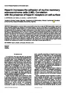
[q(8,69) ⫽ 5.962, P ⬍ 0.01; q(7,69) ⫽ 5.742, P ⬍ 0.01; and q(6,69) ⫽ 5.255, P ⬍ 0.01, respectively] and hour 1800 [q(7,69) ⫽ 5.192, P ⬍ 0.01; q(6,69) ⫽ 4.925, P ⬍ 0.01; and q(5,69) ⫽ 4.438, P ⬍ 0.01, respectively]. Within the four time points during daytime or the four time points during nighttime, there were no significant differences between NF-B activation values. In experiment I, basal NF-B activation within the other brain regions was much lower than in the cortex (data not shown); values obtained were similar to those obtained in experiment II, which are shown in Fig. 2. No diurnal variations in NF-B activation were evident in any of these other areas of brain. In experiment II, NF-B activation in the hippocampus, hypothalamus, brain stem, and cerebellum was also much lower that that observed in the cerebral cortex. Furthermore, differences in NF-B activation in the areas outside the cortex were not detectable after sleep deprivation. In contrast, DNA binding activity in the cortical sample from a sleep-deprived mouse was much higher than those from control mice. These NF-B activation patterns were consistent from animal to animal; samples from a control mouse and a sleepdeprived mouse are shown in Fig. 2. To further confirm our finding that NF-B activity increased in cerebral cortex during sleep deprivation, additional animals were examined and NF-B and DNA probe binding specificity was tested. In Fig. 3, NF-B activity obtained from four control cerebral cortical samples (lanes 1, 3, 5, and 7) was low. In contrast, cerebral cortical samples derived from four sleep-deprived mice (lanes 2, 4, 6, and 8) exhibited high NF-B activity. One of the nuclear protein samples
Fig. 2. Electrophoretic mobility shift analysis of NF-B activation in mouse brain. NF-B activity is higher in cortex than in other brain regions, and sleep deprivation further increased NF-B activity in cortex. Lanes 1–5 are nuclear proteins extracted from brain stem (BS), cerebellum (CB), cortex (CT), hippocampus (HC), and hypothalamus (HT) of control mouse, and lanes 8–12 are from sleep-deprived mouse. Lane 6 is 5 ng NF-B recombinant protein as positive control, and lane 7 is probe alone without any protein added. NF-B activity is not caused by contamination but rather a result of protein extracted from nuclei that binds to DNA probe; without nuclear protein added into binding reaction, there is no band found in lane 7. Retarded band from experimental sample is close to size of band that purified recombinant NF-B 50-kDa protein (Promega) bound to probe (lane 6). Equal amounts of nuclear extracts (20 µg/lane) and 32P-labeled double-stranded B DNA sequence (104 cpm) were incubated at 25°C in 15 µl buffer. Reaction mixture was separated on 6% polyacrylamide gel, and autograph was made by exposure to X-ray film to dried gel. In 5 brain regions from sleep-deprived and control mice, NF-B activation was limited to cortex of sleep-deprived mice.
Downloaded from www.physiology.org/journal/ajpregu by ${individualUser.givenNames} ${individualUser.surname} (139.081.195.094) on March 22, 2018. Copyright © 1999 American Physiological Society. All rights reserved.
SLEEP DEPRIVATION ACTIVATES NF-B
R1815
Fig. 3. Electrophoretic mobility shift analysis of nuclear extracts from cortex of control (Ctr) mice brains (lanes 1, 3, 5, and 7) and sleep-deprived (SD) mice brains (lanes 2, 4, 6, 8, and 9–16). Nuclear protein samples for lanes 1–8 were extracted from individual mice; nuclear protein for lanes 9–16 was from same sleep-deprived mouse. Protein samples were quantified, and equal amounts of protein (10 mg) for each lane and 104 cpm probe were mixed in 15 µl buffer. In lanes 9 and 10, 5 and 10⫻ molar excess of mouse IgK DNA fragment was added; in lanes 11–14, an increment of concentration of nitric oxide synthase (NOS)-2 gene NF-B binding sequence was added, and 2, 5, 10, and 20⫻ molar excess, respectively, was added to reaciton. Lane 15 was 150-fold molar excess of 56-bp unrelated DNA sequence, and lane 16 is nothing added.
from IgK gene promoter. Both cold unlabeled B DNA fragments from the IgK gene promoter or from the NOS-2 gene promoter effectively blocked the activated NF-B from binding to the labeled DNA oligonucleotides (Fig. 3, lanes 9–14). Results from all nine sleep-deprived mice and eight controls in experiment II are shown in Fig. 4; sleep deprivation induced a signifi-
Fig. 4. Sleep deprivation enhances NF-B activation. Nine mice were deprived of sleep for 6 h and then killed. NF-B activation in cerebral cortex was determined as described in METHODS AND MATERIALS; values shown are means ⫾ SE. After sleep deprivation, NF-B activation was significantly higher (P ⬍ 0.0001). Eight control mice were killed at same time of day.
cantincrease in NF-B activation (t15 ⫽ 5.86, P ⬍ 0.0001). DISCUSSION
Current results confirm earlier work demonstrating constitutive NF-B activity in brain (12, 13, 15–17, 34). In those studies, NF-B activation was demonstrated in neurons. Current results did not distinguish whether NF-B activity was derived from neurons, glia, or other cell types within the brain. These previous studies also demonstrated NF-B activity in the cerebral cortex, hippocampus, and the cerebellum. Results presented here clearly demonstrate NF-B activation in the cortex and show that this is dependent, in part, on the time of day and the prior sleep history of the animal. NF-B activation in the hippocampus and cerebellum was not clear in the current studies, perhaps as a result of insufficient amounts of nuclear protein used in the electrophoretic mobility shift assay. Inherent within the concept of sleep homeostasis (4) is that a consequence of wakeful neuronal activity is the production of a substance that alters input-output relationships for a population of neurons, thereby inducing an altered state. Thus the propensity for sleep, its duration, and its intensity are dependent on prior waking neuronal activity. There is ample evidence suggesting that humoral factors are involved in this state shift. For example, many studies have demonstrated that during sleep deprivation a substance(s) accumulates in the cerebrospinal fluid that, when transferred to normal animals, induces sleep (reviewed in Refs. 19 and 21). Several of these substances have now been identified and characterized as SRS, e.g., IL-1, TNF-␣, and PGD2 (reviewed in Refs. 19 and 21).
Downloaded from www.physiology.org/journal/ajpregu by ${individualUser.givenNames} ${individualUser.surname} (139.081.195.094) on March 22, 2018. Copyright © 1999 American Physiological Society. All rights reserved.
R1816
SLEEP DEPRIVATION ACTIVATES NF-B
Although each of these substances can, under appropriate experimental conditions, independently promote sleep [e.g., COX-2 inhibitors do not block IL-1-induced sleep (18)], they likely act in concert with each other in physiological sleep regulation. One likely mechanism through which they can interact is NF-B. Current results support this notion to the extent that NF-B was highest during daylight hours (the sleep period in mice) and increased after sleep deprivation; during these times, sleep propensity and IL-1 and TNF-␣ levels in cerebral cortex are also greatest. Regardless of such correlations, the role of NF-B in sleep regulation remains to be determined. NF-B is found in all nucleated cells. NF-B is activated by a variety of stimuli (reviewed in Refs. 1, 29), e.g., neuronal depolarization (reviewed in Ref. 29). NF-B also regulates or cooperates with many other transcription factors, e.g., c-Fos, c-Jun, c-Myc, Elk-1, etc., and cortical expression of c-Fos decreases during sleep (1). Furthermore, sleep per se is posited to be an emergent phenomenon resulting from the interaction of populations of neurons. How then can a nuclear transcription factor have any specificity for sleep? The answer must lie in the distribution of SRS receptors and the related specificity of neural connections activated by sleep-promoting stimuli (including SRS). If this view is correct, then NF-B does not provide any specificity for sleep regulation but does form part of the molecular mechanism leading to sleep. Nevertheless, knowledge relating sleep to NF-B activation can be useful in helping to identify cells and downstream molecular events involved in sleep generation. Regardless of such concerns, the fact that many somnogenic substances activate NF-B and many sleep-inhibitory substances inhibit NF-B suggests that this transcription factor may be important to sleep. Our results, indicating that NF-B activation in cortex is greater during daylight hours than during the night, are consistent with the previous observation that NF-B activation in rat spleen is higher during the day than during the night (8). Although it is unlikely that changes in spleen NF-B activation are related to changes in brain NF-B activation or sleep in that study, it was also shown that melatonin decreased NF-B activation. Although not measured in our study, nighttime levels of melatonin in the brain are likely higher and thus could contribute to the decreased NF-B expression at that time. Melatonin is also implicated in sleep regulation (reviewed in Ref. 22). However, its effects on sleep seem to be indirect, being the result of an effect of melatonin on the circadian pacemaker. Results are discussed within the context of sleep affecting NF-B activation; thus a few cautionary words are justified. Although NF-B activation clearly increased after sleep deprivation, factors other than sleep loss could have affected NF-B activation. Thus during sleep deprivation, several other physiological changes occur, e.g., increased brain temperature, changes in brain hormone concentrations, such as growth hormonereleasing hormone (reviewed in Ref. 21), increased
metabolism, or reduced social interactions. It is possible these changes could have affected the results observed, although these concerns would not apply to the day-night differences in NF-B activation we observed. It is also possible that values of NF-B activation determined in samples taken during the dark period were affected by the brief exposure (1 min) to light immediately before death. However, because daylight values were higher than nighttime values, this potential experimental artifact would likely reduce the differences between night and day NF-B activation observed. Furthermore, this compound would not apply to the sleep deprivation experiments. In the current studies, we relied on competitive DNA binding control experiments (Fig. 3) in combination with the electrophoretic mobility shift assay to demonstrate NF-B-like binding activity. In other studies (e.g., Refs. 2, 11), additional controls using antibodies against p50, p65, and c-Rel in supershift assays have been used to demonstrate bona fide NF-B. Because we did not perform these additional controls, we use the phrase NF-B-like activity in the title and abstract as a precautionary note. Perspectives Current results also prompt the question: How can a behavior, sleep, or lack thereof influence nuclear events such as NF-B activation? If sleep is an emergent property of a population of neurons, does there have to be a central mechanism to measure how much sleep has occurred, which in turn directs NF-B activation? If so, how can emergent properties be detected by the elements (neurons) from which they arise? Sperry (34a) acknowledged this issue in a more generic way by emphasizing that central nervous system emergent phenomena can be at the top of a hierarchical regulatory scheme. Despite the fact that other emergent properties of neurons, e.g., perception of sleepiness, obviously allow us to determine how much sleep has occurred (or not occurred), the likely answer is that neurons within highly connected groups respond to past activity and the associated changes in the humoral milieu; those changes manifest themselves at the cellular and neuronal group level, thereby inducing state changes within local small populations of neurons (20). Consistent with this view are the findings that NF-B is likely involved in signal transmission from synapses to the nucleus (22, 35) and contributes to activitydependent synaptic plasticity (23; reviewed in Ref. 29). It seems likely, therefore, that sleep, although a global phenomenon, is initiated and controlled at the local level and is neural use-dependent (reviewed in Ref. 20). Thus sleep, as defined globally, does not affect NF-B activation, but the local neural events collectively responsible for sleep do affect NF-B activation. The findings that sleep deprivation-induced and diurnal variations in NF-B occur in the cortex are consistent with the hypothesis that non-rapid eye movement sleep begins at the neuronal group level in the cortex (reviewed in Ref. 20).
Downloaded from www.physiology.org/journal/ajpregu by ${individualUser.givenNames} ${individualUser.surname} (139.081.195.094) on March 22, 2018. Copyright © 1999 American Physiological Society. All rights reserved.
SLEEP DEPRIVATION ACTIVATES NF-B This work was supported in part by National Institutes of Health Grants NS-25378, NS-31453 and HD-36520. Permanent address of J. Gardi: Endocrine Unit, Albert SzentGyo¨rgyi Medical Univ., Szeged, Hungary H6720. Address for reprint requests and other correspondence: J. M. Krueger, Dept. of VCAPP, Washington State Univ., Pullman, WA 99164–6520 (E-mail:
[email protected]). Received 29 September 1998; accepted in final form 6 April 1999. REFERENCES 1. Ballou, L. R., S. J. Laulederkind, E. F. Rosloniec, and R. Raghow. Cermide signaling and the immune response. Biochim. Biophys. Acta 1301: 273–287, 1996. 2. Bertrand, F., C. Philippe, P. J. Antoine, L. Baud, A. Groyer, J. Capeau, and G. Cherqui. Insulin activates nuclear factor B in mammalian cells through a Raf-1-mediated pathway. J. Biol. Chem. 270: 24435–24441, 1995. 3. Boissiere, F., S. Hunot, B. Faucheux, C. Duychaerts, J. J. Hauw, Y. Agid, and E. C. Hirsch. Nuclear translocation of NF-B in cholinergic neurons of patients with Alzheimer’s disease. Neuroreport 8: 2849–2852, 1997. 4. Borbe´ly, A. A., and I. Tobler. Endogenous sleep-promoting substances and sleep regulation. Physiol. Rev. 69: 605–670, 1989. 5. Carter, B. D., C. Kaltschmidt, B. Kaltschmidt, N. Offenhauser, R. Bohm-Matthaei, P. A. Baeurele, and Y. A. Barde. Selective activation of NF-B by nerve growth factor through the neurotrophin receptor p75. Science 272: 542–545, 1996. 6. Chaturveidi, M. M., M. Higuchi, and B. B. Aggarwal. Effect of tumor necrosis factors, interferons, interleukins, and growth factors on the activation of NF-B: evidence for lack of correlation with cell proliferation. Lymphokine Cytokine Res. 13: 309– 313, 1994. 7. Chen, Z., H. Fu, D. Liu, P. Hasegawa, and R. Bressan. A NaCl-regulated plant gene encoding a brain protein homolog that activated ADP-ribosyltransferase and inhibits protein kinase C. Plant J. 6: 729–740, 1994. 8. Chuang, J. I., N. Mohan, M. L. Meltz, and R. J. Reiter. Effect of melatonin on NF-B DNA binding activity in the rat spleen. Cell Biol. Int. 20: 687–692, 1996. 9. Clarke, C. J., D. A. Fishwick, A. Hales, K. Sugamura, M. Feldmann, and B. M. Foxwell. Interleukin-4 inhibits light chain expression and B but not I B degradation in 70Z/3 murine pre-B cells. Eur. J. Immunol. 25: 1961–1966, 1995. 10. Clemens, J. A., D. T. Stephenson, E. B. Smalstig, E. P. Dixon, and S. P. Little. Global ischemia activates nuclear factor-B in forebrain neurons of rats. Stroke 28: 1073–1080, 1997. 11. Friedman, W. J., S. Thakur, L. Seidman, and A. B. Rabson. Regulation of nerve growth factor mRNA by interleukin-1 in rat hippocampal astrocytes is mediated by NF B. J. Biol. Chem. 271: 31115–31120, 1996. 12. Guerrini, L., F. Blasi, and S. Denis-Donini. Synaptic activation of NF-B by glutamate in cerebellar granule neurons in vitro. Proc. Natl. Acad. Sci. USA 92: 9077–9081, 1995. 13. Guerrini, L., A. Molteni, T. Wirth, B. Kistler, and F. Blasi. Glutamate-dependent activation of NF-B during mouse cerebellum development. J. Neurosci. 17: 6057–6063, 1997. 14. Hoyos, B., D. W. Ballard, E. Bohnlein, M. Siekevitz, and W. C. Greene. B-specific DNA binding proteins: role in the regulation of human interleukin-2 gene expression. Science 244: 457–460, 1989. 15. Kaltschmidt, B., M. Uherek, B. Volk, P. Baeuerle, and C. Kaltschmidt. Transcription factor NF-B is activated in primary neurons by amyloid peptides and in neurons surrounding early plaques from patients with Alzheimer disease. Proc. Natl. Acad. Sci. USA 94: 2642–2647, 1997. 16. Kaltschmidt, C., B. Kaltschmidt, and P. A. Baeurele. Brain synapses contain inducible forms of the transcription factor NF-B. Mech. Dev. 43: 135–147, 1993. 17. Kaltschmidt, C., B. Kaltschmidt, H. Neumann, H. Wekerle, and P. Baeuerle. Constitutive NF-B activity in neurons. Mol. Cell. Biol. 14: 3981–3992, 1994.
R1817
18. Kapa´s, L., M. Opp, M. Kimura-Takeuchi, and J. M. Krueger. Peripheral prostaglandins do not mediate the hypogenic effects of interleukin-1 (Abstract). Sleep 20: 35, 1991. 19. Krueger, J. M., and J. A. Majde. Microbial products and cytokines in sleep and fever regulation. Crit. Rev. Immunol. 14: 355–379, 1994. 20. Krueger, J. M., and F. Oba´l, Jr. A neuronal group theory of sleep function. J. Sleep Res. 2: 63–69, 1993. 21. Krueger, J., M., and F. Oba´l, Jr. Sleep regulatory substances. In: Monographs in Clinical Neuroscience, edited by W. J. Schwartz. Basel, Switzerland: Karger, 1997, p. 175–194. 22. Lavie, P., and R. Luboshitzky. Melatonin: possible role in human sleep and reproduction. In: Sleep and Sleep Disorders: From Molecule to Brain, edited by O. Hayaishi and S. Inoue. New York: Academic, 1997, p. 209–222. 23. Meberg, P. J., W. R. Kinney, E. G. Valcourt, and A. Routtenberg. Gene expression of the transcription factor NF-B in hippocampus: regulation by synaptic activity. Mol. Brain Res. 38: 179–190, 1996. 24. Neri, A., C. C. Chang, L. Lombardi, M. Salina, P. Corradini, A. T. Maiolo, R. S. K. Chaganti, and R. Dalla-Favera. B-cell lymphoma-associated chromosomal translocation involved candidate oncogene lyt-10, homologous to NF-B p50. Cell 67: 1075– 1087, 1991. 25. Newton, R., L. M. Kuitert, M. Bergmann, I. M. Adcock, and P. J. Barnes. Evidence for involvement of NF-B in the transcriptional control of COX-2 gene expression by IL-1. Biochem. Biophys. Res. Commun. 237: 28–32, 1997. 26. Newton, R., D. A. Stevens, L. A. Hart, M. Lindsay, I. M. Adcock, and P. J. Barnes. Superinduction of COX-2 mRNA by cycloheximide and interleukin-1 involves increased transcription and correlates with increased NF-B and JNK activation. FEBS Lett. 418: 135–138, 1997. 27. Obata, H., S. Biro, N. Arima, H. Kaieda, T. Kahara, H. Eto, M. Miyata, and H. Tanaka. NF-B is induced in the nuclei of cultured rat aortic smooth muscle cells by stimulation of various growth factors. Biochem. Biophys. Res. Commun. 221: 27–32, 1996. 28. Oddis, C. V., and M. S. Finkel. NF-B and GTP cyclohydrolase regulate cytokine-induced nitric oxide production by cardiac myocytes. Am. J. Physiol. 270 (Heart Circ. Physiol. 39): H1864– H1868, 1996. 29. O’Neill, L. A. J., and C. Kaltschmidt. NF-B: a crucial transcription factor for glial and neuronal cell function. Trends Neurosci. 20: 252–258, 1997. 30. Park, S. K., H. L. Lin, and S. Murphy. Nitric oxide regulates nitric oxide synthase-2 gene expression by inhibiting NF-B inding to DNA. Biochem. J. 322: 609–613, 1997. 31. Ray, A., M. D. Siegel, K. E. Prefontaine, and P. Ray. Anti-inflammation: direct physical association and functional antagonism between transcription factor NF-B and the glucocorticoid receptor (Abstract). Chest 107: 139S, 1995. 32. Reddy, S. A., J. H. Huang, and W. S. Liao. Phosphatidylinositol 3-inase in interleukin 1 signaling. Physical interaction with the interleukin 1 receptor and requirement in NF-B and AP-1 activation. J. Biol. Chem. 272: 29167–29173, 1997. 33. Salzman, A. L., A. G. Deneberg, I. Ueta, M. O’Connor, S. C. Linn, and C. Szabo. Induction and activity of nitric oxide synthase in cultured human intestinal epithelial monolayers. Am. J. Physiol. 270 (Gastrointest. Liver Physiol. 33): G565–G573, 1996. 34. Schmidt-Ulrich, R., S. Me´met, A. Lilienbaum, J. Feuillard, M. Raphae¨l, and A. Israe¨l. NF-B activity in transgenic mice: developmental regulation and tissue specificity. Development 122: 2117–2128, 1996. 34a.Sperry, R. W. A unifying approach to mind and brain: ten year perspective. Prog. Brain Res. 45: 463–469, 1976. 35. Suzuki, T., S. Mitake, K. Okumura-Noji, J. P. Yang, T. Fujii, and T. Okamoto. Presence of NF-B-like and I-B-like immunoreactivities in postsynaptic densities. Neuroreport 8: 2931–2935, 1997. 36. Togashi, H., M. Sasaki, E. Frohman, E. Taira, R. R. Ratan, T. M. Dawson, and V. L. Dawson. Neuronal (type I) nitric oxide synthase regulates nuclear factor B activity and immunologic
Downloaded from www.physiology.org/journal/ajpregu by ${individualUser.givenNames} ${individualUser.surname} (139.081.195.094) on March 22, 2018. Copyright © 1999 American Physiological Society. All rights reserved.
R1818
37. 38. 39.
40.
SLEEP DEPRIVATION ACTIVATES NF-B
(type II) nitric oxide synthase expression. Proc. Natl. Acad. Sci. USA 94: 2676–2680, 1997. Unlap, M. T., and R. S. Jope. Dexamethsone attenuates NF-B DNA binding activity without inducing I B levels in rat brain in vivo. Mol. Brain Res. 45: 83–89, 1997. Unlap, T., and R. S. Jope. Inhibition of NF-B DNA binding activity by glucocorticoids in rat brain. Neurosci. Lett. 198: 41–44, 1995. Wang, P., P. Wu, M. I. Siegel, R. W. Egan, and M. M. Billah. Interleukin (IL)-10 inhbits nuclear factor B (NF-B) activation in human monocytes. IL-10 and IL-4 suppress cytokine synthesis by different mechanisms. J. Biol. Chem. 270: 9558–9563, 1995. Wood, J. H. Regulation of NF-B activity in rat dorsal root ganglia and PC12 cells by tumor necrosis factor and nerve growth factor. Neurosci. Lett. 192: 41–44, 1995.
41. Xie, Q., Y. Kashiwabara, and C. Nathan. Role of transcription factor NF-B/Rel in induction of nitric oxide synthase. J. Biol. Chem. 269: 4705–4708, 1994. 42. Xie, Q., R. Whisnant, and C. Nathan. Promoter of the mouse gene encoding calcium-independent nitric oxide synthase confers inducibility by interferon-␥ and bacterial lipopolysaccharide. J. Exp. Med. 177: 1779–1784, 1993. 43. Yamamoto, K., T. Arakawa, Y. Taketani, Y. Takahashi, Y. Hayashi, N. Ueda, S. Yamamoto, and M. Kumegawa. TNF ␣-dependent induction of cyclooxygenase-2 mediated by NF B and NF-IL6. Adv. Exp. Med. Biol. 407: 185–189, 1997. 44. Yang, K., X. S. Mu, and R. L. Hayes. Increased cortical nuclear factor-B (NF-B) DNA binding activity after traumatic brain injury in rats. Neurosci. Lett. 197: 101–104, 1995.
Downloaded from www.physiology.org/journal/ajpregu by ${individualUser.givenNames} ${individualUser.surname} (139.081.195.094) on March 22, 2018. Copyright © 1999 American Physiological Society. All rights reserved.