Nutrient Interactions and Toxicity
Aluminum Toxicity Alters the Regulation of Calbindin-D28k Protein and mRNA Expression in Chick Intestine1,2 Kimberly A. Cox and Michael A. Dunn3 Department of Human Nutrition, Food and Animal Sciences, University of Hawaii, Honolulu, HI 96822 ABSTRACT Previous studies have shown that aluminum inhibits vitamin D-dependent calcium absorption. The mechanism involves reduced sensitivity to 1,25-dihydroxycholecalciferol and reduced expression of the calcium transport protein, calbindin-D28k. Reduced expression of calbindin protein may be due to decreased levels of calbindin mRNA. To test this hypothesis, we measured calbindin mRNA levels in chicks fed diets with and without added aluminum. Groups of chicks were fed one of four diets: control, control plus aluminum, low calcium, or low calcium plus aluminum. A fifth group was fed a vitamin D-free diet as a negative control. Calbindin protein was measured by immunoblotting. Serum calcium and inorganic phosphorus were determined. Intestinal mRNA was isolated and assayed by slot-blot hybridization to a fluorescein-conjugated oligonucleotide probe complementary to calbindin-D28k mRNA. Antifluorescein antibodies conjugated to alkaline phosphatase were used to detect hybrids and mRNA levels were quantified by densitometry. Specificity of the probe was verified by Northern analysis. Intestinal calbindin protein was greater in the control plus aluminum group than in controls, but no difference in calbindin mRNA was observed. These changes were associated with small decreases in serum phosphorus and calcium, suggesting a postranscriptional effect of aluminum. Chicks fed the low calcium diet had greater intestinal calbindin protein and mRNA levels relative to the control group in association with a 45% decrease in serum calcium. In contrast, no difference in calbindin protein, and significantly less mRNA were found in the low calcium plus aluminum group compared with controls, despite a decrease in serum calcium similar to that of chicks fed the low calcium diet without aluminum. These results show that in chicks fed a low calcium diet, aluminum intake decreases transcription and/or stability of intestinal calbindin mRNA, and that aluminum may inhibit the expression of vitamin D-dependent genes. J. Nutr. 131: 2007–2013, 2001. KEY WORDS:
●
aluminum
●
calbindin mRNA
●
vitamin D
Aluminum can accumulate in the body whenever uptake exceeds the disposal of this metal via urinary or biliary excretion (1). Toxic accumulation most often occurs from large oral intakes, contaminated parenteral solutions or in individuals with renal insufficiency. Aluminum toxicity has been clearly demonstrated in patients with renal failure (2). It has also been reported in preterm infants fed intravenously (3), healthy premature infants (4), healthy adults chronically consuming aluminum containing antacids and their offspring (5,6). The symptoms of aluminum toxicity include neurodegenerative disorders, mycrocytic anemia unresponsive to iron and bone disease (vitamin D-resistant osteomalasia). The molecular mechanisms causing these symptoms are not well understood. Many biochemical processes have been shown to be affected by aluminum, but there is little agreement as to which are relevant to its toxic effects. Some proposed mechanisms include disrupting membrane function, inducing oxidative
●
intestine
●
chicks
stress, altering G-protein function, interfering with gene expression and disrupting mineral metabolism, especially iron, phosphate and calcium (7–9). In this article, we extend our studies into the effects of dietary aluminum on vitamin Ddependent processes regulating calcium metabolism (10,11). This area may be of particular relevance to individuals with compromised vitamin D status as occurs with damaged or immature kidneys or in the elderly. Aluminum is known to affect at least two vitamin D target tissues, intestine and bone, and within these tissues to inhibit the expression of at least two vitamin D-dependent proteins involved in calcium metabolism, calbindin and osteocalcin. In bone, the metal has been shown to inhibit mineralization and bone formation by decreasing both osteoblast numbers and their functional activity (7,12). Osteocalcin is an abundant bone matrix protein secreted by osteoblasts and is widely accepted as an indicator of osteoblast activity. The synthesis of osteocalcin is upregulated at the transcriptional and translational levels by 1,25-dihydroxycholecalciferol [1,25(OH)2D]4, the active form of vitamin D, in cooperation with other
1 Presented in part at Experimental Biology 2000, April 2000, San Diego, CA by [Cox, K. A. & Dunn, M. A. (2000) Upregulation of calbindin-D28k mRNA in chick intestine is inhibited by dietary aluminum. FASEB J. 14: A563 (abs. 407.7)]. 2 Supported in part by Grant 95-372000-1627 from the U.S. Department of Agriculture, National Research Initiative Competitive Grants Program. 3 To whom correspondence should be addressed. E-mail:
[email protected]
4 Abbreviations used: 1,25(OH)2D, 1,25-dihydroxycholecalciferol; TBS, Trisbuffered saline; TTBS, TBS plus Tween 20; SSC, standard saline citrate; FITC, fluorescein isothiocyanate.
0022-3166/01 $3.00 © 2001 American Society for Nutritional Sciences. Manuscript received 22 November 2000. Initial review completed 22 December 2000. Revision accepted 27 March 2001. 2007
COX AND DUNN
2008
regulatory factors (13,14). Cell-culture studies have shown that aluminum inhibits 1,25(OH)2D stimulation of osteocalcin synthesis and release from osteoblast-like osteosarcoma cells, suggesting that aluminum inhibits osteoblast activity by inhibiting cellular responsiveness to 1,25(OH)2D (15,16). Aluminum toxicity has also been shown to decrease the responsiveness of the intestine to 1,25(OH)2D. Intestinal calbindin is a calcium-binding protein involved in the transcellular movement of calcium across the enterocyte (17). Its concentration is also thought to be regulated at both transcriptional and posttranscriptional levels by 1,25(OH)2D, in cooperation with other regulatory factors (18,19). In both rats and chicks, aluminum toxicity inhibits the classic ability of injected 1,25(OH)2D to upregulate the concentration of intestinal calbindin (11,20). In a more practical setting, we have shown that dietary aluminum also inhibits the upregulation of intestinal calbindin when chicks are placed on a low calcium diet (10). Low calcium diets are a physiologic stimulus to increase circulating levels of 1,25(OH)2D, enhance calbindin gene expression and, in cooperation with other vitamin D-dependent processes, increase the efficiency of calcium absorption (17,21). Consistent with these inhibitory effects of aluminum on intestinal calbindin levels, studies have shown that aluminum inhibits the ability of vitamin D to increase the efficiency of intestinal calcium absorption in experimental animals (20,22–24). The evidence cited above indicates that the ability of 1,25(OH)2D to regulate the expression of important functional proteins in target tissues is inhibited by aluminum. The mechanism for this inhibition is not known. Fanti et al. (16) showed that in rat osteosarcoma cells stimulated with 1,25(OH)2D, osteocalcin mRNA levels were not affected by aluminum, nor were rates of osteocalcin degradation. These findings led to the conclusion that aluminum inhibits osteocalcin synthesis at a posttranscriptional step. No similar studies have been conduced on the regulation of intestinal calbindin. The purpose of the present study is to determine whether dietary aluminum intake inhibits the expression of calbindin mRNA in the intestine. In particular, we wanted to determine whether aluminum inhibits the increase in intestinal calbindin levels in chicks fed low calcium diets, by preventing the 1,25(OH)2-dependent increase in calbindin mRNA. To test this hypothesis, groups of chicks were fed control and low calcium diets, with and without added aluminum. Intestinal calbindin and calbindin mRNA were measured by immunoblotting and a nonradioactive oligonucleotide-based hybridization assay, respectively. MATERIALS AND METHODS Animals and diets. One-day-old White Leghorn cockerels (Asagi Hatchery, Honolulu, HI.) were assigned to five pens of 15 birds each so that mean body weight within pens was similar (35 ⫾ 1 g). Chicks were housed in a stainless steel brooder in a room free of sunlight with a 12-h light:dark cycle. Four pens of chicks were fed a control diet for 1 wk. For the following 2 wk, one of the pens was maintained on the control diet, and three were switched to one of the following diets: low calcium, low calcium plus aluminum or the control diet plus aluminum. The fifth pen was fed a vitamin D-free diet from d 1 as a negative control. All chicks had free access to the diets and deionized water. The composition of the experimental diets is shown in Table 1. All diets were mixed in our laboratory. The control diet composition was based on the reference soy isolate diet for chicks defined by the National Research Council (25). Aluminum was added in the form of AlCl3-6H2O to a final concentration of 0.3 g of aluminum/100 g diet.
Mineral content of the diets was verified by analysis as described previously (11). The experimental protocol was approved by the University of Hawaii Animal Care Advisory Committee. Serum and tissue sampling. After 3 wk, individual body weights and feed consumption per pen were measured. Chicks were euthanized by CO2 asphyxiation and 10 cm of duodenum just distal to the gizzard was immediately excised and flushed with ice-cold saline. The intestinal segment was slit open and divided in two, lengthwise. One half was immediately frozen in liquid nitrogen and held at ⫺80°C until pooled (see below) for mRNA extraction. The other half was scraped with a glass slide to collect the mucosal layer, which, after pooling (see below), was frozen in liquid nitrogen and stored at ⫺80°C until used for protein extraction. Within a dietary treatment, samples of intestine or mucosal scrapings from three chicks were pooled, resulting in five samples for extraction of calbindin mRNA or protein, respectively. Blood samples were obtained by heart puncture after excision of the intestine. Stainless steel needles and vacutainers without additives were used for collection. After allowing the blood to clot for 30 – 60 min, the vacutainers were centrifuged for 10 min at 2000 ⫻ g. Serum was transferred to microfuge tubes and stored at ⫺20°C until analysis. Analysis of serum calcium and phosphorous. For calcium analysis, serum samples were diluted 1:20 with 120 mmol/L HCl (trace metal grade). The diluted serum was analyzed via inductively coupled plasma emission spectroscopy (model 6500; Perkin Elmer, Norwalk, CT). A sample of deionized water, passed through the same handling procedures and containers as the serum, was used as a blank. Serum inorganic phosphorus was analyzed using a commercial kit based on the ultraviolet absorbance of a phosphomolybdate complex (Sigma, St. Louis, MO). Calbindin extraction and immunoblotting. Pooled samples of mucosal scrapings (⬃ 0.5 g) were homogenized (Polytron; Brinkman Instruments, Westbury, NY) in two volumes of cold homogenization buffer (10 mmol/L Tris, 1.5 mmol/L ethylene glyco-bis(-aminoethyl ester)-N,N,N⬘,N⬘-tetraacetic acid, 120 mmol/L NaCl, 1 mmol/L phenylmethysulfonyl fluoride, 1 mg/L aprotinin). The homogenate was centrifuged at 18,000 ⫻ g for 20 min at 4°C. The supernatant was heated at 60°C for 5 min, cooled on ice and centrifuged as above. Protein concentration of the heat-stable supernatants was determined (Bio-Rad Protein Assay, Richmond, CA) and they were stored at ⫺80°C until used for the assay of calbindin by slot immunoblots, and for verification of calbindin antibody specificity by Western immunoblotting. For Western immunoblotting, 20-g samples of heat-stable supernatant protein were subjected to discontinuous sodium dodecyl sulfate-polyacrylamide gel electrophoresis on 13% T separating gels using standard procedures (Hoefer Scientific Instruments, San Francisco, CA). Molecular weights were estimated by comparison to low-molecular-weight protein standards (Bio-Rad Laboratories, Hercules, CA). Duplicate gels were run, one was stained in Coomassie Blue R-250 for screening of protein quantity and quality, and the other was processed for protein transfer to nitrocellulose membranes. For protein transfer, gels were equilibrated in transfer buffer (25 mmol/L Tris, 192 mmol/L-glycine, 20% methanol, pH 8.5) and electroblotted for 40 min at 0.5 Amp. After transfer, the nitrocellulose was rinsed in deionized water, air dried, and stored at 4°C until developed by immunodetection of calbindin-D28k. For slot immunoblots, 1.5 g of heat-stable supernatant protein was diluted in 100 L of homogenization buffer and applied to nylon membranes using a slot-blot apparatus following manufacturer’s procedures (Bio-Rad Laboratories). The membrane was air dried and stored at 4°C until immunodevelopment. For immunodevelopment, the membrane from either the Western or slot-blot was equilibrated in Tris-buffered saline [TBS (20 mmol/L Tris, 500 mmol/L NaCl, pH 7.5)], then blocked in 10 g/L bovine serum albumin in TBS for 0.5 h. After blocking, the membrane was washed in TBS plus 0.05% Tween 20 (TTBS) and incubated for 2 h with monoclonal anticalbindin-D28k (Sigma) diluted 1:1000 in TTBS. The membrane was then rinsed in TTBS and incubated for 1 h with anti-mouse immunoglobulin G-alkaline phosphatase conjugate (Sigma) diluted 1:30,000 in TTBS. The membrane was then
ALUMINUM ALTERS CALBINDIN-D28K
MRNA
EXPRESSION
2009
TABLE 1 Composition of experimental diets Diet
Ingredient
Control
Control ⫹ aluminum
Low calcium
Low calcium ⫹ aluminum
Vitamin D-free
nc nc nc nc 554 nc nc nc nc 0 60 1.0 16 27
nc nc nc nc nc nc nc 0 10 nc nc nc nc nc
g/kg diet Soy protein isolate1 DL-Methionine Glycine Corn oil3 Dextrose Cellulose Choline chloride Vitamin mix4 Vitamin D-free mix5 Mineral mix6 Calcium-free mineral mix7 CaCO3 KH2PO4 AlCl3 䡠 6H2O
250 6.0 4.0 40 598 30 2.0 10 0 60 0 0 0 0
nc2 nc nc nc 571 nc nc nc nc nc nc nc nc 27
nc nc nc nc 581 nc nc nc nc 0 60 1.0 16 nc g/100 g diet
Calculated analyses Calcium Available phosphorus8 Aluminum
1.2 0.6 09
1.2 0.6 0.3
0.1 0.6 09
0.1 0.6 0.3
1.2 0.6 09
1 2 3 4
From ICN Biomedicals, Cleveland, OH (92 g protein/kg). nc ⫽ no change from control diet composition. Corn oil contains 0.2% BHT ⫽ butylated hydroxytoluene. Formulated to match the National Research Council (25) reference soy isolate diet for chicks when added as 10 g/kg of diet (ICN Biomedicals). Provides the following amounts (mg/kg diet): thiamin HCl, 15; riboflavin, 15; calcium pantothenate, 20; niacin, 50; pyridoxine HCl, 7.8, folic acid 6; biotin, 0.6; vitamin B-12, 0.02; retinyl acetate, 9; cholicalciferol, 0.53; all rac-␣-tochopheryl acetate, 200; menadione, 1.5. 5 Same vitamin mix as described in footnote 4, except cholicalciferol was replaced with sucrose. 6 Formulated to match the National Research Council (25) reference soy isolate diet for chicks when added as 60 g/kg of diet (ICN Biomedicals). Provides the following amounts (mg/kg diet): NaCl, 6000; CaCO3, 14,800; CaHPO4 䡠 2H2O, 20,700; MgSO4 䡠 7H2O, 6000; KH2PO4, 10,000; KCl, 1,000; MnSO4 䡠 H2O, 350; ZnCO3, 150; Fe2(SO4)3 䡠 7H2O, 500; CuSO4 䡠 5H2O, 30; Na2SeO3, 0.2; KIO3, 2; CoCl2 䡠 6H2O, 1.7, Na2MoO4 䡠 2H2O, 8.3. 7 Same mineral mix as described in footnote 6, except the calcium sources, i.e., CaCO3, and CaHPO4 䡠 2H2O, were replaced with an equal mass of sucrose. 8 Calculated as all inorganic phosphorous in the mineral mix plus added KH2PO4. 9 Aluminum concentration determined by chemical analysis was 0.001 g/100 g diet.
washed twice in TTBS and once in TBS, followed by color development using standard procedures (Sigma Fast alkaline phosphatase substrate tablets; Sigma). Northern and slot-blot analysis of calbindin mRNA. mRNA was extracted directly from pooled intestinal tissue (⬃1 g) using an oligo-dT-cellulose-based commercial kit (PolyAPure; Ambion, Austin, TX). Absorbance at 260 nm was used to determine the concentration of mRNA. The mRNA (5 g) was denatured in formaldehyde and formamide and size-fractionated by electrophoresis in 1% agarose:1.5% formaldehyde gels following standard procedures (Life Technologies, Rockville, MD). The samples were size-fractionated with ethidium bromide added (0.05 g/L) to allow visual comparison of mRNA quantity and integrity, and size comparison to RNA molecular weight markers (Life Technologies). All mRNA was screened in this way before slot-blot analysis (described below). Northern analysis was performed on a random sample from both the control and vitamin D-free groups to verify the specificity of the calbindin hybridization probe (see Results). Samples used for Northern analysis had no ethidium bromide added during electrophoresis. For Northern transfer, excess formaldehyde was rinsed out of the gel with water, and the mRNA was blotted onto a nylon membrane (Tropilon Plus; Chemicon, Temecula, CA) via capillary transfer overnight in 20 ⫻ standard saline citrate [SSC (3 mol/L NaCl, 0.3 mol/L sodium citrate, pH 7.0)]. After transfer, the membrane was rinsed in 2 ⫻ SSC, air-dried, and ultraviolet cross-linked at 120 mJ/cm2 (GS Gene Linker; Bio-Rad Laboratories).
For slot-blots, mRNA was denatured in formaldehyde and formamide, diluted in 10 ⫻ SSC, and applied to a nylon membrane using a slot-blot device following recommended procedures (Bio-Rad Laboratories). Each mRNA sample was applied twice, 1 g was applied on one half of the membrane for analysis of calbindin mRNA, and 0.5 g was applied on the other half for analysis of B-actin mRNA. These amounts were determined in pilot experiments to fall within the linear range of the hybridization assays for the respective mRNA. After sample application, the membrane was air-dried and ultraviolet cross-linked at 120 mJ/cm2 (GS Gene Linker; Bio-Rad Laboratories). Membranes from both the Northern and slot-blots were prehybridized, hybridized and washed under the same conditions. The hybridization probes for calbindin-D28k and B-actin mRNA were synthetic oligonulceotides double-end-labeled with fluorescein isothiocyanate (FITC) purchased from Biognostik (Gottingen, Germany). The calbindin probe (5⬘-ATT TTC CTC AGC ACA GAG AAT GAG AGC CAG TTC TGC TCG GTA-3⬘) was complementary to 42 bases of the chicken intestinal calbindin-D28k message coding for amino acids 249 –262 at the carboxy-terminus (26). A -actin probe complementary to bases 1092–1120 of the human sequence (100% homologus to chicken B-actin) was used as a control probe. Membranes were prehybridized for 3 h at 30 –33°C in hybridization buffer (3 mol/L NaCl, 200 mmol/L NaH2PO4, 20 mmol/L EDTA, pH 7.4, 5 ⫻ Denhardt’s solution, 5 g/L sodium dodecyl sulfate, 0.1 g/L denatured herring sperm DNA, 50% formamide). The slot-blot membrane was cut in half for separate calbindin and -actin
COX AND DUNN
2010
TABLE 2
TABLE 3
Weight gain, feed intake and feed efficiency in chicks fed various levels of calcium, aluminum and vitamin D
Effects of dietary calcium, aluminum and vitamin D on serum calcium and inorganic phosphorus levels in chicks1
Dietary treatment
Weight gain1
Feed intake2
g/chick Control Control ⫹ aluminum Low calcium Low calcium ⫹ aluminum Vitamin D-free
Feed efficiency3
Dietary treatment
mg/dL2
g gain/g feed
185 ⫾ 14a 137 ⫾ 15b 125 ⫾ 15c
265.6 222.5 213.9
0.69 0.61 0.58
101 ⫾ 15d 104 ⫾ 17d
217.7 275.1
0.46 0.38
1 Values are means of individual weight gains of all chicks in a
treatment group ⫾ SD (n ⫽ 15). Values in a column without a common superscript differ, P ⬍ 0.05. 2 Values are average intake per chick (i.e., total intake of treatment group divided by 15 chicks per group). 3 Ratio of mean weight gain/mean feed intake.
hybridizations. Hybridizations were performed overnight at 30 –33°C in hybridization buffer containing either 10 nmol/L of heat-denatured calbindin-D28k probe or 21 nmol/L -actin probe. After hybridization, the membranes were washed for 30 s in 2 ⫻ SSC, followed by two more washes for 15 min each at room temperature, and two stringent washes in 0.8 ⫻ SSC for 15 min each at 40°C. The chemiluminescent detection of hybridized FITC-labeled probe was carried out using a commercial anti-FITC immunodetection kit (Southern Light; Tropix, Bedford, MA) and X-ray film. Quantification of calbindin protein and mRNA. Slot immunoblots and autoradiographed films from mRNA slot-blots were analyzed using scanning densitometry (Molecular Dynamics, Sunnyvale, CA). Quantification of calbindin-D28k protein was reported as densitometry units and mRNA as the ratio of densitometry units of calbindin-D28k mRNA:-actin mRNA. Statistical analysis. Data are reported as means ⫾ SD. Means for weight gain, serum calcium and inorganic phosphorous were compared using one-way analysis of variance followed by the WallerDuncan K-ratio t test (SAS Institute, Cary, NC). Values for calbindin protein and the ratio of calbindin mRNA:B-actin mRNA were analyzed by two-way analysis of variance and the means compared using the Waller-Duncan K-ratio t test (SAS Institute). A value of P ⬍ 0.05 was considered significant.
Inorganic phosphorus
Calcium
Control Control ⫹ aluminum Low calcium Low calcium ⫹ aluminum Vitamin D-free
11.4 ⫾ 0.4a 10.9 ⫾ 0.4b 6.3 ⫾ 0.9c 6.2 ⫾ 1.1c 6.6 ⫾ 0.8c
7.1 ⫾ 1.0a 6.1 ⫾ 1.7b 7.5 ⫾ 1.6a 6.9 ⫾ 1.3a 5.6 ⫾ 1.1b
1 Values are means ⫾ SD (n ⫽ 15). Means in a column without a common superscript differ, P ⬍ 0.05. 2 To convert mg/dL to mmol/L multiply calcium values by 0.249 and inorganic phosphorus values by 0.323.
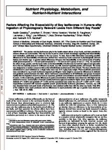
Calbindin protein levels. The effect of dietary treatments on calbindin protein concentrations in the intestine are demonstrated visually in a representative Western blot (Fig. 1). The lack of a calbindin band in the vitamin D-free group and the increase in band density in the low calcium group relative to the control validate the specificity of the antibody for calbindin-D28k protein and the ability of the assay to detect physiological changes in calbindin concentrations. Also apparent is that dietary aluminum greatly inhibited the upregulation of calbindin concentrations by low calcium intakes. To quantify the changes in calbindin protein with dietary treatment, slot immunoblots performed on five samples from each group were analyzed by scanning densitometry, and the means were compared (Fig. 2 black bars). There was significantly more (⬃40%) calbindin protein in the low calcium group relative to controls. In the low calcium plus aluminum group, however, calbindin protein levels did not differ from the controls. Surprisingly, calbindin protein was greater in the control plus aluminum group than in the control group. Calbindin mRNA. Specificity of the oligonucleotide used as a probe for calbindin-D28k mRNA is demonstrated in the Northern blot shown in Figure 3. Two bands were visible
RESULTS Body weight gain, food intake and feed efficiency. Adding aluminum to either control or low calcium diets significantly reduced body weight gain relative to chicks fed the same diets without aluminum (Table 2). In the control plus aluminum group, this seemed to be due in large part to reduced food intake relative to the control group. In the low calcium plus aluminum group, the reduced weight gain relative to the low Ca group seemed to result from the inability to use food for weight gain rather than a reduction in food intake. Reduced feed efficiency seemed even more pronounced in the vitamin D-free group. Serum calcium and inorganic phosphorus. Chicks fed a low calcium diet had significantly lower serum calcium concentrations than did controls, but serum inorganic phosphorus was unaffected (Table 3). Adding aluminum to the low calcium diet did not alter the already low serum calcium concentrations and did not significantly affect serum phosphorus levels. Aluminum in the control diet, however, caused small but significant decreases in both serum calcium and inorganic phosphorus levels relative to controls.
FIGURE 1 Representative Western immunoblot showing the effect of dietary calcium, aluminum and vitamin D on intestinal calbindinD28k levels in the chick. Except for the vitamin D-free group, chicks were fed an adequate control diet for 1 wk and then shifted to the dietary treatments indicated at the top of the figure for 2 wk (lane 5 is low calcium plus aluminum). The vitamin D-free group received the control diet without vitamin D added throughout. Heat-stable proteins were isolated from pooled (n ⫽ 3) duodenal mucosal scrapings. Equal amounts of protein (20 g) from each treatment group were size fractionated by sodium dodecyl sulfate-polyacrylamide gel electrophoresis, electroblotted onto nitrocellulose and immunodeveloped with an anticalbindin-D28k antibody.
ALUMINUM ALTERS CALBINDIN-D28K
FIGURE 2 Quantification of calbindin protein and calbindin mRNA in the duodenum of chicks fed control, control plus aluminum, low calcium, or low calcium plus aluminum diets as described in Figure 1. Calbindin protein was measured by slot immunoblots, and mRNA by slot blot hybridization assays. Values are means ⫾ SD (n ⫽ 5 for protein; n ⫽ 3 for mRNA). Units are image density units for calbindin protein, and the ratio of calbindin mRNA:-actin mRNA image density units for mRNA. Like-colored bars without a common letter differ (P ⬍ 0.05 by Waller-Duncan K-ratio t test). The vitamin D-free group was analyzed, but gave no signal for either calbindin protein or mRNA and was not included in the analysis. Two-way analysis of variance P values for overall effects on calbindin protein were: calcium, P ⫽ not significant (NS); aluminum, P ⫽ NS; calcium ⫻ aluminum interaction, P ⫽ 0.0003. P values for overall effects on the ratio of mRNA were: calcium, P ⫽ NS; aluminum, P ⫽ 0.0004; calcium ⫻ aluminum, P ⫽ 0.0005.
(indicated by arrows) representing the predominant 2.0-kb and 2.8-kb species of calbindin-D28k mRNA (26,27). Specificity was established by the lack of signal in the vitamin D-free group (negative control). Increasing the amount of mRNA used on the blot from 5 to 7 g produced a corresponding increase in signal intensity, demonstrating that the assay could detect small changes in calbindin mRNA levels. To determine whether the treatment effects on calbindin
FIGURE 3 Representative Northern blot demonstrating the specificity of the hybridization probe for intestinal calbindin-D28k mRNA. mRNA was extracted from pooled (n ⫽ 3) duodenal samples from chicks fed either the control diet or the vitamin D-free diet (negative control) for 3 wk. The mRNA (5 or 7 g) was size-fractionated by electrophoresis on a 1% agarose, 1.5% formaldehyde gel, transferred to a nylon membrane and hybridized to a 42 base, double FITC-labeled, synthetic oligonucleotide complementary to the bases coding for amino acids 249 –262 of the carboxy-terminus of chicken intestinal calbindin-D28k (26). Arrows indicate the position of the two predominant calbindin mRNA bands (2.0 and 2.8 kb).
MRNA
EXPRESSION
2011
FIGURE 4 Representative slot-blot hybridization assay for the ratio of calbindin mRNA:-actin mRNA in the duodenum of chicks fed either the control, control plus aluminum, low calcium, or low calcium plus aluminum diet as described in Figure 1. mRNA was extracted from pooled (n ⫽ 3) duodenal samples and an aliquot from each treatment group was blotted onto nylon membranes using a slot-blot device (1 g mRNA for calbindin blots and 0.5 g for -actin). Calbindin mRNA and -actin mRNA were analyzed using double FITC-labeled synthetic oligonucleotide probes, followed by chemiluminescent immunodetection. The ratios of image density units for calbindin mRNA:-actin mRNA are listed at the right of the figure.
protein concentrations were related to changes in the amounts of calbindin mRNA, slot-blot hybridization assays were performed on mRNA isolated from the duodenal segments. Representative slot-blots are shown in Figure 4, which illustrates a large treatment effect on calbindin mRNA in contrast to stable expression of -actin in the same samples, thus, validating the use of -actin as an internal control. The ratios of calbindin mRNA to -actin mRNA for the different treatments can be compared to determine relative changes in calbindin message levels. For example, the low calcium sample had approximately three times as much calbindin mRNA relative to the control sample. In sharp contrast, the low calcium plus aluminum sample had less than half the calbindin mRNA as the controls. To statistically analyze the relative changes in calbindin mRNA, the mean ratios determined from three slot-blots from each treatment group were compared. The results are presented with the calbindin protein data in Figure 2. As expected, chicks consuming the low calcium diet had more calbindin mRNA than the controls. This is consistent with an associated increase in calbindin protein and the decrease in serum calcium. However, when aluminum was consumed with the low calcium diet, there was no such increase in calbindin mRNA or calbindin protein. In fact, in the low calcium plus aluminum group, calbindin mRNA concentrations were less than in the controls. This occurred even though serum calcium had decreased to the same level as in the low calcium group (Table 3). Interestingly, calbindin protein levels were not lower than the controls in the low calcium plus aluminum group, even though mRNA levels were lower. In contrast to the effects seen with a low calcium diet, when chicks consumed aluminum in the control diet, calbindin mRNA levels were not different than in controls (Fig. 2). Yet, there were reductions in both serum calcium and phosphorus in this group (Table 3), suggesting that mRNA levels should have increased. These findings indicate that aluminum inhibited the upregulation of calbindin mRNA in the control diet
COX AND DUNN
2012
as well. It is interesting that aluminum increased calbindin protein in this group relative to the controls, even though mRNA levels were unaffected. DISCUSSION The normal adaptive response of the intestine to low calcium intakes is to increase the efficiency of calcium absorption. This process involves an increase in calbindin mRNA and calbindin protein mediated by circulating 1,25(OH)2D. We have shown that dietary aluminum toxicity interferes with this adaptation by inhibiting the expression of calbindin mRNA. This result demonstrates for the first time that aluminum intake can inhibit gene expression in vivo and supports the hypothesis that aluminum interferes with the biological actions of vitamin D in the intestine. These conclusions are consistent with studies demonstrating that aluminum inhibits vitamin D-dependent calcium absorption in experimental animals (20,22–24,28). Whether physiological functions in other tissues expressing calbindin-D28k, such as the kidney, bone or pancreatic  cells (29), are also affected by aluminum at the level of mRNA expression is not known. The mechanism(s) by which aluminum interferes with calbindin mRNA expression was not resolved by this study. At present, we can say that reduced expression was not due to aluminum-induced changes in serum calcium or phosphorous levels, because these were the same in the low calcium and low calcium plus aluminum groups. Aluminum could have inhibited expression by causing reductions in serum levels of 1,25(OH)2D because previous studies have indicated that aluminum may decrease renal conversion of vitamin D to 1,25(OH)2D (23). In that study and others, however, it was also shown that aluminum inhibits the ability of physiological injections of 1,25(OH)2D to increase calbindin concentrations in the intestine (11,20,23). Therefore, aluminum inhibited calbindin expression even in the presence of physiological levels of the active hormone. This finding indicates that aluminum directly interferes with the ability of the intestinal cell to respond to 1,25(OH)2D. Our results suggest that this inability to respond is due to inhibition of vitamin D-dependent transcription of the calbindin gene and/or a decrease in the stability of calbindin mRNA. In bone, aluminum toxicity also causes decreased responsiveness to the active form of vitamin D, but the mechanism seems to differ from that in the intestine. Studies in cultured rat ostoblast-like osteosarcoma cells have shown that the ability of 1,25(OH)2D to stimulate osteocalcin synthesis is inhibited by aluminum (15,16). Although the mechanism is not fully understood, Fanti et al. (16) showed that osteocalcin mRNA expression in these cells was not affected. They concluded that aluminum did not decrease the transcription or stability of the mRNA but rather, it had a direct posttranscriptional effect on osteocalcin production. Because osteocalcin protein degradation was not affected, they suggested that aluminum altered some aspect of osteocalcin synthesis, such as mRNA translation or processing of the synthesized protein. These findings differ from our results, showing decreased calbindin mRNA levels, and indicate that aluminum affects the expression of calbindin and osteocalcin at different biochemical sites. This argues against a common effect of aluminum on the fundamental genomic actions of vitamin D across tissues. There are several possibilities for how aluminum could affect the expression of different vitamin D-dependent genes by different mechanisms. Tissue-specific aspects of aluminum metabolism, such as the concentration and/or species of aluminum in different cell types, could result in different sites of
action. The level of vitamin D exposure under different experimental conditions could be a factor, because high concentrations of 1,25(OH)2D are protective against some actions of aluminum (15). It is also possible that the different molecular aspects of the transcriptional and translational events that occur during the expression of calbindin versus osteocalcin could have different susceptibilities to the action of aluminum. It is conceivable then, that calbindin-D28k is affected at the level of mRNA expression, but that osteocalcin is affected posttranscriptionally. Our own data suggest that the expression of calbindin is affected posttranscriptionally as well, but the effect seems to be stimulatory. A posttranscriptional effect was most evident in the control plus aluminum group, where relative to controls, calbindin protein was increased, but calbindin mRNA was not. This finding suggests that aluminum either increased the translation of calbindin mRNA and, hence, calbindin synthesis or that it stabilized the calbindin protein and reduced its rate of degradation. This hypothesis is supported by the greater ratio of calbindin protein to calbindin mRNA in both groups exposed to aluminum relative to their respective controls (Fig. 2). Potential mechanisms for posttranscriptional effects of aluminum are only hypothetical at present. There is evidence that aluminum binds to nucleic acids and calcium-binding proteins (9,30), but little is known about how this may alter their function or turnover. There is also evidence that 1,25(OH)2D and calcium can upregulate posttranscriptional steps in calbindin production (18), but the mechanisms are unclear and no studies have been performed to determine whether aluminum can influence this regulation. It is possible that the decrease in serum calcium and phosphorous induced by aluminum intake in the control group stimulated calbindin synthesis without a corresponding increase in calbindin mRNA. Whatever the mechanism, the net effect of aluminum on the concentration of calbindin in the intestine seems to depend on the relative effects of this metal on decreasing mRNA expression versus stimulating calbindin production. Apparently the inhibitory effect on mRNA expression was dominant in the low calcium diets, but not in the control diets. The reason for the different effects of aluminum depending on the concentration of calcium in the diet is not clear. It may be related to a lower concentration of aluminum in the intestinal cell in chicks fed the control diet because calcium has been shown to inhibit mucosal uptake of aluminum (31). Diets adequate in calcium, then, may be protective against the inhibitory effects of aluminum on calbindin mRNA expression. We have shown that high intakes of aluminum can inhibit the expression of calbindin mRNA, thus, inhibiting the intestinal adaptation to low calcium diets mediated by 1,25(OH)2D. The mechanism for this effect is not fully understood. Comparisons with studies on osteocalcin expression indicate that aluminum may not universally inhibit a fundamental step in the genomic regulation of vitamin D-dependent genes. Rather, aluminum can affect multiple sites in the transcriptional and posttranscriptional control of protein expression depending on the tissue involved, the calcium content of the diet and possibly other dietary and physiological factors. ACKNOWLEDGMENT We thank Soccoro Tauyan for technical support and assistance with the care and feeding of the chicks in this study.
ALUMINUM ALTERS CALBINDIN-D28K
LITERATURE CITED 1. Greger, J. L. & Sutherland, J. E. (1997) Aluminum exposure and metabolism. Crit. Rev. Clin. Lab. Sci. 34: 439 – 474. 2. Flaten, T. P., Alfrey, A. C., Birchall, J. D., Savory, J. & Yokel, R. A. (1996) Status and future concerns of clinical and environmental aluminum toxicology. J. Toxicol. Environ. Health 48: 527–541. 3. Bishop, N. J., Morley, R., Chir, B., Day, J. P. & Lucas, A. (1997) Aluminum neurotoxicity in preterm infants receiving intravenous-feeding solutions. N. Engl. J. Med. 337: 1557–1561. 4. Bougle, D., Sabatier, J. P., Bureau, F., Laroche, D., Brouard, J., Guillois, B. & Duhamel, J. F. (1998) Relationship between bone mineralization and aluminum in the healthy infant. Eur. J. Clin. Nutr. 52: 431– 435. 5. Woodson, G. C. (1998) An interesting case of osteomalcia due to antacid use associated with stainable bone aluminum in a patient with normal renal function. Bone 22: 695– 698. 6. Gilbert-Barness, E. & Barness, L. (1998) Aluminum toxicity. Arch. Pediatr. Adolesc. Med. 15: 511–512. 7. Jeffery, E. H., Abreo, K., Burgess, E., Canmata, J. & Greger, J. L. (1996) Systemic aluminum toxicity: effects on bone, hematopoietic tissue and kidney. J. Toxicol. Environ. Health 48: 649 – 665. 8. Exley, C. (1999) A molecular mechanism of aluminum-induced Alzheimer’s disease? J. Inorg. Biochem. 76: 133–140. 9. Lukiw, W. J., LeBlanc, H. J., Carver, L. A., McLachlan, D. R. C. & Bazan, N. G. (1998) Run-on gene transcription in human neocortical nuclei. J. Mol. Neurosci. 11: 67–78. 10. Dunn, M. A., Johnson, N. E., Liew, M. Y. B. & Ross, E. (1993) Dietary aluminum chloride reduces the amount of intestinal calbindin-D28k in chicks fed low calcium or low phosphorus diets. J. Nutr. 123: 1786 –1793. 11. Dunn, M. A., Ishizakie, A. S., Liew, M. Y. B., Too, S. L. & Johnson, N. (1995) Dietary aluminum chloride inhibits the ability of vitamin D to regulate intestinal calbindin-D28k levels in chicks. J. Trace Elem. Exper. Med. 8: 47–57. 12. Goodman, W. G. & Duarte, M. E. L. (1991) Aluminum: effects on bone and role in the pathogenesis of renal osteodystrophy. Miner. Electrolyte Metab. 17: 221–232. 13. Lian, J. B., Stein, G. S., Stein, J. L. & Van Wijnen, A. J. (1999) Regulated expression of the bone-specific osteocalcin gene by vitamins and hormones. Vitam. Horm. 55: 443–509. 14. Mosavin, R. & Mellon, W. S. (1996) Post-transcriptional regulation of osteocalcin mRNA in clonal osteoblast cells by 1,25-dihydroxyvitamin D3. Arch. Biochem. Biophys. 332: 142–152. 15. Lajeunesse, D., Moreau, R., Hobbs, W., Qui, W., Lafond, J. & Guggino, S. E. (1998) Influence of aluminum on the regulation of PTH and 1,25(OH)2D3dependent pathways in the rat osteosarcoma cell line ROS 17/28. J. Bone Miner. Res. 13: 962–969. 16. Fanti, P., Kindy, M. S., Mohapatra, S., Klein, J., Columbo, G. & Malluche,
MRNA
EXPRESSION
2013
H. H. (1992) Dose-dependent effects of aluminum on osteocalcin synthesis in osteoblast like ROS 17/2 cells in culture. Am. J. Physiol. 263: E1113–E1118. 17. Bronner, F. (1992) Current concepts of calcium absorption: an overview. J. Nutr. 122: 641– 643. 18. Duflos, C., Bellaton, C., Baghdassarian, N., Gadoux, M., Pansu, D. & Bronner, F. (1996) 1,25-dihydroxycholecalciferol regulates rat intestinal calbindin-D9k post-transcriptionally. J. Nutr. 126: 834 – 841. 19. Meyer, J., Galligan, M. A., Jones, G., Komm, B. S., Haussler, C. A. & Haussler, M. R. (1995) 1,25(OH)2D3-Dependent regulation of calbindin-D28k mRNA requires ongoing protein synthesis in chick duodenal organ culture. J. Cell. Biochem. 58: 315–327. 20. Orihuela, D., Favre, C., Monti, J. A., Carnovale, C. E. & Carrillo, M. C. (1999) Aluminum effects upon calbindin D9k-linked duodenal calcium transport in diabetic male rats. Toxicol. Lett. 104: 211–219. 21. Meyer, J., Fullmer, C. S., Wasserman, R. H., Komm, B. S. & Haussler, M. R. (1992) Dietary restriction of calcium, phosphorous, and vitamin D elicits differential regulation of the mRNAs for avian intestinal calbindin-D28k and the 1,25-dihydroxyvitamin D3 receptor. J. Bone Miner. Res. 7: 441– 448. 22. Adler, A. J., Zara, C. & Berlyne, G. M. (1989) Effect of aluminum on bidirectional calcium flux in rat everted intestinal sacs. Am. J. Physiol. 257: G433–G437. 23. Henry, H. L. & Norman, A. W. (1985) Interactions between aluminum and the actions and metabolism of vitamin D3 in the chick. Calcif. Tissue Int. 37: 484 – 490. 24. Blahos, J., Care, A. D., Abbas, S. K. & Corlette, S. C. (1991) Aluminuminduced decrease in osteocalcin levels in the chick. Horm. Metab. Res. 23: 50 –51. 25. National Research Council. (1994) Nutrient Requirements of Poultry, 9th revised ed, pp. 81. National Academy Press, Washington, D.C. 26. Clemens, T. L., McGlade, S. A., Garrett, K. P., Horiuchi, N. & Hendy, G. N. (1998) Tissue-specific regulation of avian vitamin D-dependent calcium binding protein 28kDa mRNA by 1,25-dihydroxyvitamin D3. J. Biol. Chem. 263: 13112–13116. 27. Huang, Y. C. & Christakos, S. (1988) Modulation of rat calbindin-D28k gene expression by 1,25-dihydroxyvitamin D3 and dietary alteration. Mol. Endocrinol. 2: 928 –935. 28. Merke, J., Lucas, P. A., Szabo, A., Helbing, F., Hugel, U., Drueke, T. & Ritz, E. (1987) 1,25(OH)2D3 Receptors and end organ response in experimental aluminum intoxication. Kidney Int. 32: 204 –211. 29. Sooy, K., Schermerhorn, T., Noda, M., Surana, M., Rhoten, W. B., Meyer, M., Fleischer, N., Sharp, G.W.G. & Christakos, S. (1999) Calbindin-D28k controls [Ca2⫹]I and insulin release. J. Biol. Chem. 274: 34343–34349. 30. Adler, A. J., Caruso, C. & Berlyne, G. M. (1991) Effect of aluminum on calcium-binding to bovine intestinal calcium-binding protein. Miner. Elect. Metab. 17: 141–146. 31. van der Voet, G. B. & de Wolff, F. A. (1998) Intestinal absorption of aluminum: effect of sodium and calcium. Arch. Toxicol. 72: 110 –114.