In Well-Fed Young Rats, Lactose-Induced Chronic Diarrhea Reduces the .... in accordance with 18 USC section 1734 solely to indicate this fact. .... Total body fat was determined gravimetrically after ex- ..... village. Am. J. Clin. Nutr. 30: 1215â1227. National Research Council (1996) Guide for the Care and Use of Laboratory.
Nutrient Requirements and Interactions
In Well-Fed Young Rats, Lactose-Induced Chronic Diarrhea Reduces the Apparent Absorption of Vitamins A and E and Affects Preferentially Vitamin E Status1,2,3 Juan P. Liuzzi, Anna M. Cioccia and Patricio Hevia4 Laboratorio de Nutricio´n Universidad Simo´n Bolı´var, Apartado Postal 89000, Caracas Venezuela ABSTRACT To establish the effect of lactose-induced diarrhea on the apparent absorption and status of vitamins-A and E in well-fed young rats, we fed Sprague Dawley rats a balanced diet or a lactose diet (350 g/kg). A group of rats fed the control diet equal to the level measured in the lactose-fed rats (pair-fed) was also included. The experiment lasted 23 d and feces were collected on days 4 – 6, 10 –12, 14 –16 and 20 –22. Samples of serum and tissues were taken on days 10 and 23. Lactose caused a significant reduction in food intake, had no effect on body weight and produced a diarrhea that persisted during the whole experiment. The severity of diarrhea decreased with time, indicating that the rats partially adapted to lactose feeding. At the onset of diarrhea, the apparent absorption of vitamins A and E in the rats with diarrhea was significantly lower than in the control or pair-fed rats, but the rats with diarrhea recovered gradually, and in the case of vitamin E was normalized by day 15. At day 10 the rats with lactose-induced diarrhea had serum and liver concentrations of vitamins A and E that did not differ from the control or pair-fed rats. However, at day 23 the lactose-fed rats with diarrhea had significantly lower serum and liver concentrations of vitamin E than the control or pair-fed rats. Measured at that time, diarrhea had no effect on liver vitamin A, but lower serum concentrations of this vitamin were detected in both the lactose-fed rats and in the pair-fed rats. In general, in well-nourished rats, the chronic diarrhea associated with excessive dietary lactose reduced the apparent absorption of vitamin A and E and particularly compromised the nutritional status of vitamin E. J. Nutr. 128: 2467–2472, 1998. KEY WORDS:
●
lactose-induced diarrhea
●
rats
●
vitamin A
●
vitamin E
●
episodes of diarrhea further deteriorating their nutritional status (Guerrant et al. 1992). Balance studies (Bowie et al. 1975, Cioccia et al. 1995, Gonza´lez et al. 1992, Romer et al. 1989) have shown that hospitalized children with acute diarrhea consume substantially less food than healthy children, and they absorb only 40 – 60% of the energy, carbohydrates, protein and fat present in the diet. The macronutrient absorption in these children, with the exemption of fat, which remains relatively constant, is inversely related to the severity of the diarrhea. In the case of persistent diarrhea, the situation is similar, but its effect on macronutrients absorption is less pronounced (Cioccia et al. 1995). Persistent diarrhea, however, affects the patient for a longer time and is more prevalent in undernourished children. Therefore, its negative effects on nutritional status are very important. The main causes of the nutritional deficiencies associated with diarrhea are a reduction in food consumption and in the utilization of the nutrients consumed. In addition, the increase in the nutritional requirements of the patients, which were caused by infection and fever, further contributes to their nutritional deficit. This combination of negative factors emphasize the importance of providing appropriate nutrition during diarrhea (Guerrant et al. 1992). Although the effects of diarrhea on macronutrient availability are fairly well established (Bowie et al. 1975, Cioccia et al. 1995, Gonza´lez et al. 1992, Romer et al. 1989), there is
Diarrheal diseases claim the lives of some 3 million children a year. They rank fourth among the leading causes of death in the world, and their incidence is higher than any other disease condition worldwide (WHO 1995). Childhood mortality due to diarrhea is particularly frequent in developing countries. In those countries it has been estimated that about one-half of the cases are related to malnutrition (Yoon et al. 1997). Nutrition is critical in the treatment of diarrhea (Brown et al. 1990) because diarrhea in malnourished children and animals is more difficult to treat (Ghademi et al. 1973, Zijlstra et al. 1997) and because one of the consequences of diarrhea is malnutrition (Mata et al. 1977). The association between nutrition and diarrhea has been described as a vicious cycle. According to this description, diarrhea causes malnutrition. This impairs the immune response and alters the intestinal mucosa, making the patient more prone to new or longer
1 Abstract presented at the Summer meeting of the Nutrition Society : Genes, Nutrition and Health. University of Newcastle. England 9 –11 July 1997. Hevia, P., Luizzi, J.P. & Cioccia, A.M. (1998) Micronutrient intestinal balance in growing rats with lactose induced diarrhoea. Proc. Nutr. Soc. 57: 70A. 2 Funded by CONICIT. Grant S-1 2265. 3 The costs of publication of this article were defrayed in part by the payment of page charges. This article must therefore be hereby marked ‘‘advertisement’’ in accordance with 18 USC section 1734 solely to indicate this fact. 4 To whom correspondance should be addresses.
0022-3166/98 $3.00 © 1998 American Society for Nutritional Sciences. Manuscript received 30 March 1998. Initial review completed 13 May 1998. Revision accepted 18 August 1998. 2467
Downloaded from https://academic.oup.com/jn/article-abstract/128/12/2467/4724246 by guest on 01 March 2018
absorption
LIUZZI ET AL.
2468
little information regarding its effect on the availability of the micronutrients. Recently there has been increased interest regarding the effect on vitamin A (retinol) status and supplementation with retinol on the incidence, duration and treatment of diarrheal disease. The results of this interest indicate that diarrhea has a negative effect on vitamin A nutritional status (Alvarez et al. 1995, Buyukgebiz et al. 1990, SalazarLindo and Alvarez 1993, Sommer et al. 1987, Usha et al. 1991), that children with mild xerophthalmia or rats deficient in vitamin A have an increased risk for diarrheal disease (Sommer et al. 1984, Warden et al. 1997) and that vitamin A suplementation reduces the mortality associated with diarrhea (Barreto et al. 1994, Filteau et al. 1995, Glasziou and Mackerras 1993). An interesting finding in this area is the observation that during diarrhea and infection there is an increased urinary excretion of vitamin A (Alvarez et al. 1995). A possible relationship between diarrhea and vitaminE was also suggested. Thus, an increased excretion of vitaminE and lower circulation levels of the vitamin as well as a significant degree of shortening of erythrocyte survival were reported in patients with enteropathies leading to chronic steatorrea (Farrell and Robertson 1994). Gastrointestinal lipid peroxidation due to vitamin E deficiency was implicated in the etiology of persistent diarrhea and malnutrition (Darmon et al. 1993, Saverwein et al. 1997), altered circulating concentrations of antioxidants (Hoffenberg et al. 1997) and oxidants and free radicals (Grisham 1994) were associated with inflammatory bowel disease. Based on the previous information, the objective of this study was to establish in well-nourished rats with lactoseinduced chronic diarrhea the availability of vitamins A and E and also the effect of diarrhea on circulating and tissue levels of these vitamins. Lactose was previously used for inducing a chronic osmotic diarrhea in rats. Bueno et al. (1994) described in rats with lactose-induced diarrhea changes in the morphological structure of the small intestine similar to those found in children with diarrhea. Because infection has a profound effect on markers of the nutritional status of vitamin A and other micronutrients, this noninfectious model was used here to study the effect of diarrhea on vitamins A and E (Warden et al. 1997). We used well-nourished rats because vitamin A deficiency was a confounding factor in the results of the already available studies describing the relationships between infant diarrhea and vitamin A (Warden et al. 1997). The concentration of dietary lactose used in this study was lower than the concentration used by Bueno et al. (1994). The reason for this modification was that preliminary studies showed that the total replacement of dietary carbohydrates by lactose caused mortality and a severe reduction in food intake. MATERIALS AND METHODS Experimental protocol. Forty-two 3–wk– old male Sprague Dawley rats from the University colony were fed the control diet shown in Table 1 for 7 d prior to the experiment. They were then randomly divided into three groups of 14 rats each and kept in hanging wire individual stainless steel cages. Two of these groups continued receiving the control diet, and the third was fed the lactose diet shown in Table 1. Because the inclusion of lactose in the diet causes diarrhea and a reduction in food consumption, one of the groups assigned to the control diet was pair-fed with the lactose group. This pair-fed control group was offered daily an amount of food that was equal to the mean food consumed by the lactose group on the previous day. All rats had free access to water throughout the experiment. During the experiment, which lasted 23 d, food intake was registered daily, body weight every other day, and feces were collected on Downloaded from https://academic.oup.com/jn/article-abstract/128/12/2467/4724246 by guest on 01 March 2018
TABLE 1 Diet composition Ingredient
Control
Lactose g/100g
Casein Corn oil Mineral Mix1 Vitamin Mix1 Choline bitartrate L-Methionine Corn starch Lactose
16.3 5.0 3.5 1.0 0.2 0.3 73.7 0
16.3 5.0 3.5 1.0 0.2 0.3 38.7 35.0
1 AIN - 76A Mineral and Vitamin mixtures (American Institute of Nutrition 1977).
Days 4 to 6; 10 to 12; 14 to 16 and 20 to 22 for the determination of apparent vitamin A and vitamin E absorption. During the fecal collections, which lasted 48 h each, feces were separated from the urine by a with a separatory funnel and a narrow stainless steel sieve (20 mesh) that was provided in the individual stainless steel metabolic cages. This sieve allowed the collection of feces in the group with diarrhea. The wet fecal weight was determined as the difference between the weight of the sieve before and after the collection. To diminish the deterioration of the vitamins in the fecal material, the 48 h collections were divided into two separate 24 h collections. The feces collected from the sieves immediately after the termination of each 24-h collection period were freeze dried, finely ground and kept at 220°C until analyzed. On Day 10 one half of the rats assigned to each group were killed by decapitation for the determination of circulating and liver levels of vitamins A and E, total body fat and adipose tissue vitamin E. The remaining rats were killed on the last day of the experiment for similar reasons. At day 10 and 23, blood samples were obtained by heart puncture of rats under ether anesthesia after overnight food deprivation. The livers and a sample of fat taken from the back of the rats were dissected and kept frozen at 220°C, and the rat carcasses were weighed and dried in a forced air oven (80°C). This study was approved by the Universidad Simo´n Bolı´var and followed the principles set forth in the Guide for the Care and Use of Laboratory Animals (National Research Council 1996). Vitamins A and E were determined in the diets, plasma, liver and feces and vitamin E and total lipids were determined in the sample of adipose tissue taken from the back of the rat. The plasma concentration of these vitamins was determine by the method recommended by Chow and Omaye (1983), whereas in the case of the diets, feces and livers the vitamins were determined after extraction and saponification essentially as recommended by Zahar and Smith (1990). Vitamin E in dorsal adipose tissue was determined after saponification using the procedure of Ueda and Igarashi (1990). In all cases, the vitamins were determined by high pressure liquid chromatography (HPLC). The HPLC equipment (Water Associates, Milford, MA) had a Novapack C18 column, a 510 solvent delivery pump, a U6K injector, a 486 UV detector and a 476 data module. The vitamins were separated in 100% Methanol (flow rate 0.8 mL/min) and detected at 296 nm. The apparent absorption of vitamins A and E was calculated as 100 x (vitamin consumed [minus] vitamin excreted in feces)/vitamin consumed. Total body fat was determined gravimetrically after extraction using the method recommended by Blight and Dyer (1959). The results were analyzed using one-way ANOVA, correlation and regression. Duncan9s multiple range test was used to comparetreatment means when the F test from ANOVA was significant. In all cases the BMDP Statistical Package was used with the level of significance fixed at 5%. (Dixon et al. 1990).
RESULTS Food intake, growth and fecal mass. The incorporation of 350 g lactose/kg in the diet was associated with a significant
VITAMINS A AND E ABSORPTION AND STATUS IN DIARRHEA
2469
TABLE 2 Total food intake, body weight gain, liver weight and body fat measured after 10 and 23 d in young rats fed control and lactose diets1. Control
Pair-fed
Lactose
g Day 10 Food intake Body weight gain Liver weight Total body fat Day 23 Food intake Body weight gain Liver weight Total body fat
86.3 6 7.10a 37.58 6 2.44 4.30 6 0.22a 6.90 6 0.54a
73.8 6 0.83b 31.37 6 2.75 4.33 6 0.27a 5.75 6 0.45a
72.1 6 9.12b 35.12 6 2.88 3.54 6 0.19b 3.28 6 0.22b
247.5 6 7.10a 65.80 6 4.28 5.84 6 0.26 7.50 6 0.43a
217.9 6 0.83b 61.97 6 5.53 5.47 6 0.34 5.97 6 0.53b
220.46 6 9.12b 54.40 6 4.47 5.19 6 0.34 5.12 6 0.45b
1 Values are means 6 SD, n 5 14 rats per group until Day 10, and 7 rats per group thereafter. At Day 10, seven rats per group were killed for body composition determination. Values in a row with a different superscript letter are significantly different, P # 0.05 (Duncan’s multiple range test).
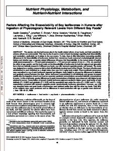
reduction in food consumption (Table 2). Thus, the food consumed by the lactose group was 85.5 and 88.0% of that measured in the controls at days 10 and 23, respectively. The pair-fed group had a food intake similar to the lactose group. The reduction in food intake seen both in the lactose and the pair-fed groups had no effect on body weight. However, at day 10 this reduction in food intake caused a significant reduction in total body fat and in the weight of the liver exclusively in the lactose group, whereas at day 23 both the lactose and the pair-fed groups had significantly less body fat than the control rats. Rats consuming lactose produced significantly more feces than the control or pair-fed rats throughout the experiment (Figure 1). The feces of these rats were yellowish and soft, but not watery. The diarrheic appearance of the feces produced by the lactose-fed rats contrasted with the well-formed appearance of the feces of the control or pair-fed groups. Figure 1 also shows that the higher fecal output associated with lactose consumption was more pronounced at the beginning of the experiment, and it became less apparent with its progression. The negative slopes of the regression lines (Fig. 1), showing the changes in fecal weight (dry or wet) with time in the lactose group, further describe the adaptation to the consumption of lactose in this group. Despite this adaptation, the feces of the lactose group had a diarrheic appearance during the whole experiment. Apparent absorption of Vitamin A and E. Both in the control and in the pair-fed rats the apparent absorption of vitamin A remained high and constant throughout the experiment (Fig. 2, upper panel). In contrast, in the lactose-fed rats, there was a severe reduction in the apparent absorption of this vitamin, which was very pronounced at the initial collection periods and became less apparent thereafter. The positive slope of the regression line describing the changes in vitamin A apparent absorption with time (Fig. 2) indicated that the lactose-fed rats absorbed more vitamin A with the progression of the experiment. However, despite this significant improvement in the absorption of this vitamin with time, the apparent absorption of vitamin A measured in the lactose-fed animals was significantly lower than in the controls throughout the whole experimental period. The apparent absorption of vitamin E was lower in the lactose-fed group than in the control or the pair-fed groups (Fig. 2, lower panel). However, this reduction was significant Downloaded from https://academic.oup.com/jn/article-abstract/128/12/2467/4724246 by guest on 01 March 2018
only at the first and second collection periods. The vitamin E apparent absorption measured in the lactose-fed rats at Days 15 and 22 was not different from that measured in the control or pair-fed groups on those same days. The positive slope of the regression line showing the changes in the apparent absorption
FIGURE 1 Fecal weight of rats fed control and lactose diets for 23 d. The concentration of lactose in the diet was 350 g/kg diet. Pair–fed rats were offered the mean amount of food that the lactose group ate the day before. Feces were collected on Days 4 – 6, 10 –12, 14 –16 and 20 –22. The x-axis shows the middle point for each of these collection periods. The feces of the lactose group had a diarrheic appearance. Values are means 6 SD of 7 rats. * Significantly different from the control and pair-fed control. P # 0.05 (Duncan9s multiple range test). Lactose group fecal weight (wet) y 5 20.283x, 18.195, r 5 20.58. Lactose group fecal weight (dry) y 5 20.051x, 12.068, r 5 20.53. For both equations the intercept, slope and correlation coefficient were significant (P , 0.05)
LIUZZI ET AL.
2470
FIGURE 2 Apparent absorption of retinol and a -tocopherol measured in rats fed control and lactose diets for 23 d. The concentration of lactose in the diet was 350 g /kg diet. Pair–fed rats were offered the mean amount of food, which the lactose group ate the day before. Feces were collected on Days 4 – 6,10 –12,14 –16 and 20 –22. The x-axis shows the middle point for each of these collection periods. The feces of the lactose group had a diarrheic appearance. Values are means 6 SD of 7 rats. * Significantly different from the control and pair-fed control. P # 0.05 (Duncan9s multiple range test). Lactose group retinol apparent absorption y 5 1.32x 1 47.91, r 5 0.44. Lactose group a–Tocopherol apparent absorption y 5 3.11x 1 27.12, r 5 0.91. For both equations the intercept, slope and correlation coefficient were significant (P , 0.05)
of vitamin E with time, measured in the lactose-fed rats (Fig. 2), further indicates that these rats progressively adapted to the consumption of this disaccharide. In contrast to the absorption of vitamin A, which remained low during the whole experiment, the absorption of vitamin E, at the end of the experiment, recovered completely. Because Figure 2 shows that the apparent absorption of vitamins A and E increased with time and Figure 1 shows that with the progress of the experimental time there was a reduction in fecal mass, we correlated the absorption of the vitamins with fecal mass. The results showed significant (P , 0.05)
negative correlation coefficients between fecal mass and the apparent absorption for both vitamins (Vit A r 5 2 0.36 ; Vit E r 5 2 0.66). This suggests that the severity of diarrhea negatively affected the absorption of both liposoluble vitamins. Serum concentration of Vitamin A and E. After 10 d of diarrhea, the serum levels of vitamins-A and E did not differ significantly from the levels measured either in the control or the pair-fed groups (Table 3). However, the lactose-fed group, which had diarrhea for 23 d, had significantly lower levels of circulating vitamin E than the control or pair-fed groups, and serum vitamin A level, which were lower than the control but not different from the pair-fed rats. The observed effect of lactose-induced diarrhea on the circulating levels of these vitamins was not related to changes in hemodinamics because the hematocrit did not differ among groups. Reserves of vitamins-A and E. At Day 10, lactose-induced diarrhea had no effect on the concentration of vitamins-A in liver or vitamin E in liver and adipose tissue. However, after 23 d, the liver vitamin E measured in the group with lactoseinduced diarrhea was significantly lower (33.27 6 2.09 nmol /g, P , 0.05) than that measured in the control (43.37 6 2.83 nmol/g) or the pair-fed group (48.18 6 1.74 nmol/g). In contrast, the concentration of liver vitamin A measured after 23 d was not different among groups. Identical results were obtained when the total liver contents of these vitamins was compared (data not shown). In contrast to the effect of long-standing diarrhea on liver vitamin E, total contents of this vitamin in adipose tissue, estimated from the concentration of Vitamin E in a sample of adipose tissue dissected from the back of the rats, was not affected by lactose-induced diarrhea of 10 or 23 d of duration. DISCUSSION Bueno et al. (1994) reported that totally replaceing the corn starch with lactose in the diets of young rats produces chronic diarrhea and malnutrition. In their work, they indicate that lactose-induced diarrhea is associated with changes in the small intestines’ structure similar to those found in children with acute gastroenteritis or chronic diarrhea. They suggest that, in rats with lactose-induced diarrhea, nutrient malabsorption and malnutrition may have partially explained the changes seen in the intestinal mucosa. Accordingly, the purpose of this study was to further characterize this model of diarrhea and also to determine if a long-standing diarrhea, produced in young rats by lactose, could affect the apparent absorption of vitamins A and E as well as the nutritional status of these two liposoluble vitamins.
TABLE 3 Serum retinol, a-Tocopherol and hematocrit of young rats fed control or lactose diets for 10 or 23 d1 Control Day 10 Retinol, mmol/L a-Tocopherol, mmol/L Hematocrit Day 23 Retinol, mmol/L a-Tocopherol, mmol/L Hematocrit
Pair-fed
Lactose
1.84 6 0.06 18.47 6 1.07 0.45 6 0.01
1.78 6 0.17 15.71 6 0.48 0.48 6 0.02
1.65 6 0.10 16.43 6 1.39 0.43 6 0.01
2.55 6 0.15a 22.65 6 1.58a 0.49 6 0.02
2.18 6 0.06ab 19.54 6 1.56a 0.44 6 0.01
1.84 6 0.24b 14.70 6 1.17b 0.48 6 0.02
1 Values are means 6 SD, n 5 7 rats. Values in a row with a different superscript letter are significantly different, P # 0.05 (Duncan’s multiple range
test) Downloaded from https://academic.oup.com/jn/article-abstract/128/12/2467/4724246 by guest on 01 March 2018
VITAMINS A AND E ABSORPTION AND STATUS IN DIARRHEA
In this study, the concentration of lactose used was lower than that reported by Bueno et al. (1994). The carbohydrates in the lactose diet included a mixture of corn starch and lactose. In this mixture, lactose represented 47.5% of the total carbohydrates and 35% of the total diet. This is ;1/2 of the lactose used by Bueno et al. (1994). This lower concentration of lactose was used because preliminary studies showed that the total replacement of the carbohydrates by lactose in the diet caused a high rat mortality and a marked reduction in food intake. The incorporation of lactose at 35% of the diet caused a chronic diarrhea that had a small but significant effect on food intake. It did not affect growth, but caused a significant reduction in body fat content. An additional feature of the lactose induced diarrhea seen in this study was that it was severe at the beginning of the exposure to lactose but it became less and less severe with the progression of the experiment. This indicates that the rats adapted to the utilization of this carbohydrate. This adaptation resulting from prolonged lactose feeding has also been reported in humans who have difficulties in digesting dietary lactose (Jiang and Saviano 1997) . Factors that may have contributed to the adaptation to chronic lactose feeding include a possible induction in intestinal lactase in rats, which has been observed by some investigators (Bolin et al. 1971). In humans (Jiang and Saviano 1997), this adaptation has been associated with a progressive modification of the intestinal microflora. This modification results in an increased microbial glucosidase activity and a better capacity of fermentation of malabsorbed lactose into short chain fatty acid. These effects may mitigate the lactoseinduced diarrhea by reducing the osmotic load produced by the unabsorbed lactose and also by stimulating the absorption of the short chain fatty acids, which promotes water and sodium uptake by the colonic mucosa (Jiang and Saviano 1997). From a practical point of view, in this experiment this adaptation was considered advantageous because it allowed us to monitor the effect of diarrheas of different severities on the apparent absorption of the studied vitamins. The apparent absorption of both vitamins A and E was reduced by lactose-induced diarrhea, and in both cases the reduction was inversely proportional to the severity of diarrhea. This is similar to the effect of diarrhea on macronutrient apparent absorption (Cioccia et al. 1995, Gonza´lez et al. 1992) and indicates that if the diarrhea is severe, as it usually happens in hospitalized children with diarrhea, the reduction in the absorption of these vitamins may be substantial. These results agree with a reduction in the absorption of vitamin A seen in children with diarrhea (Reddy et al. 1986) and also with the observation that in patients with steatorrea of different etiologies the losses of vitamin E were proportional to the severity of steatorrea (Farrell and Roberts 1994) . It is important to state that although the apparent absorption of vitamin A increased as the severity of the diarrhea decreased, it always remained significantly lower than in the control rats. At day 23, serum and liver vitamin E concentrations in lactose-fed rats were lower than in controls. This difference may be associated with the absorption mechanisms of these vitamins because vitamin E is absorbed by passive diffusion (Sokol 1996), whereas the absorption of vitamin A requires a specific protein carrier (Blomhoff et al. 1991). In nutrient balance studies, as in the one reported here, there are uncontrolled factors that can affect the results of apparent absorption. The most important are, on the one hand, the possible production or secretion of the nutrient into the lumen of the intestine and on the other hand, its possible Downloaded from https://academic.oup.com/jn/article-abstract/128/12/2467/4724246 by guest on 01 March 2018
2471
utilization for purposes not associated with absorption. These factors could alter the concentration of the nutrient in the feces and thus, the magnitude of its apparent absorption. Furthermore, they could also have an effect on the nutritional status of the nutrient under study. The intestinal microflora is one of these factors. Thus, its contribution in the endogenous production of biotin or vitamin K is well established, and its role in the utilization of lactose in the alleviation of the osmotic diarrhea seen in this experiment was referred previously. The role of the microflora as a possible cause for the time changes in the apparent absorption of vitamins-A and E seen here in the rats with lactose-induced diarrhea can not be ruled out. However, a more plausible explanation for these observations may be that they were associated with the alleviation of the diahrreic condition seen in these rats. This in turn may have resulted in a longer transit time and a less affected intestinal mucosa, thus improving the absorption of these vitamins. Even though the apparent absorption of vitamin A was affected for a longer time than vitamin E, the nutritional status of vitamin E was more negatively affected than that of vitamin A. Thus, the only effect that lactose-induced diarrhea had on vitamin A was a reduction in the circulating levels of the vitamin. However, this was also seen in the pair-fed group, indicating that it could have been associated with changes in food intake rather than to reduced absorption. In contrast, in the case of vitamin E at day 23 of the experiment in the group with diarrhea, there was a substantial reduction in both the circulating levels and the liver reserves. These results suggest that, in the group with diarrhea, in addition to reduced intake and absorption there was higher utilization of vitamin E. These observations coincide with the increasing evidence that diarrhea and other gastrointestinal disorders are associated with an elevated oxidative stress and lipid peroxidation in conjunction with a lower levels of antioxidant defenses (Darmon et al. 1993, Farrell and Roberts 1994, Grisham 1994, Hoffenberg et al. 1997, Saverwein et al 1997). In this study, oxidative stress may have resulted from the same intestinal inflammation seen by Bueno et al. (1994) in mucosal specimens obtained from rats with lactose-induced diarrhea as well as to a reduced absorption of other micronutrient antioxidants, such us Zinc, not measured in this study . Adipose tissue vitamin E was not affected by the diarrhea probably due to its more difficult mobilization from this tissue (Sokol 1996). In general, the results of this experiment show that in well-nourished rats chronic diarrhea reduces the apparent absorption of vitamins A and E and compromised the nutritional status of vitamin E. From a practical point of view these data suggest that in the formulation of foods designed for the dietary treatment of diarrhea it may be advisable to increase the use of vegetable oils rich in vitamin E. This will provide both additional calories to compensate for the reduction in food intake and absorption seen during diarrhea (Brown et al. 1990, Cioccia et al. 1995, Gonza´lez et al. 1992) and also the vitamin E necessary to compensate for the increased requirement for the vitamin suggested by this and other studies. LITERATURE CITED Alvarez, J. O., Salazar-Lindo, E., Kohatsu, J., Miranda, P. & Stephensen, C. B. (1995) Urinary excretion of retinol in children with acute diarrhea. Am. J. Clin. Nutr. 61: 1273-1276. American Institute of Nutrition (1977) Report of the American Institute of Nutrition ad hoc committee on standards for nutritional studies. J. Nutr. 107: 1340-1348.
2472
LIUZZI ET AL.
Barreto, M., Santos, L.M.P., Assis, A.M.O., Araujo, M. P., Farenzena, G. G., Santos, P. A. & Fiaccone, R. L. (1994) Effect of Vitamin A supplementation on diarrhea and acute lower respiratory tract infections in young children in Brazil. Lancet 344: 228 –231. Blight. E. G. & Dyer, W. J. (1959) A rapid method of total lipid extraction and purification. Can. J. Biochem. Physiol. 32: 911–917. Blomhoff, R., Green, M. H., Green, J. B., Berg, T. & Norum, K. R. (1991) Vitamin A metabolism: New perspectives on absorption, transport and storage. Physiol. Rev. 71: 951–990. Bolin, T. D., McKern, A. & Davis, A. E. (1971) The effect of diet on lactase activity in the rat. Gastroenterology 60: 432– 437. Bowie, M. D., Mann, M. D. & Hill, I. D. (1975) Effect of lactose induced diarrhea on absorption of nitrogen and fat. Arch. Dis. Child 50: 363–366. Brown, K. H., Stalling, R. Y., de Kamashiro, H. C., de Romana, G. L. & Black, R. E. (1990) Effects of common illness and infant9s energy intakes from breast milk and other foods longitudinal community-based studies in Huascar (Lima) Peru´. Am. J. Clin. Nutr. 52: 1005–1013. Bueno, J., Torres, M., Almendros, A., Carmona, R., Nun˜ez, M. C., Rios, A. & Gil, A. (1994) Effect of dietary nucleotides on small intestinal repair after diarrhoea. Histological and ultrastructural changes. Gut 33: 926 –933. Buyukgebiz, B., Ozalp, Y & Oran,O. (1990) Investigation of serum vitamin A levels of children who had a history of recurrent diarrhoea and acute respiratory infections in Ankara. J. Trop. Ped. 36: 251–255. Chow, F. Y. & Omaye, S. T. (1983) Use of antioxidants in analysis of vitamin A and E in mammalian plasma by high performance liquid chromatography. Lipids 18: 837– 841. Cioccia, A. M., Gonza´lez, E., Pe´rez, M., Mora, J., Romer, H., Molina, E. & Hevia, P. (1995) Application of a colorimetric method to the determination of the protein content of comercial foods, mixed human diets and nitrogen losses in infantile diarrhea. Int. J. Food Sci. Nutr. 46: 21–29 Darmon, N., Pellisier, M. A., Heyman, M., Albrecht, R. & Desjeux, J. F. (1993) Oxidative stress contribute to the intestinal dysfuction of weanling rats fed a low protein diet. J. Nutr. 123: 1068 –1075. Dixon, W. J., Brown, M. B., Engelman, L. & Jennrich, R. Y. (1990) BMDP Statistical Software Manual. University of California Press, Berkeley, CA. Farrell, P. M. & Roberts, R. J. (1994) Vitamin E. In: Modern Nutrition in Health and Disease (Shils, M. E., Olson, J. A. and Shike, M., eds.) Vol.1, pp. 326 –341. Lea & Febiger, Philadelphia, PA. Filteau, S. M., Morris, S. S., Raynes, J. G., Arthur, P., Ross, D. A., Kirkwood, B. R., Tomkins, A. M. & Gyapong, J. O. (1995) Vitamin A supplementation, morbidity, and serum acute phase proteins in young Ghanaian children. Am. J. Clin. Nutr. 62: 434 – 438. Ghademi, H., Kumar, S. & Abaci, F. (1973) Endogenous amino acids loss and its significance in infantile diarrhea. Pediatr. Res. 7: 161–168. Glasziou, P. P. & Mackerras, D.E.M. (1993) Vitamin A supplementation in infectious diseases: a meta- analysis. Br. Med. J. 306: 366 –370. Gonza´lez, E., Pin˜ero, D., Romer, H., Guerra, M. & Hevia, P. (1992) Alternativas para la alimentacio´n durante la diarrea aguda. Arch. Venezolanos de Puericultura y Pediatria 55: 16 –19. [Spanish] Grisham, M. B. (1994) Oxidants and free radicals in inflammatory bowel disease. Lancet 344:859 – 861. Guerrant, R. L., Schorling, J. B., McAnliffe, F. & De Souza, M. A. (1992) Diarrhea as a cause and effect of malnutrition.: Diarrhea prevents catch-up growth
Downloaded from https://academic.oup.com/jn/article-abstract/128/12/2467/4724246 by guest on 01 March 2018
and malnutrition increases frecuency and duration. Am. J. Trop. Med. Hyg. 47: 28 –35. [Suppl.] Hoffenberg, E. F., Deutsh, J., Smith, S. & Sokal, R. J. (1997) Circulating antioxidants in children with inflammatory bowel disease. Am. J. Clin. Nutr. 65: 1482–1488. Jiang, T. & Saviano, D. (1997) In vitro lactose fermentation by colonic bacteria is modified by Lactobacilus acidophilus supplementation. J. Nutr. 127: 1489 – 1495. Mata, L. J., Kromad, R. A., Urrutia, J. J. & Garcia, B. (1977) Effect of infection on food intake and its nutritional state: perspectives as viewed from the village. Am. J. Clin. Nutr. 30: 1215–1227. National Research Council (1996) Guide for the Care and Use of Laboratory Animals. National Academy Press, Washington DC. Reddy, V., Raghuramulu, A., Arunjyoti, M. & Underwood, B. (1986) Absorption of vitamin A by children with diarrhea during treatment with oral rehydration salt solution. Bull. WHO 64: 721–724. Romer, H., Paez, M., Hevia, P., Pin˜a, J. M., Urrestaza, Y. & Perez-Shael, I. (1989) Estudio comparativo de las pe´rdidas de nitro´geno, lı´pidos y energı´a en nin˜os deshidratados por diarrea aguda debida a rotavirus y otros agentes. GEN 43: 23–27. [Spanish] Salazar-Lindo, E. & Alvarez, J. O. (1993) Association of diarrhea and low serum retinol in Peruvian children. Am. J. Clin. Nutr. 58: 110 –113. Saverwein, R. W., Mulder, J. A., Mulder, L., Lowe, B., Peshv, N., Demacker, J., Meer, W. N. & Marsh, K. (1997) Inflammatory mediators in children with protein-calorie malnutrition. Am J. Clin. Nutr. 65: 1534 –1539. Sokol, J. R. (1996) Vitamin E. In: Present Knowledge in Nutrition (Ziegler, E. E. & Filer, L. J., eds.), pp. 130 –136. ILSI Press, Washington DC. Sommer, A., Katz, J. & Tawotjo, Y. (1984) Increased risk of respiratory disease and diarrhea in children with preexisting mild vitamin A deficiency. Am. J. Clin. Nutr. 40: 1090 –1095. Sommer, A., Tarwotjo, Y. & Katz, J. (1987) Increased xeroftalmia risk following diarrhea and respiratory disease. Am. J. Clin. Nutr. 61: 1273–1276. Ueda, T. & Igarashi, O. (1990) . Determination of vitamin E in biological specimens and foods by HPLC. Pretreatment of samples and extraction of tocopherols. J. Micronutr. Anal. 7 :79 –96. Usha, N., Sankaranarayanan, A., Walia, B.N.S. & Ganguly, N. K. (1991) Assesment of preclinical vitamin A deficiency in children with persistant diarrhea. J. Pediatr. Gastroenterol. Nutr. 13: 168 –175. Warden, R. A., Noltorp, R. S., Francis, J. L., Dunkley, P. R. & O9Loghlin, E. V. (1997) Vitamin A deficiency exacerbates methotrexate-induced jejunal injury in rats. J. Nutr. 127: 770 –776. WHO (1995) The World Health Report. Bridging the Gaps. Report of the Director General, Geneva, Switzerland. Yoon, P. W., Black, R. E., Moulton, L. H. & Becker, S. (1997) The effect of malnutrition on the risk of diarrheal and respiratory mortality in children , 2 of age in Cebu, Philippines. Am. J. Clin. Nutr. 65: 1070 –1077. Zahar, M. & Smith, D. E. (1990) Vitamin A quantification in fluid dairy products: Rapid method for vitamin A extraction for high performance liquid chromatography. J. Dairy Sci. 73: 3402–3407. Zijlstra, R. T., Donovan, S. M., Odle, J., Gelberg, H. B., Petschow, B. W. & Gaskins, H. R. (1997) Protein-energy malnutrition delays small-intestine recovery in neonatal pigs infected with rotavirus. J. Nutr. 127: 1118 –1127.