Purified rabbit eEF2 was donated generously by Dr. Nick. Redpath (University of Leicester, Leicester, U.K.). REFERENCES. 1 Kleijn, M., Scheper, G. C., Voorma, ...
433
Biochem. J. (1999) 344, 433–441 (Printed in Great Britain)
Nutrients differentially regulate multiple translation factors and their control by insulin Linda E. CAMPBELL1, Xuemin WANG1 and Christopher G. PROUD2 Department of Anatomy and Physiology, Medical Sciences Institute, University of Dundee, Dundee DD1 5EH, Scotland, U.K.
Eukaryotic initiation factor eIF2B and eukaryotic elongation factor eEF2 each mediate regulatory steps important for the overall regulation of mRNA translation in mammalian cells and are activated by insulin. Here, we demonstrate that their activation by insulin requires the presence, in the medium in which the cells are maintained, of both amino acids and glucose : insulin only induced activation of eIF2B and the dephosphorylation of eEF2 when cells were exposed to both types of nutrient. Other translational regulators, e.g. the 70 kDa ribosomal protein S6 kinase (p70 S6 kinase) and the eIF4E binding protein 1, 4E-BP1, are also regulated by insulin but their control does not require glucose, only amino acids. The effects of nutrients on the activation of eIF2B do not reflect changes in the phosphorylation of eIF2 (and, by inference, operation of a kinase analogous to yeast Gcn2p), or a requirement for nutrients for inactivation of glycogen synthase kinase-3 or dephosphorylation of eIF2B.
Nutrients did not affect the ability of insulin to activate protein kinase B. These data show that activation by insulin of p70 S6 kinase, which modulates the translation of specific mRNAs, depends on the availability of amino acids whereas regulation of factors involved in overall activation of translation (eIF2B, eEF2) requires both amino acids and glucose. These results add substantially to the emerging evidence that nutrients themselves modulate functions of mammalian cells and indicate that (i) nutrients modulate the activation of eIF2B and eEF2 through asyet unidentified mechanisms and (ii) regulation of p70 S6 kinase and 4E-BP1 by insulin requires other inputs in addition to protein kinase B.
INTRODUCTION
over-expressing the insulin receptor (CHO.T cells), the activation of eIF2B by insulin requires phosphatidylinositol 3-kinase but not mitogen-activated protein kinase (MAP kinase, also termed Erk) or the rapamycin-sensitive mammalian target of rapamycin (mTOR) pathway [15,16]. Recent data from this laboratory have shown that eIF2B is phosphorylated by glycogen synthase kinase3 (GSK-3) on its ε-subunit and that this decreases the activity of eIF2B. Insulin (and other agents which activate eIF2B) bring about the inactivation of GSK-3 via a signalling pathway which requires phosphatidylinositol 3-kinase (see, e.g. [15,17–20]). Furthermore, insulin causes the dephosphorylation of the GSK-3 site in eIF2Bε (Ser-540) [16]. These data support the operation of a signalling pathway which leads from the insulin receptor through phosphatidylinositol 3-kinase and which (via protein kinase B, PKB, which phosphorylates and inactivates GSK-3) brings about the dephosphorylation and concomitant activation of eIF2B. A number of other regulatory components in translation are under the control of signalling pathways involving the mTOR [21]. The function of this protein is inhibited by the immunosuppressant rapamycin. One major mTOR-regulated signalling pathway involves the 70 kDa ribosomal protein S6 kinase (p70 S6 kinase), which appears to control the translation of a set of mRNAs that possess 5h terminal tracts of pyrimidines (so-called 5h-TOP mRNAs) and encode ribosomal proteins and elongation
In mammalian cells mRNA translation is activated by a variety of stimuli. In particular, hormones, such as insulin, and growth factors acutely stimulate protein synthesis by activating key regulatory translation factors, primarily by altering their states of phosphorylation, and hence their intrinsic activities or their interactions with one another [1,2]. Much work has focused on the translation factors (eukaryotic initiation factors, eIFs) that mediate the initiation phase of translation, since initiation appears to be a major site at which translation is regulated. Recent data from several laboratories, including this one, have shown that nutrients also play an important role in controlling the activity and\or activation of certain proteins involved in regulating mRNA translation [3–7]. eIF2 mediates the attachment of the initiator methionyl-tRNA to the ribosome. The regulation of eIF2 activity is an important control point in translation initiation, and is modulated under a wide range of different conditions [8,9]. eIF2 is active when bound to GTP and the GTP is hydrolysed late in initiation to yield the inactive eIF2–GDP complex. Recycling of eIF2–GDP to the active eIF2–GTP species requires a guanine nucleotideexchange factor, eIF2B, which is a heteropentamer of subunits α–ε [8,10]. The activity of eIF2B is stimulated by insulin in a number of cell types [11–14]. In Chinese hamster ovary cells
Key words : amino acid, elongation, glucose, initiation, phosphorylation.
Abbreviations used : 2-DOG, 2-deoxy-D-glucose ; 3-OMG, 3-O-methyl-D-glucose ; eIF, eukaryotic initiation factor ; 4E-BP, eIF4E-binding protein ; CHO.T cells, Chinese hamster ovary cells over-expressing the human insulin receptor ; eEF, eukaryotic elongation factor ; GSK-3, glycogen synthase kinase3 ; MAP kinase, mitogen-activated protein kinase ; mTOR, mammalian target of rapamycin ; p70 S6 kinase, 70 kDa ribosomal protein S6 kinase ; PKB, protein kinase B. 1 These authors contributed equally to the studies described in this article. 2 To whom correspondence should be addressed (e-mail c.g.proud!dundee.ac.uk). # 1999 Biochemical Society
434
L. E. Campbell, X. Wang and C. G. Proud
factors [22,23]. Such mRNAs are poorly translated in serumstarved cells but shift into polyribosomes upon stimulation of the cells. This effect, like the activation of p70 S6 kinase (reviewed in [21,22]), is blocked by pretreatment of the cells with rapamycin (for a review see [24]). A second family of translational regulators downstream of mTOR are the eIF4E-binding proteins (4E-BPs) [24]. These low-molecular-mass proteins interact with eIF4E (the protein that binds to the 5h cap of cellular mRNAs) and prevent it from interacting with a scaffolding protein termed eIF4G, which is required for assembly of the translation-factor complex eIF4F [24]. Insulin and other stimuli that activate translation bring about the phosphorylation of 4E-BP1, via a pathway that is inhibited by rapamycin and thus involves mTOR, causing it to dissociate from eIF4E and making eIF4E available to bind to eIF4G. The third translation factor that is known to be regulated by the mTOR pathway is eukaryotic elongation factor (eEF) 2, which mediates the translocation step of elongation and is therefore required for the translation of all mRNAs. The activity of eEF2 is inhibited by phosphorylation. Insulin brings about the dephosphorylation of eEF2, and the activation of peptide-chain elongation, and these effects are prevented by rapamycin [25]. These rapamycin-sensitive regulatory steps in translation are activated by agents (insulin, growth factors, serum) that stimulate protein synthesis [26]. Recent data have shown that amino acids can also stimulate p70 S6 kinase [3–6], increase the phosphorylation of 4E-BP1 [4–6,27] and the formation of eIF4F [6], and regulate the phosphorylation of eEF2 [6]. These data are consistent with the idea that the mTOR pathway serves to couple the availability of certain nutrients (in this case amino acids) to the regulation of proteins involved in the regulation of mRNA translation. This is broadly analogous to the role of the TOR proteins in yeast, which are also involved both in the control of mRNA translation and in cellular responses to nutrient availability [21]. In the yeast Saccharomyces cereisiae it is well established that amino acid availability can also regulate mRNA translation through a protein kinase, Gcn2p [the product of the GCN2 gene (general amino acid control, non-derepressing), an eIF2 kinase], which phosphorylates the α-subunit of initiation factor eIF2, leading to inhibition of eIF2B [28,29]. Phosphorylation of eIF2α also inhibits eIF2B in mammalian cells [10]. Structural homologues of Gcn2p have been identified in Drosophila melanogaster [30] and in mammals [31], suggesting that similar mechanisms may operate in higher eukaryotes (see [9]). Here we show, for the first time, that the ability of insulin to activate two translation factors required for overall mRNA translation (eIF2B and eEF2) requires the presence in the medium of both glucose and amino acids. In contrast, the activation of p70 S6 kinase, which regulates the translation of a subset of mRNAs, does not require glucose but only amino acids. Amino acids also play the major role in regulating 4E-BP1 and the assembly of the eIF4F complex. These findings further extend the recently emerging data showing that nutrients directly regulate the responses of mammalian cells.
EXPERIMENTAL Materials Unless otherwise stated, chemicals and biochemicals were obtained as indicated previously [32,33]. Antisera against eEF2, phosphorylated eEF2, eIF4E, 4E-BP1, phosphorylated eIF2α, eIF2Bε and phospho-eIF2Bε have been described previously [16,33–38]. # 1999 Biochemical Society
Culture and treatment of cells CHO.T cells were maintained in Ham’s F12 medium (with 10 % foetal calf serum and 400 µg\ml G418) and treated as described previously [39], except where modified as indicated in the text. All cells used in this study were starved of serum overnight (18 h) prior to use. For studies of the effects of nutrient withdrawal, cells were transferred to Dulbecco’s PBS supplemented, where indicated, with -glucose or glucose analogues (at 5 mM) and\or the standard amino acid mixture (as described in [6]) for 60 min prior to addition of insulin (20 nM). In all cases, insulin treatment was for 15 min.
Assays for translation-factor phosphorylation, association and activity The state of phosphorylation of 4E-BP1 (determined by mobility on SDS\PAGE), and the association of 4E-BP1 and\or eIF4G with eIF4E were assessed as described in [6,32,40]. The states of phosphorylation of the α-subunits of eIF2 and eEF2 were assessed using antisera specific for the phosphorylated forms of these proteins [6,35,38] in conjunction with an antiserum for each protein that was not sensitive to its phosphorylation state, to determine the total level of each protein. The activity of eIF2B was determined by the standard guanine nucleotide-exchange reaction [41], while its phosphorylation state was examined using the site-specific anti-phosphopeptide antibody described in [16].
Protein-kinase assays p70 S6 kinase and PKB were assayed as described previously, after immunoprecipitation with appropriate antisera (for PKB a mixture reacting with all three isoforms, α–γ, was used), and using specific peptide substrates [6,42,43]. GSK-3 was assayed as described in [42,44] after immunoprecipitation with specific antiGSK-3 antisera recognizing the β-isoform of the enzyme (the major one present on CHO cells). The activity of eEF2 kinase was determined as described earlier [25] using purified rabbit eEF2 as substrate. The activation states of MAP kinase (Erk), p38 MAP kinase and c-Jun N-terminal kinase were assessed by Western blotting using antisera specific for the activated, phosphorylated, forms of these enzymes.
RESULTS AND DISCUSSION Regulation of eIF2B by insulin is dependent upon nutrient conditions In order to test whether the ambient nutrient conditions affected the ability of insulin to activate eIF2B, CHO.T cells were starved of serum overnight and then either treated directly with insulin or incubated for a further 60 min in PBS containing or lacking glucose and\or a mixture of fourteen amino acids [6], and then treated with insulin. Insulin activated eIF2B approximately 2fold in the control (serum-starved) CHO.T cells (as previously reported [15] ; here, P 0.005 versus minus-insulin control), but failed to stimulate eIF2B significantly in cells incubated without amino acids or glucose or without both (Figure 1A, P 0.5 versus minus-insulin control in each case). In contrast, in cells that had been preincubated in the presence of glucose and amino acids, insulin was able to activate eIF2B (Figure 1A) to at least the same extent as in control cells (P 0.05 versus the corresponding minus-insulin control). Thus both glucose and amino acids must
Modulation of translation factors by nutrients
Figure 1
435
Nutrients modulate the ability of insulin to activate eIF2B
(A) Serum-starved CHO.T cells were incubated for 60 min in PBS or PBS with the addition of glucose (G), the standard amino acid mixture (AA) or both (AA/G). Insulin was then added where indicated (j) and the cells were extracted. Controls were serum-starved CHO.T cells (i.e. maintained in Ham’s F12 medium) with or without insulin treatment, as indicated. Extracts were assayed for eIF2B activity. The Figure shows eIF2B activity as a percentage of the control (being the activity of serum-starved CHO.T cells without insulin treatment). (B) CHO.T cells were incubated for 60 min in serum-free medium (Ham’s F-12) or in PBS with added D-glucose (D-Glc), L-glucose (L-Glc), 2-deoxy-D-glucose (2-DOG) or 3-O-methyl-D-glucose (3-OMG) and the standard amino acid mixture or Leu (4 mM), as indicated. Insulin was then added and the cells were extracted. Extracts were assayed for eIF2B activity. The Figure shows eIF2B activity in extracts of cells incubated under differing conditions and then stimulated with insulin expressed as a percentage of the control (control being the activity of the corresponding non-insulin treated CHO.T cells). In both panels, the number of experiments performed is given for each condition and the data are expressed as meanspS.E.M.
be present in the external medium for insulin to be able to activate eIF2B. Incubation of the cells without glucose and\or amino acids had little or no effect on the basal activity of eIF2B (Figure 1A, see also below). Thus the effect of nutrients appears primarily to be a permissive one on the ability of insulin to stimulate eIF2B. Insulin was still able to activate MAP kinase (Erk) in the absence of nutrients (results not shown), showing that their removal does not adversely affect the overall function of the insulin receptor and associated signalling events. Removal of nutrients did not result in activation of either of the two stressactivated protein-kinase cascades (p38 MAP kinase and c-Jun Nterminal kinase, results not shown), indicating that these signalling pathways are unlikely to be involved in the effects of nutrient withdrawal on the regulation of translation-factor activity. The non-physiological glucose isomer, -glucose, was unable to support insulin activation of eIF2B (Figure 1B). We also tested the abilities of the glucose analogues 2-deoxy--glucose (2DOG) and 3-O-methyl--glucose (3-OMG) to support activation of eIF2B : these two compounds can both enter cells but are either not metabolized further (3-OMG) or are only metabolized as far as glucose 6-phosphate (2-DOG). In the presence of either compound, insulin was unable to activate eIF2B significantly (Figure 1B), suggesting that metabolism of glucose beyond glucose 6-phosphate is required to facilitate activation of eIF2B by insulin.
How do nutrients modulate the activation of eIF2B by insulin ? It was clearly important to establish whether any of the previously identified mechanisms for the regulation of eIF2B accounted for the ability of nutrients to modify its activation by insulin, and we therefore investigated four different ways in which glucose and amino acids might do this.
First, nutrient availability might interfere with activation of eIF2B through changes in the phosphorylation state of eIF2α, since if nutrient withdrawal increased the phosphorylation of eIF2α this could affect eIF2B activity. We made use of an antibody specific for the phosphorylated form of eIF2α [35] to examine whether nutrient withdrawal affected eIF2α phosphorylation. In all cases, additional gels were run in parallel to check the loading and developed with an antibody that detects eIF2α irrespective of its phosphorylation state. When corrected for the loading, it was clear that there was no significant change in the level of phosphorylation of eIF2α following nutrient withdrawal in four separate experiments [the level of eIF2α phosphorylation in nutrient-deprived cells was 104p11 % (meanpS.E.M., n l 4) of that in controls maintained in Ham’s F12 medium, see also Figure 2A]. Although there appears to be a small increase in eIF2α phosphorylation after insulin treatment in Figure 2(A), this was not seen in two other experiments. Overall, in the three experiments performed, the level of phosphorylation in insulin-treated cells was 98p14 % of that in untreated control cells. The finding that nutrient withdrawal did not significantly affect eIF2α phosphorylation is consistent with the observation that the basal level of eIF2B activity was not significantly altered under these conditions (Figure 1A). As a positive control for this detection procedure, we treated cells growing in serum with sorbitol (hyperosmolar stress), which led clearly to increased phosphorylation of eIF2α, as ascertained using the anti-phospho-eIF2α antibody (Figure 2A). Consistent with this increase in eIF2α phosphorylation, sorbitol caused a modest inhibition of the activity of eIF2B in CHO.T cells. In two separate experiments, the activity of eIF2B in extracts of sorbitoltreated cells was approximately 80 % of that in control cell extracts. The observation that amino acid withdrawal did not cause a rise in eIF2α phosphorylation suggests that it does not elicit # 1999 Biochemical Society
436
L. E. Campbell, X. Wang and C. G. Proud
A
D
Figure 2
Effects of nutrient withdrawal on the phosphorylation states of eIF2α and eIF2Bε and the activities of PKB and GSK-3
(A) Serum-starved CHO.T cells were incubated for 60 min in PBS. Insulin was then added where indicated (j) and the cells were extracted. Controls are serum-starved CHO.T cells in Ham’s F12 medium with or without insulin treatment, as indicated. Equal amounts of protein from each extract were subjected to SDS/PAGE and Western blotting using an antiserum specific for the phosphorylated form of eIF2α, eIF2α(P). Results typical of five experiments are shown. Also shown, as a positive control, are the results from serum-maintained CHO.T cells treated with 300 mM sorbitol (Sor, 30 min) and an appropriate control cell extract (Con, on the right). Sorbitol significantly increases eIF2α phosphorylation in these cells. (Equal amounts of protein were applied to each lane and samples were normalized for eIF2α content using a monoclonal antibody that recognises eIF2α irrespective of its state of phosphorylation [67]). (B–D) CHO.T cells were incubated for 60 min in PBS, alone or with addition of glucose (G), the standard amino acid mixture (AA) or both, as shown. Insulin (INS) was then added where indicated (j) and the cells were extracted. Controls are serum-starved CHO.T cells with or without insulin treatment, as indicated. (B) Extracts were subjected to immunoprecipitation with a mixture of antisera that reacts with all three isoforms of PKB (α–γ) and the immunoprecipitates were assayed for PKB. Shown is PKB activity as a percentage of untreated control cells (100 %) in each case (meanspS.E.M., n is indicated in each case). (C) As (B), but extracts were subjected to immunoprecipitation with antiserum to GSK-3β and immunoprecipitates were assayed for GSK-3 activity [44]. GSK-3 activity as a percentage of untreated control cells (100 %) is shown (meanspS.E.M., n l 3 in each case). (D) Equal amounts of protein from each extract were subjected to SDS/PAGE and Western blotting using an antiserum specific for the phosphorylated form of eIF2Bε (phosphorylated at the GSK-3 site, Ser-540). Shown are results typical of four experiments. # 1999 Biochemical Society
Modulation of translation factors by nutrients activation of a Gcn2-like kinase and that activation of mammalian Gcn2 is not involved in the effects of amino acid withdrawal on proteins such as p70 S6 kinase and 4E-BP1 (see below), under the conditions used here, although it may play a role in other situations (see, e.g. [45]). Increased levels of eIF2α phosphorylation have also been observed when cells are treated with drugs that block tRNA charging or in cells harbouring conditional mutations in amino acyl-tRNA synthetases [46–48], and in Ehrlich ascites tumour cells deprived of certain amino acids, especially glutamine [49–51]. The data of Rowlands et al. [50] suggested that the effects of glutamine withdrawal involved a direct effect on the activity of eIF2B. While our studies were in progress, Kimball et al. [7] reported that, in L6 myoblasts, leucine and histidine affect the basal activity of eIF2B, although they were unable to study their effects on the activation of eIF2B by insulin in these cells. We also examined whether serum starvation itself affected eIF2α phosphorylation : in these cells, no significant change was seen in four separate experiments performed (all involving controls for the loading of total eIF2α). In serum-starved cells, the level of phosphorylation was 105p4 % (meanpS.E.M., n l 4) of the level in serum-fed cells. Secondly, nutrients might act by affecting the ability of insulin to inactivate GSK-3 or to activate the upstream kinase PKB, or in another way act to modulate the dephosphorylation of the regulatory GSK-3 site in eIF2Bε. We therefore tested these possibilities. In cells deprived of glucose, amino acids or both, insulin was able to activate PKB to at least the same extent as in control cells (Figure 2B). The small decline in basal PKB activity seen when nutrients were withdrawn was not statistically significant (in all cases P 0.2 versus serum-starved control maintained in normal medium). Consistent with this, insulin caused similar inactivation of GSK-3 in control cells, cells incubated with glucose and amino acids or cells incubated without either (Figure 2C). Finally, we made use of our antibody that detects the form of eIF2B phosphorylated at the GSK-3 site [16]. In addition to ensuring that identical amounts of total protein were loaded on to each lane of the gel, we also used developed parallel Western blots using an antibody which detects the γ-subunit of eIF2B to assess the total amount of eIF2B applied to the gel. Insulin was able to decrease the phosphorylation of Ser-540 (as assessed from the signal seen with the anti-phospho-eIF2Bε antiserum), and to similar extents, irrespective of the incubation conditions of the cells (Figure 2D). Similar observations were made in four independent experiments. Thus the absence of nutrients does not prevent the operation of the PKB\GSK-3 pathway, which leads to the dephosphorylation of Ser-540 in eIF2Bε. This observation is important as it shows that other inputs, in addition to the control of GSK-3, are involved in the regulation of eIF2B by insulin. Thirdly, earlier studies reported that casein kinase-2 could phosphorylate eIF2Bε and activate eIF2B [52,53]. However, we found that insulin has only a small effect on the activity of casein kinase-2 in CHO.T cells and that this is not affected by the nutrient status of the cells (results not shown ; see also [39]). Fourthly, the effects of nutrients might reflect the action of an allosteric regulator on the activity of eIF2B. For example, the effect of dephosphorylation of Ser-540 on the activity of eIF2B might only be fully apparent in the presence of an allosteric activator whose concentration was diminished in cells deprived of nutrients. There were two principal candidates for the role of the relevant allosteric modulator of eIF2B, reduced nicotinamide dinucleotides and fructose 1,6-bisphosphate. Purified eIF2B is activated in itro by NADH or NADPH [54,55]. Inclusion of up to 1 mM NADPH in the eIF2B assay had no effect whatsoever
437
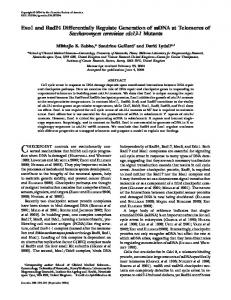
Figure 3 Requirement for nutrients for the insulin-induced dephosphorylation of eEF2 Serum-starved CHO.T cells were incubated for 60 min in PBS with addition of glucose (G), the standard amino acid mixture (AA), or both, as indicated. In some cases, insulin was then added (j), after which cells were extracted and the protein concentration was determined. Controls are serum-starved CHO.T cells (Con, maintained in Ham’s F12 medium) with or without insulin treatment, as indicated. Equal amounts of cell protein were applied to an SDS/polyacrylamide gel and the phosphorylation state of eEF2 was assessed by Western blotting using an antiserum specific for the phosphorylated form of the protein. Data are typical of four independent experiments. The differences in intensity of the signals in the last four lanes is due to the fact that, for purely technical reasons, the last four were run on a separate gel from the others and reflect differences in the transfer or development of the blot : identical amounts of cell protein were loaded into each lane and, on three other occasions, when fewer samples were analysed in parallel, so that PBS/G/AApinsulin samples were run on the same gel as the others, these differences in intensity were not observed. The position of phosphorylated eEF2 [eEF2(P)] is indicated.
on the observed activity of eIF2B under any conditions, and thus did not reveal a ‘ latent ’ activation of the factor, e.g. in extracts of insulin-treated cells incubated without nutrients (results not shown). Singh and Wahba [56] have reported that purified eIF2B can be activated by fructose 1,6-bisphosphate. However, addition of fructose 1,6-bisphosphate oat 0.5 mM, ten times the reported A . (the concentration of ligand required for half-maximal !& activation) [56]q had only small and inconsistent effects on eIF2B activity in cell extracts and, in particular, did not ‘ restore ’ activation by insulin. Thus none of the established allosteric regulators of eIF2B appears to be responsible for the effects of nutrients on its activation by insulin.
The insulin-induced dephosphorylation of eEF2 also requires both glucose and amino acids In control cells (maintained in Ham’s F12 medium), insulin induced the complete dephosphorylation of eEF2, as judged using an antiserum which specifically detects the phosphorylated form of this protein [38] (Figure 3) in agreement with our earlier observations [25]. When we tested whether the nutrient status of the cells affected this response, it was clear that in cells incubated without amino acids or glucose, or in the presence of only one of these, insulin was unable to bring about the dephosphorylation of eEF2 (a consistent result of four separate experiments). In contrast, when cells were incubated in PBS containing both glucose and amino acids, insulin did bring about substantial dephosphorylation of eEF2, albeit not to the full extent seen in cells maintained in Ham’s F12 medium (Figure 3 ; similar data were obtained in four independent experiments, in all of which control gels and immunoblots were carried out to confirm equal loading of total eEF2 in each lane). In cells kept in both nutrients, the level of phosphorylation of eEF2 fell to 49.5p9 % of the untreated control level following insulin treatment. # 1999 Biochemical Society
438
L. E. Campbell, X. Wang and C. G. Proud
The decrease seen in response to insulin in glucose\amino acid-maintained cells was always less than that observed in cells maintained in Ham’s F12, suggesting that factors other than nutrients may also affect the regulation of eEF2. Thus like the activation of eIF2B, the ability of insulin to regulate eEF2 (in this case to bring about its dephosphorylation and hence its activation) is dependent upon the presence in the incubation medium of both amino acids and glucose. None of the glucose analogues tested (-glucose, 3-OMG and 2-DOG) showed a consistent ability to permit the insulin-induced dephosphorylation of eEF2 (when used in combination with the amino acid mixture, results not shown). In our earlier studies, we showed that insulin induced a decrease in the activity of eEF2 kinase [25], although this was quantitatively much smaller than the large change in the level of phosphorylation of its substrate, eEF2. In the present series of experiments, the effects of insulin on eEF2 kinase were again small. In control cells, insulin decreased eEF2 kinase activity to 82.7p2.5 % of the non-insulin-treated control (n l 6). eEF2 kinase activity was slightly elevated in extracts of cells incubated in PBS (122p11 %, n l 3) and insulin reduced this, but to a lesser extent that in control cells (to 115p7 %, n l 3). In cells incubated in the presence of both glucose and amino acids, where insulin was able to induce partial dephosphorylation of eEF2, kinase activity was 109p2.3 % (n l 3) of control before insulin treatment and fell to 80p0.8 % after insulin treatment (n l 3). Thus the presence of the nutrients in the medium allowed insulin to bring about a more marked reduction in eEF2 kinase activity than in their absence, but the magnitude of the changes in kinase activity was small. This suggests that the activity of the protein phosphatase acting on eEF2 (most likely a form of protein phosphatase-2A [57]) may also be regulated by insulin. It is therefore clear from the above data that the ability of insulin to activate both eIF2B and eEF2 in CHO.T cells requires the presence of both glucose and amino acids. Both factors are involved primarily in the overall regulation of mRNA translation rather than the control of the translation of specific transcripts and it thus makes physiological ‘ sense ’ for their activation to be modulated by the availability of amino acids (precursors for protein biosynthesis) and glucose (important energy source). These data are of particular interest in the light of the early observations of van Venrooij et al. [58], who found that glucose positively regulated protein synthesis in Ehrlich ascites tumour cells through a mechanism which involved stimulation of peptidechain initiation and elongation.
Effects of amino acids and glucose on the regulation of p70 S6 kinase and 4E-BP1 by insulin In view of the above data showing that glucose modulates the regulation of eEF2, which is controlled through an mTORdependent signalling pathway, we considered it important to test whether the presence in the medium of glucose is also required for the effects of insulin on p70 S6 kinase and 4E-BP1. Preincubation of cells in nutrient-deficient medium brought about a decrease in p70 S6 kinase activity, as reported earlier [6]. The presence of amino acids partially prevented this (Figure 4A, P 0.15 versus cells incubated without nutrients) but the ability of leucine alone to maintain a higher basal p70 S6 kinase activity was not statistically significant (P � 0.2). Analogous effects were also seen for 4E-BP1 (Figure 4B). When cells were incubated in the absence of glucose or amino acids, 4E-BP1 underwent dephosphorylation, appearing as the less-phosphorylated α and β species, whereas when cells were kept in medium containing # 1999 Biochemical Society
amino acids, it remained more highly phosphorylated, and none of the α form was evident. The presence of glucose together with amino acids resulted in a higher proportion of the protein being in the slowest-migrating, most highly phosphorylated, γ form (which does not bind eIF4E). In the absence of amino acids and glucose, insulin was unable to activate p70 S6 kinase (Figure 4A) or to increase 4E-BP1 phosphorylation (Figure 4B). In the presence of glucose alone, insulin elicited only a very small effect on either p70 S6 kinase activity (P 0.05) or 4E-BP1 phosphorylation (Figures 4A and 4B). In contrast, in the presence of amino acids, insulin substantially activated p70 S6 kinase and enhanced the phosphorylation of 4E-BP1 (Figures 4A and 4B), in the latter case apparently to levels seen following insulin treatment of cells maintained in culture medium. In cells kept in medium containing glucose and amino acids, insulin activated p70 S6 kinase and induced 4E-BP1 phosphorylation to at least the same extent as in cells maintained in growth medium (Figure 4A and 4B). Thus unlike the situation for eIF2B and eEF2, the availability of glucose is not essential for the regulation of p70 S6 kinase and 4E-BP1 by insulin : amino acids appear to the be the major regulator here. In the case of p70 S6 kinase, it can be argued that this is again a physiologically reasonable situation. This enzyme is involved in the upregulation of the translation of mRNAs encoding components of the translational machinery [23] : the ability of amino acids to activate it allows them to function as feed-forward activators of the synthesis of these proteins, and their permissive role for the effects of insulin would ensure that synthesis of these proteins is only turned on when amino acids are available. Earlier studies have suggested that PKB is involved in the upstream regulation of mTOR, p70 S6 kinase and 4E-BP1 [59–63]. The present data demonstrate clearly that the activation of PKB is not sufficient for the regulation of p70 S6 kinase or 4EBP1 by insulin. Thus further inputs provided by the prevailing nutrient levels (primarily those of amino acids) are essential for insulin to regulate p70 S6 kinase or 4E-BP1.
Effects of nutrients on formation of the eIF4E–eIF4G complex We also examined the effect of nutrients upon the association of eIF4E with 4E-BP1 and with eIF4G. In control cells, very little, if any, 4E-BP1 was bound to eIF4E, even in the absence of insulin and, consistent with this, insulin scarcely affected the association of eIF4G with eIF4E (Figure 4D). These data show that the eIF4E–eIF4G complex is already fully formed in cells maintained in our normal medium. Transfer of the cells to PBS resulted in a marked fall in the amount of eIF4G bound to eIF4E and treatment of the cells with insulin failed to increase this. Similarly, shifting the cells to PBS markedly increased the amount of 4E-BP1 bound to eIF4E and insulin was unable to decrease this (Figure 4D). This is consistent with both the marked dephosphorylation of 4E-BP1 in the absence of nutrients (Figure 4B) and with the inability of insulin to enhance 4E-BP1 phosphorylation under this condition (Figure 4B). In cells supplied with glucose, the basal level of the eIF4E–eIF4G complex was still low but insulin was now able to bring about a modest increase in eIF4G binding, and to promote a slight release of 4EBP1 from eIF4E. Thus the small change in the phosphorylation of 4E-BP1 in response to insulin does appear to result in a reduction in its binding to eIF4E (Figure 4B). In cells incubated in the presence of amino acids, the basal level of eIF4F formation was similar to that in control cells and was not significantly enhanced by insulin. Under this condition, the basal amount of
Modulation of translation factors by nutrients
439
D.
Figure 4
Regulation of p70 S6k, 4E-BP1 and eIF4F by insulin under different conditions
Serum-starved CHO.T cells were incubated for 60 min PBS with addition of glucose (G), the standard amino acid mixture (AA), leucine (4 mM, i.e. five times the concentration present in the AA mixture) or combinations of these, as indicated. Insulin (Ins) was then added where shown (j) and the cells were extracted. Controls are serum-starved CHO.T cells (maintained in Ham’s F12 medium) with or without insulin treatment, as indicated. (A) Samples of extract containing equal amounts of total protein were subjected to immunoprecipitation with antisera to p70 S6 kinase and assayed for activity. Data are presented as a percentage of the control (serum-starved cells without insulin) ; meanspS.E.M. are shown, n l 6 unless otherwise indicated. (B and C) Samples of cell extract were subjected to SDS/PAGE and Western blotting using an antiserum against 4E-BP1. The positions of the three forms of 4E-BP1 separated in this gel system (α, β and γ, in order of increasing state of phosphorylation) are indicated. (D) Samples of extract (containing equal amounts of protein) were subjected to affinity chromatography on 7-methyl GTP–Sepharose and the bound material was analysed by SDS/PAGE and Western blotting using antisera for eIF4G (upper panel) or eIF4E and 4E-BP1 (lower panel). The positions of migration of each of the three proteins are shown. Numbers below each lane show the ratio of the signals for 4E-BP1 and eIF4E in this experiment, as determined by densitometric analysis of the blot. Con, control.
4E-BP1 bound to eIF4E was low and insulin treatment brought about its complete dissociation from eIF4E (Figure 4D). Consistent with this, 4E-BP1 phosphorylation is increased substantially in the presence of amino acids alone, relative to the situation in PBS, so that the least-phosphorylated α form of 4EBP1 is undetectable (Figure 4B). These data show that amino
acids themselves exert a marked effect upon the basal levels of formation of the eIF4E–eIF4G complex and the phosphorylation and binding of 4E-BP1, and that glucose is not required for complex formation. Nonetheless, glucose alone does have a small, but significant, positive effect upon the ability of insulin to promote formation of the eIF4E–eIF4G complex. # 1999 Biochemical Society
440
L. E. Campbell, X. Wang and C. G. Proud
Efficacy of leucine in the regulation of translation-factor activation Recent data for p70 S6 kinase and 4E-BP1 [4–7,64] have shown that leucine by itself can exert substantial effects and have suggested that it might play a particularly important role in regulating translation. We therefore compared the effects of leucine and the amino acid mixture on p70 S6 kinase activity and the phosphorylation of 4E-BP1 (Figures 4A and 4C). Leucine facilitated the activation of p70 S6 kinase by insulin, but to a lesser extent that the mixture of amino acids, even when used at five times the concentration present in that mixture (Figure 4A). As was the case for the mixture, the effect of leucine was enhanced by the presence of -glucose (Figure 4A). Neither of the other branched chain amino acids (valine, isoleucine) or αketoisocaproic acid (a product of leucine metabolism) significantly affected basal or insulin-stimulated p70 S6 kinase activity (results not shown). Whereas the full amino acid mixture largely prevented the dephosphorylation of 4E-BP1 (Figures 4B and 4C), leucine had only a small effect upon the basal level of 4EBP1 phosphorylation (Figure 4C). Nonetheless, leucine could support insulin-induced phosphorylation of 4E-BP1 in the absence of glucose, albeit to a significantly lesser extent than the amino acid mixture under the same conditions (Figure 4C). We therefore tested whether leucine alone was able to support the activation of eIF2B by insulin, in the presence of glucose. It did so only very modestly (Figure 1B) compared with the complete mixture of amino acids, and markedly less so than was observed for the insulin-stimulated activation of p70 S6 kinase (see Figure 4A). Even in the presence of glucose, leucine was unable to support the ability of insulin to bring about the dephosphorylation of eEF2 (Figure 3). These data define another significant difference between the regulation of p70 S6 kinase and 4E-BP1 on one hand (where leucine alone has both an effect in its own right and also acts to facilitate the regulation of these proteins by insulin) and eEF2 and eIF2B (see Figure 1B) on the other hand (where leucine alone has neither effect).
Conclusions Our data, and the earlier work of others, suggest : (i) that amino acids, through as-yet-unknown mechanisms, modulate the activity of proteins controlled through the mTOR signalling pathway ; (ii) that glucose, via a mechanism which requires its metabolism beyond glucose 6-phosphate, acts to permit the activation of eIF2B and eEF2 by insulin ; and (iii) that amino acids, through an additional mechanism not related to mTOR of the phosphorylation of eIF2α, exert a permissive effect on the activation of eIF2B by insulin. Unravelling these mechanisms responsible for these effects in mammalian cells may be facilitated by the very recent advances in understanding nutrient signalling processes in budding yeast [65,66]. This work was supported by a Programme Grant (to C. G. P.) from the Wellcome Trust. We thank Dr. Graham Pavitt, Dr. Peter Taylor and Dr. Harinder Hundal (all of Dundee University) for helpful discussions, and Ms. Amanda MacKenzie (Dundee University) who performed some of the studies on the regulation of p70 S6 kinase by amino acids. We are grateful to Dr. Jackie Vandenheede (Katholieke Universiteit te Leuven, Leuven, Belgium), Dr. Dario Alessi (Dundee University), Dr. Gary Krause (Wayne State University, Detroit, MI, U.S.A.) and Dr. Angus Nairn (Rockefeller Institute, New York, NY, U.S.A.) for antisera to GSK-3, PKB, phosphorylated eIF2α and eEF2, respectively. Purified rabbit eEF2 was donated generously by Dr. Nick Redpath (University of Leicester, Leicester, U.K.).
2 3 4 5 6 7 8 9
10 11 12 13 14 15 16 17 18 19 20 21 22 23
24 25 26 27 28 29 30 31 32 33 34 35
36 37 38 39 40 41 42
REFERENCES 1
Kleijn, M., Scheper, G. C., Voorma, H. O. and Thomas, A. A. M. (1998) Eur. J. Biochem. 253, 531–544
# 1999 Biochemical Society
43 44
Pain, V. M. (1996) Eur. J. Biochem. 236, 747–771 Fox, H. L., Kimball, S. R., Jefferson, L. S. and Lynch, C. J. (1998) Am. J. Physiol. 43, C206–C213 Patti, M.-E., Brambilla, E., Luzi, L., Landaker, E. J. and Kahn, C. R. (1998) J. Clin. Invest. 101, 1519–1529 Hara, K., Yonezawa, K., Weng, Q.-P., Kozlowski, M. T., Belham, C. and Avruch, J. (1998) J. Biol. Chem. 273, 14484–14494 Wang, X., Campbell, L. E., Miller, C. M. and Proud, C. G. (1998) Biochem. J. 334, 261–267 Kimball, S. R., Horetsky, R. L. and Jefferson, L. S. (1998) J. Biol. Chem. 273, 30945–30953 Webb, B. L. J. and Proud, C. G. (1998) Int. J. Biochem. Cell Biol. 29, 1127–1131 Clemens, M. J. (1996) in Translational Control (Hershey, J. W. B., Mathews, M. B. and Sonenberg, N., eds.), pp. 139–172, Cold Spring Harbor Press, Cold Spring Harbor Price, N. T. and Proud, C. G. (1994) Biochimie 76, 748–760 Kimball, S. R. and Jefferson, L. S. (1988) Biochem. Biophys. Res. Commun. 156, 706–711 Karinch, A. M., Kimball, S. R., Vary, T. C. and Jefferson, L. S. (1993) Am. J. Physiol. 264, E101–E108 Jeffrey, I. W., Kelly, F. J., Duncan, R., Hershey, J. W. and Pain, V. M. (1990) Biochimie 72, 751–757 Welsh, G. I. and Proud, C. G. (1992) Biochem. J. 284, 19–23 Welsh, G. I., Stokes, C. M., Wang, X., Sakaue, H., Ogawa, W., Kasuga, M. and Proud, C. G. (1997) FEBS Lett. 410, 418–422 Welsh, G. I., Miller, C. M., Loughlin, A. J., Price, N. T. and Proud, C. G. (1998) FEBS Lett. 421, 125–130 Welsh, G. I., Foulstone, E. J., Young, S. W., Tavare! , J. M. and Proud, C. G. (1994) Biochem. J. 303, 15–20 Moule, S. K., Welsh, G. I., Foulstone, E. J., Heesom, K., Edgell, N., Proud, C. G. and Denton, R. M. (1997) J. Biol. Chem. 272, 7713–7719 Cross, D. A. E., Alessi, D. R., Vandenheede, J. R., McDowell, H. E., Hundal, H. S. and Cohen, P. (1994) Biochem. J. 303, 21–26 Kleijn, M., Welsh, G. I., Scheper, G. C., Voorma, H. O., Proud, C. G. and Thomas, A. A. M. (1998) J. Biol. Chem. 273, 5536–5541 Thomas, G. and Hall, M. N. (1998) Curr. Opin. Cell Biol. 9, 782–787 Proud, C. G. (1996) Trends Biochem. Sci. 21, 181–185 Meyuhas, O., Avni, D. and Shama, S. (1996) in Translational Control (Hershey, J. W. B., Mathews, M. B. and Sonenberg, N., eds.), pp. 363–388, Cold Spring Harbor Press, Cold Spring Harbor Lawrence, J. C. and Abraham, R. T. (1997) Trends Biochem. Sci. 22, 345–349 Redpath, N. T., Foulstone, E. J. and Proud, C. G. (1996) EMBO J. 15, 2291–2297 Proud, C. G. and Denton, R. M. (1997) Biochem. J. 328, 329–341 Xu, G., Marshall, C. A., Lin, T.-A., Kwon, G., Munivenkatappa, R. B., Hill, J. R., Lawrence, J. C. and McDaniel, M. L. (1998) J. Biol. Chem. 273, 4485–4491 Hinnebusch, A. G. (1997) J. Biol. Chem. 272, 21661–21664 Price, N. T., Mellor, H., Craddock, B. L., Flowers, K. M., Kimball, S. R., Wilmer, T., Jefferson, L. S. and Proud, C. G. (1996) Biochem. J. 318, 637–643 Santoyo, J., Alcalde, J., Mendez, R., Pulido, D. and de Haro, C. (1997) J. Biol. Chem. 272, 12544–12550 Sood, R. and Wek, R. C. (1997) FASEB J. 11, 2196 Flynn, A. and Proud, C. G. (1996) FEBS Lett. 389, 162–166 Flynn, A. and Proud, C. G. (1996) Eur. J. Biochem. 236, 40–47 Wang, X., Flynn, A., Waskiewicz, A. J., Webb, B. L. J., Vries, R. G., Baines, I. A., Cooper, J. and Proud, C. G. (1998) J. Biol. Chem. 273, 9373–9377 DeGracia, D. J., Sullivan, J. M., Neumar, R. W., Alousi, S. S., Hikade, K. R., Pittman, J. E., White, B. C., Rafols, J. A. and Krause, G. S. (1997) J. Cereb. Blood Flow Metab. 17, 1291–1302 Oldfield, S., Jones, B. L., Tanton, D. and Proud, C. G. (1994) Eur. J. Biochem. 221, 399–410 Redpath, N. T. (1992) Anal. Biochem. 202, 340–343 Marin, P., Nastiuk, K. L., Daniel, N., Girault, J., Czernik, A. J., Glowinski, J., Nairn, A. C. and Premont, J. (1997) J. Neurosci. 17, 3445–3454 Dickens, M., Chin, J. E., Roth, R. A., Ellis, L., Denton, R. M. and Tavare! , J. M. (1992) Biochem. J. 287, 201–209 Diggle, T. A., Moule, S. K., Avison, M. B., Flynn, A., Foulstone, E. J., Proud, C. G. and Denton, R. M. (1996) Biochem. J. 316, 447–453 Kleijn, M., Korthout, M. M. R., Voorma, H. O. and Thomas, A. A. M. (1996) FEBS Lett. 396, 165–171 Moule, S. K., Edgell, N. J., Welsh, G. I., Diggle, T. A., Foulstone, E. J., Heesom, K. J., Proud, C. G. and Denton, R. M. (1995) Biochem. J. 311, 595–601 Walker, K. S., Deak, M., Paterson, A., Hudson, K., Cohen, P. and Alessi, D. R. (1998) Biochem. J. 331, 299–308 Welsh, G. I., Patel, J. C. and Proud, C. G. (1997) Anal. Biochem. 244, 16–21
Modulation of translation factors by nutrients 45 Iiboshi, Y., Papst, P. J., Kawasome, H., Hosoi, H., Abraham, R. T., Houghton, P. J. and Terada, N. (1999) J. Biol. Chem. 274, 1092–1099 46 Kimball, S. R., Antonetti, D. A., Brawley, R. M. and Jefferson, L. S. (1991) J. Biol. Chem. 266, 1969–1976 47 Pollard, J. W., Galpine, A. R. and Clemens, M. J. (1989) Eur. J. Biochem. 182, 1–9 48 Clemens, M. J., Galpine, A., Austin, S. A., Panniers, R., Henshaw, E. C., Duncan, R., Hershey, J. W. and Pollard, J. W. (1987) J. Biol. Chem. 262, 767–771 49 Scorsone, K. A., Panniers, R., Rowlands, A. G. and Henshaw, E. C. (1987) J. Biol. Chem. 262, 14538–14543 50 Rowlands, A. G., Montine, K. S., Henshaw, E. C. and Panniers, R. (1988) Eur. J. Biochem. 175, 93–99 51 Pain, V. M., Lewis, J. A., Huvos, P., Henshaw, E. C. and Clemens, M. J. (1980) J. Biol. Chem. 255, 1486–1491 52 Singh, L. P., Aroor, A. R. and Wahba, A. J. (1994) Biochemistry 33, 9152–9157 53 Dholakia, J. N. and Wahba, A. J. (1988) Proc. Natl. Acad. Sci. U.S.A. 85, 51–54 54 Dholakia, J. N., Mueser, T. C., Woodley, C. L., Parkhurst, L. J. and Wahba, A. J. (1986) Proc. Natl. Acad. Sci. U.S.A. 83, 6746–6750 55 Oldfield, S. and Proud, C. G. (1992) Eur. J. Biochem. 208, 73–81 56 Singh, L. P. and Wahba, A. J. (1998) Biochem. Biophys. Res. Commun. 217, 616–623
441
57 Redpath, N. T. and Proud, C. G. (1990) Biochem. J. 272, 175–180 58 van Venrooij, W. J. W., Henshaw, E. C. and Hirsch, C. A. (1970) J. Biol. Chem. 245, 5947–5953 59 Scott, P. H., Brunn, G. J., Kohn, A. D., Roth, R. A. and Lawrence, J. C. (1998) Proc. Natl. Acad. Sci. U.S.A. 95, 7772–7777 60 Burgering, B. M. T. and Coffer, P. J. (1995) Nature (London) 376, 599–602 61 Ueki, K., Yamamoto-Honda, R., Kaburagi, Y., Yamauchi, T., Tobe, K., Burgering, B. M. T., Coffer, P. J., Komuro, I., Akanuma, Y., Yazaki, Y. and Kadowaki, T. (1998) J. Biol. Chem. 273, 5315–5322 62 Kitamura, T., Ogawa, W., Sakaue, H., Hino, Y., Kuroda, S., Takata, M., Matsumoto, M., Maeda, T., Konishi, H., Kikkawa, U. and Kasuga, M. (1998) Mol. Cell. Biol. 18, 3708–3717 63 Gingras, A.-C., Kennedy, S. G., O’Leary, M. A., Sonenberg, N. and Hay, N. (1998) Genes Dev. 12, 502–513 64 Fox, H. L., Pham, P. T., Kimball, S. R., Jefferson, L. S. and Lynch, C. J. (1998) Am. J. Physiol. 44, C1232–C1238 65 Ozcan, S., Dover, J. and Johnston, M. (1998) EMBO J. 17, 2566–2573 66 Iraqui, I., Vissers, S., Bernard, F., de Craene, J.-O., Boles, E., Urrestarazu, A. and Andre, B. (1999) Mol. Cell. Biol. 19, 989–1001 67 Montine, K. S. and Henshaw, E. C. (1989) Biochim. Biophys. Acta 1014, 282–288
Received 6 April 1999/17 August 1999 ; accepted 9 September 1999
# 1999 Biochemical Society