To gain a better knowledge about reservoir petroleum biodegradation, it is ... forming gram-positive bacteria were isolated from the rock of an oil reservoir ...
OIL BIODEGRADATION BY BACILLUS STRAINS ISOLATED FROM THE ROCK OF AN OIL RESERVOIR LOCATED IN A DEEP-WATER PRODUCTION BASIN IN BRAZIL Claudia Cunha1, Alexandre S. Rosado1, Gina V. Sebastián2, Lucy Seldin1 and Irene von der Weid1* 1
Instituto de Microbiologia Prof. Paulo de Góes, UFRJ, CCS, Bloco I, Ilha do Fundão, Rio de Janeiro – RJ 2
CENPES, Petrobrás, Ilha do Fundão, Rio de Janeiro – RJ
Abstract. To gain a better knowledge about reservoir petroleum biodegradation, it is essential to associate hydrocarbon degradation with specific organisms and metabolic processes. In this study, eighteen sporeforming gram-positive bacteria were isolated from the rock of an oil reservoir located in a deep-water production basin in Brazil. These strains were identified as using classical biochemical techniques and API 50CH kits and their identity was confirmed by sequencing of part of their 16S rRNA gene. All strains were tested for their oil degradation ability in microplates using Arabe Leve and Marlin oils and only seven strains showed positive results in both kinds of oils. They were further tested to their capability to grow in the presence of carbazol, quinoline and n-hexadecane as the only carbon source. These strains showed positive results to carbazol and n-hexadecane, but not to quinoline. The production of key enzymes involved with biodegradation process by Bacillus strains (catechol 1,2 dioxygenase and catechol 2,3 dioxygenase) was verified spectrophotometrically by detection of cis, cis-muconic acid and 2-hydroxymuconic semialdehyde. Preliminary results showed that the ortho ring cleavage pathway is preferential. Biodegradation tests using Arabe Leve oil were carried out and only one strain (B. licheniformis T4.2) presented a good result, with 94% of n-alkanes reduction. Furthermore, the seven Bacillus strains were screened for the presence of catabolic genes encoding alkane monooxygenase, catechol 1,2 dioxygenase and catechol 2,3 dioxygenase by using primers specific for these functional genes and PCR products were obtained with the DNA of different strains of B. licheniformis and B. subtilis. After DNA sequencing of the PCR products, they will be compared to previously well-known degradative genes.
Keywords: Biodegradation, Bacillus, Oil reservoir
1. Introduction Biodegradation of crude oil in the reservoir is an important alteration process with major economic consequences. While the effects of biodegradation on the molecular composition and physical properties of crude oil are relatively well known (Hunt, 1979, Connan, 1984), the actual processes taking place during biodegradation of crude oil in deep reservoirs remain obscure. Oxidation of oil during biodegradation leads to a systematic decrease in paraffin content with increasing degradation and an increase in oil density, sulphur content, acidity, and viscosity (Connan, 1984) with huge negative economic consequences on oil production and refining operations.
1
The subsurface is defined as terrestrial habitats below 8 m and marine sediments below 10 cm. Few direct enumerations of subsurface prokaryotes have been made, largely because of the difficulty in obtaining uncontaminated samples. Nevertheless, circumstantial evidence suggests that the subsurface biomass of prokaryotes is enormous (Gold, 1992). For instance, groundwater from deep aquifers and formation water from petroleum deposits contain 103–106 prokaryotic cells/ ml (Nilsen et al., 1996). For deeper sediments to 4 km, the number of prokaryotes was extrapolated from the formula of Parkes et al. (1994), considering that at 4 km, the average temperature reaches 125°C (Garland, 1971), which is close to the upper temperature limit for prokaryotic life. Despite the large apparent mass and possible biogeochemical effects of subseafloor life, the magnitude of its metabolic activity in situ remains largely unknown. During the past 15 years, studies of Ocean Drilling Program (ODP) cores have consistently identified abundant prokaryotes in deeply buried oceanic sediments (Parkes et al., 2000). Microorganisms have been recovered from depths as great as 800 m below the seafloor (Taylor et al., 1999). The number and mass of prokaryotes in subseafloor sediments have been estimated by extrapolation from direct counts of sedimentary microorganisms at a small number of ODP sites (Parkes et al., 1994, Whitman et al., 1998). Acridine orange direct bacterial counts decrease from about 109 cells/ ml near the sediment surface to 104 cells/ ml in the deep layers (Parkes et al., 1994; Reed et al., 2002; Wellsbury et al., 2002). However, the extent of the diversity, as well as the activity, cell density and productivity of prokaryotes in deep marine sediments is not well known, primarily because of poor cultivability. Enrichments from deep sediments have enabled novel organisms to be isolated (Barnes et al ., 1998), but most probable number counts of bacteria have been very low, with viability down to 0.0000087% (Cragg et al., 1990). Aromatic compounds, including chlorinated ones, are frequently a major fraction of petroleum hydrocarbons. Further, most of the aromatic compounds, including polyaromatic, are known to be metabolized to a common intermediated, catechol, which is then oxidized through the two ring-cleavage pathways, ortho and meta cleavage pathways, catalyzed by C12O and C23O, respectively. Considering that, it has been proposed that catechol dioxigenases genes are good markers for the detection of a whole range of aromatic compounddegrading bacteria (Sei et al., 1999). On the other hand, degenerated primers have also been described to identify n-alkane degrading bacteria belonging to different genera without cultivation (Luz et al., 2004). The aims of our study were to isolate and identify endospore forming bacteria present in the rock of an oil reservoir located in a deep-water production basin in Brazil and determine the ability of oil degradation by these strains, which would help in predicting biodegradation potential in these environmental conditions. For general detection of indigenous bacterial strains capable of degrading aromatic compounds as well as n-alkanes, some molecular detection methods based on the genotype were applied together with chemical detection of these enzymes in seven Bacillus strains isolated from the rock at about 1,658 to 3,067 meters deep in a deep-water production basin in Brazil.
2. Materials and Methods 2. 1. Field sampling and bacterial isolation The core samples used in this study were taken from a virgin field located in the Atlantic Ocean, Rio de Janeiro, Brazil, prior to any production or seawater injection, using all facilities developed in PETROBRAS
2
Research Center for accessing deep-subsurface oil reservoirs. The temperature of the reservoir was 60oC, the saturation pression varied from 243.09 to 270.60 kgf/ cm2 and the salinity was equivalent to 55 g/L of NaCl. As the core emerged to surface, it was immediately removed from the core barrel and kept into the liner (fiberglass inner sleeve). Samplings were obtained from the marine subsurface at different levels of depth (from 2,658 to 3,067m). End caps were placed on the core liner and the cores samples were transported to the laboratory preserved in anaerobic jars filled with CO2 and N2 and kept in dry ice. In the laboratory, the anaerobic jars were maintained in a pre-sterilized anaerobic box and the core samples were removed aseptically. The outer portion of the rock was removed and inner portions of the core, which have not been contaminated with drilling mud, were then sampled by using a sterilized sampler developed specially for that purpose. Six core samples were obtained in different depth levels. All samplings were kept in sterile flasks and preserved on refrigeration before pasteurization and inoculation into growth media. Inside the anaerobic chamber, samples of core (10 g/100 mL distilled water) were pasteurized (10 min, 80oC) and plated onto BS agar (glucose 10 g, tryptone 10 g, yeast extract 5 g, NaCl 55 g/L) and incubated at 30oC for 7 days. Further isolation and purification were performed as described before (Seldin et al., 1983). 2. 2. Identification of the isolates In order to identify the 18 bacterial isolates, most cultural and biochemical tests were performed by using the methods and media of Gordon et al. (1973). Some strains, that presented an unprecise biochemical profile, were also characterized by using API tests (API 50CH kit, Appareils et Procédés d'Identification, bioMérieux sa, Lyon, France) as described by the manufacturer. Sixteen strains, identified using API 50CH as belonging to B. subtilis, B. cereus or B. licheniformis species, and isolated from different levels of depth, were first analyzed by ARDRA (Amplified Ribosomal DNA Restriction Analysis) using three restriction enzymes. PCR reaction for 16S rDNA gene were performed as described by Massol-Deya et al. (1995) and the products obtained were digested with RsaI, HinfI and MspI, separately. Then, five representative strains belonging to different ARDRA patterns were selected for 16S rDNA sequencing. The results of PCR digestion with three endonucleases were collected into a matrix indicating the presence or absence of specific bands. Pairs of strains were compared using the simple matching coefficient and a dendrogram was constructed using the unweighted pair group method with arithmetic mean (UPGMA). For this analysis, the NTSYS software package (version 2.02, Exeter Software, Setauket, NY, USA) was used. 2. 3. Biodegradation experiments 2. 3. 1. Microplate assay Microplates containing 24 cavities were filled with an appropriate volume of three different growth medium: 1. Bushnell Haas medium, 2. Bushnell Haas medium supplemented with 0.3% (W/V) of yeast extract, 3. LB (1% tryptone, 0.5% NaCl and 0.5% yeast extract) medium. The inoculum of each Bacillus strain and a drop of the respectively oil (Arabe Leve or Marlin) were added in each cavity. At the end of an incubation time of 7 days at 300C, the biodegradation was determined visually considering any alteration in oil film.
3
2. 3. 2. Flask assay The potential of the isolated bacteria for biodegradation of Arabe Leve oil was examined by gas chromatography, quantifying remaining TPH in batch cultures. For each tested strain, two Erlenmeyer flasks were filled with 100 mL of Bushnell Haas medium containing 0.1% (V/V) of crude oil. Cells were precultured in 20 mL of LB medium for 24 h, washed twice and ressuspended in sterile saline solution (0.7% W/V). Inoculum corresponding to 10% (V/V) was transferred to each experimental flask. The batch experiment was performed on a rotary shaker at 300C and 150 rpm for 7 days. 2. 4. Growth in carbazol, quinoline and n-hexadecane Growth of seven Bacillus strains, previously selected, was determined using flasks containing 20 mL of BH medium amended with 300mg/ L of quinoline, 300mg/ L of carbazole or 770mg/ L of n-hexadecane as unique carbon source. The batch experiment was performed on a rotary shaker at 300C and 150 rpm for 7 days. Cell density was determined spectrophotometrically using a Shimadzu model (UV-1601) spectrophotometer. Absorbance (A450) was measured using mineral medium at a wavelength of 450 nm. 2. 5. Production of enzymes involved in catechol biodegradation Growth of each Bacillus strain was performed in 2 L Erlenmeyer flask containing 1 L of LB medium at 300C for 24 h. The cells were harvested by centrifugation at 5000 rpm for 20 min and washed twice using sterile saline solution. For the induction experiments, cells were cultivated for 18 h at 300C in Bushnell Haas medium supplemented with 77 mg/L of naphthalene as inductor. The cells were harvested by centrifugation at 5000 rpm for 20 min at 40C, washed twice and ressuspended in potassium phosphate buffer (pH 7.5). In enzyme assays, Bacillus cells obtained as described above were suspended in 2.5 mL of 10 mM phosphate buffer, broken by sonication (three cycles of 30 s each) and then centrifuged at 10.000 g for 10 min at 40C. The absorption changes during enzymatic conversion was performed spectrophotometrically at 250C using a Shimadzu model (UV-1601) spectrophotometer with quartz cuvettes. The reaction was started in a cuvette containing in a final volume of 3 mL, 95 µM of catechol and suitable amount of enzyme and phosphate buffer (pH 7.5). Spectra were recorded during enzymatic conversion between 400 and 220 nm at periodic intervals. 2. 6. Detection of catabolic genes by PCR The presence of catabolic genes encoding alkane monooxygenase, catechol 1,2 dioxygenase and catechol 2,3 dioxygenases was determined by PCR using primers specific for these functional genes. Eight strains that were able to modify the oil film in the microplate assays were then tested for the presence of these genes. PCR reactions for n-alkane monooxygenase were performed using a set of degenerated primers as described by Luz et al. (2004). For catechol 1,2 and 2,3 dioxygenases genes, two sets of primes were used in PCR reactions as described by Sei et al. (1999). The PCR products were analyzed in 1.2% agarose gels at 80V for 4h at room temperature.
4
3. Results and Discussion The new methodology described in this paper for isolation of bacteria from deep sub-surface had shown that despite the very low number of cells present in this habitat, the core sample could be aseptically removed and indigenous bacteria could be isolated and characterized. This method allowed the isolation of eighteen bacterial strains from the core at different levels of depth. These strains were first identified by classical biochemical methods as belonging to Bacillus subtilis (n=2), B. licheniformis (n=14) and Bacillus sp. (n=2). API tests were performed with some of the isolates in order to identify the Bacillus sp. at species level and to confirm the identity of the others. In addition, 16 of these strains were used in ARDRA experiments. The dendrogram was generated by using the combined data from digestion analysis of the amplified 16S rDNA with RsaI, HinfI and MspI (Figure 1). Using this approach the 16 strains could be separated into two main clusters at less than 30% similarity, indicating that at least 2 different species were present in the sample. Moreover, the strain T4.3 was separated from the others, forming a third cluster, suggesting that this strain could belong to a different species. Considering the three clusters obtained in ARDRA analysis, five representative strains were then selected for sequencing, as indicated in Figure 1. The results obtained after sequencing suggest that B. licheniformis and B. cereus were the main species isolated in the core sample. However, the identification of the strain T4.3 remains unclear, since it showed high similarity with both B. subtilis and B. licheniformis (data not shown).
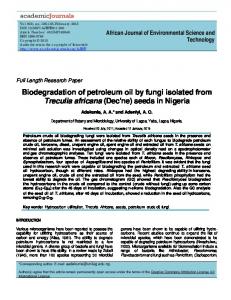
Fig. 1. Dendrogram (UPGMA) of genetic relationships among Bacillus strains, isolated from a deep sub-surface rock in an oil reservoir, estimated by ARDRA using RsaI, HinfI and MspI. Biodegradation experiments From eighteen strains tested for their oil degradation ability in microplates using Arabe Leve and Marlin oil, only seven showed positive results in both kind of oils. It was possible to verify alteration in superficial film oil which indicates possible biodegradation process (Figure 2). Some observed alterations suggest the capability of
5
bioemulsifier production by these strains through the formation of oil drops coalescence. Experiments of biosurfactant production are being developed to confirm and characterize the biomolecule structure. The biodegradation tests using agitated flasks showed that strain T4.2 presented high n-alkane reduction and could play an important role in reducing the paraffin content of oil, altering the quality of petroleum (Table 1).
BH medium
BH medium + Yeast ext.
BH medium
LB medium
BH medium + Yeast ext.
LB medium
T6.1
Cont.
T4.3
T7.0
T4.2
T6.5
T3.3
T6.2
Fig. 2. Biodegradation experiments using microplates containing Arabe Leve oil in 7 days. (strains T3.3, T4.2, T4.3, T6.1, T6.2, T6.5, T7.0 and control without cells) Table 1. Percentage values of n-alkanes degradation by Bacillus strains n-alKanes n-C11 n-C12 n-C13 n-C14 n-C15 n-C16 n-C17 Pri n-C18 Phy n-C19 n-C20 n-C21 n-C22 n-C23 n-C24 n-C25 n-C26 n-C27 n-C28 n-C29 n-C30 n-C31 n-C32
Strains T3.3
T4.2
T4.3
T6.1
T6.2
T6.5
T7.0
0,00 0,00 0,00 0,00 0,00 2,02 2,48 6,94 2,54 6,39 4,52 1,28 1,82 3,11 2,09 0,00 0,00 0,00 0,00 5,08 0,00 0,00 0,00 0,00
99,59 96,33 95,49 97,30 96,78 95,84 95,02 71,29 96,03 66,76 95,97 98,22 95,07 93,92 93,58 85,90 82,46 84,04 80,45 85,10 85,06 71,31 79,33 79,14
0,00 0,00 0,00 0,00 0,00 0,00 0,00 0,00 0,00 0,00 0,00 0,00 0,00 0,00 0,00 0,00 0,00 0,00 0,00 0,00 0,00 0,00 0,00 0,00
0,00 15,60 6,49 2,71 0,00 6,42 3,99 13,50 5,45 11,84 7,38 5,24 5,80 4,81 4,13 0,00 0,00 0,00 0,00 0,00 0,00 0,00 0,00 0,00
0,00 0,00 7,72 16,84 19,67 20,68 19,91 26,26 20,67 28,51 21,75 19,57 21,51 20,77 19,46 11,52 1,48 0,00 17,84 19,53 13,53 14,13 0,00 4,13
0,00 0,00 0,68 7,89 8,72 8,58 7,72 13,04 8,48 11,55 7,90 7,61 7,16 7,07 6,58 0,00 0,00 0,00 0,00 0,00 0,00 0,00 0,00 0,00
0,00 0,00 0,00 0,00 0,00 0,00 0,00 24,09 0,00 6,67 0,00 0,00 0,00 0,00 0,00 0,00 0,00 0,00 0,00 0,00 0,00 0,00 0,00 0,00
A comparison of the GC profiles 7 days after incubation of strain T4.2 revealed an appreciable decrease in nalkane peaks reaching 98% of degradation. Generally, the straight chain alkanes are easier to be degraded than
6
the aromatic ones. Microbial degradation of oil has been shown to occur primarily by attack on aliphatic or light aromatic fraction of oil, according to the results observed in our experiments. Similar investigation was made by Grishchenkov et al. (2000) using nitrate-reducing bacterial strains (Pseudomonas and Brevibacillus) isolated from petroleum contaminated soil. The strains degraded 20-25% of the total extractable matrice (TEM), including up to 90-95% of the alkanes analyzed (n-C10-C35) in a 10 days experiment in liquid media, under aerobic conditions. Growth in Carbazole, Quinoline and n-hexadecane The results of growth in different hydrocarbon sources by selected Bacillus strains have shown that all strains were able to grow in the presence of carbazole and n-hexadecane, but not in quinoline. Carbazole is one of the most predominant poliaromatic N-heterocyclic compounds in crude oil (Loh and Yu 2000) and n-hexadecane belong to the n-alkane fraction whose reduction alters petroleum quality. Molecular analysis of catabolic genes Considering the analysis of functional genes related to oil degradation, seven strains that were able to change the oil film in microplate assays were tested for the presence of n-alkane monooxigenase and catechol dioxigenases by PCR using three sets of primers. None of the PCR reactions provided fragments of the predicted size. However, in some cases, as for n-alkane monoxigenase and catechol 1,2 dioxigenase, more than one fragments could be observed, indicating that the described primers and PCR conditions are not so specific for these genes. On the other hand, three strains (T4.3, T6.5 and T7) presented a singular fragment amplified by PCR using the catechol 2,3 dioxigenase primers. Despite the small difference between the expected and obtained size of the PCR product, the result suggests that these strains could have related genes with different sizes or new genes that are not known yet. Enzyme Assay Cleavage of the aromatic ring is a key reaction in the oxidation of aromatic compounds. One of the most frequently encountered key dihydroxyaromatic species before ring cleavage occurs is catechol. The meta or the ortho ring cleavage are catalysed by catechol 2,3 –dioxygenase and catechol 1,2 –dioxygenase yielding 2hydroxi muconic semialdehyde (λmax at 255 nm) and cis,cis –muconate (λmax at 375 nm), respectively. To study the enzymatic conversion of catechol in the naphthalene-grown Bacillus strains, the absorption changes were recorded from 400 to 220 nm. In such conversion, the product formed was identified spectrophotometrically. Figure 3 shows enzymatic conversion of catechol by lysate of T3.3 and T7.0 of Bacillus strains. It was possible to observe for strains T3.3 and T7.0 the formation of a product with an absorption maximum at 260 nm and 264 nm, respectively. On the other hand no PCR products of the anticipated sizes were generated using the C12DO and /or C23DO primers. These results suggest that these isolates (T4.3 and T7.0) may possess neither C12DO nor C23DO genes, but have genes encoding a novel catechol catabolic enzyme. Hamzah and Al-Baharna in similar experiments using P. cepacia ATCC 29351 showed that this strain possesses the genetic capacity for enzymes of both ortho and metha cleavage pathway of benzoate degradation, although the phenotypic expression for the ortho pathway was higher.
7
1,60
1,6
1,40
1,4
1,00
275 nm
T7.0 (lysate)
0,80
catechol (0 time)
0,60
Absorbance
264 nm Absorbance
260 nm
1,2
1,20
1 catechol (0 time)
0,8
T3.3 (lysate)
0,6
275 nm
0,4 0,40
0,2
0,20 0,00 245 255 265
0 275
285
295
305
315
325
335
345
355
365 375
385
395
225
Wavelength (nm)
275
325
375
Wavelength (nm)
Fig. 3. Enzymatic conversion of catechol by lysate of naphthalene grown Bacillus cells. Spectra was recorded at 10 min (end of reaction). (a) T7.0 strain (b) T3.3 strain.
4. Conclusions In this study we showed that Bacillus strains can be recovered from deep sub-surface rock samples in oil reservoir environment at about 3.000 meters deep. Eighteen isolated strains were identified as B. cereus, B. subtilis and B. licheniformis. Some of these strains were able to modify the superficial film oil which indicates a possible biodegradation process. It was observed a probable biosurfactant production by these strains through coalescence of oil drops. Two of the Bacillus strains also demonstrated the genetic capacity for enzymes involved in cleavage pathways of aromatic degradation. In addition, it was found that 94% of n-alkanes were degraded by T4.2 strain after 7 days, suggesting that a possible alteration in petroleum composition could be attributed to the presence of these Bacillus strains in oil reservoir.
5. References Barnes, S.P., Bradbrook, S.D., Cragg, B.A., Marchesi, J.R., Weightman, A.J., Fry, J.C., Parkes, R.J. (1998). Isolation of sulfate-reducing bacteria from deep sediment layers of the Pacific Ocean. Geomicrobiol J., 15, 67. Connan, J. (1984). Biodegradation of crude oils in reservoirs. In: Brooks, J., Welte, D.H. (Eds.), Advances in Petroleum Geochemistry 1. Academic Press, London, 299. Cragg, B.A., Parkes, R.J., Fry, J.C., Herbert, R.A., Wimpenny, J.W.T., Getliff, J.M. (1990). Bacterial biomass and activity profiles within deep sediment layers. Proc. Ocean Drill Prog. Sci. Results, 112, 607. Garland, G. D. (1971). Introduction to Geophysics: Mantle, Core,and Crust (Saunders, Philadelphia). Gold, T. (1992). The deep, hot biosphere. Proc. Natl. Acad. Sci,. 89, 6045. Gordon, R. E., Haynes, W. C.,Pang, H.-N. (1973). The genus Bacillus. Agriculture Hhandbook 427. Agricultural Research Service, U. S. Department of Agriculture, Washington, DC. Grishchenkov, V. G., Townsend, R. T., McDonald, T. J., Autenrieth, R. L., Bonner, J. S., Boronin, A. M. (2000). Degradation of petroleum hydrocarbons by facultative anaerobic bacteria under aerobic and anaerobic conditions. Process Biochemistry, 35, 889. Hamzah, R. Y., Al-Baharna, B. S. (1994). Catechol ring-cleavage in Pseudomonas cepacia: the simultaneous induction of ortho and metha pathways. Appl. Microbiol. Biotechnol, 41, 250. Hunt, J.M. (1979). Petroleum Geochemistry and Geology. Freeman and Co., 617. Loh, K-C. ,Yu, Y-G. (2000). Kinetics of carbazole degradation by Pseudomonas putida in presence of sodium salicylate. Wat. Res., 34, 4131.
8
Luz, A. P., Pellizari, V. H., Whyte, L. G., Greer, C. W. (2004). A survey of indigenous microbial hydrocarbon degradation genes in soils from Antarctica and Brazil. Can. J. Microbiol., 50, 323. Massol-Deya, A. A., Odelson, D. A., Hichey, R. P., Tiedje, J. M. (1995). Bacterial community fingerprinting of amplified 16S and 16-23S ribosomal DNA genes sequences and restriction endonuclease analysis (ARDRA). In: Molecular Microbial Ecology Manual (eds. Akkermans et al. ). Kluwer Academic, 3.3.2., 1. Nilsen, R. K., Beeder, J., Thorstenson, T. ,Torsvik, T. (1996). Distribution of Thermophilic Marine Sulfate Reducers in North Sea Oil Field Waters and Oil Reservoirs Appl. Environ. Microbiol,. 62, 1793. Parkes, R.J., Cragg, B.A., Wellesbury, P. (2000). Recent studies on bacterial populations and processes in subseafloor sediments: a review. Hydrogeol. J., 8, 11. Parkes, R.J., Cragg, B.A., Bale, S.J., Getliff, J.M., Goodman, K., Rochelle, P.A., Fry, J.C., Weightman, A.J.,Harvey, S.M. (1994). Deep bacterial biosphere in Pacific Ocean sediments. Nature, 371, 410. Reed, D.W., Fujita, Y., Delwiche, M.E., Blackwelder, D.B., Sheridan, P.P., Uchida, T., Colwell, F.S. (2002). Microbial communities from methane hydrate-bearing deep marine sediments in a forearc basin. Appl. Environ. Microbiol., 68, 3759. Sei, K., Asano, K-I., Tateishi, N., Mori, K., Ike, M., Fujita, M. (1999). Design of PCR primers and gene probes for the general detection of bacterial populations capable of degrading aromatic compounds via catechol cleavage pathways. J. Biosc. Bioengineering., 88, 542. Seldin, L., van Elsas, J. D., Penido, E. C. G. (1983). Bacillus nitrogen fixers from brasilian soils. Plant soil., 70, 243. Taylor, B., Huchon, P., Klaus, A. (1999). Shipboard Scientific Party, Proc. ODP Init. Rep. 180,: 4. Wellsbury, P., Mather, I., Parkes, R.J. (2002). Geomicrobiology of deep, low organic carbon sediments in the Woodlark Basin, Pacific Ocean. FEMS Microbiol. Ecol., 42, 59. Whitman, W. B., Coleman, D. C., Wiebe, W. J. (1998). Prokaryotes: the unseen majority. Proc. Natl. Acad. Sci., 95, 6578.
9