1
Biol. Rev. (2018), pp. 000–000. doi: 10.1111/brv.12441
Ommochromes in invertebrates: biochemistry and cell biology Florent Figon and J´erˆome Casas∗ Institut de Recherche sur la Biologie de l’Insecte, UMR CNRS 7261, Universit´e de Tours, 37200 Tours, France
ABSTRACT Ommochromes are widely occurring coloured molecules of invertebrates, arising from tryptophan catabolism through the so-called Tryptophan → Ommochrome pathway. They are mainly known to mediate compound eye vision, as well as reversible and irreversible colour patterning. Ommochromes might also be involved in cell homeostasis by detoxifying free tryptophan and buffering oxidative stress. These biological functions are directly linked to their unique chromophore, the phenoxazine/phenothiazine system. The most recent reviews on ommochrome biochemistry were published more than 30 years ago, since when new results on the enzymes of the ommochrome pathway, on ommochrome photochemistry as well as on their antiradical capacities have been obtained. Ommochromasomes are the organelles where ommochromes are synthesised and stored. Hence, they play an important role in mediating ommochrome functions. Ommochromasomes are part of the lysosome-related organelles (LROs) family, which includes other pigmented organelles such as vertebrate melanosomes. Ommochromasomes are unique because they are the only LRO for which a recycling process during reversible colour change has been described. Herein, we provide an update on ommochrome biochemistry, photoreactivity and antiradical capacities to explain their diversity and behaviour both in vivo and in vitro. We also highlight new biochemical techniques, such as quantum chemistry, metabolomics and crystallography, which could lead to major advances in their chemical and functional characterisation. We then focus on ommochromasome structure and formation by drawing parallels with the well-characterised melanosomes of vertebrates. The biochemical, genetic, cellular and microscopic tools that have been applied to melanosomes should provide important information on the ommochromasome life cycle. We propose LRO-based models for ommochromasome biogenesis and recycling that could be tested in the future. Using the context of insect compound eyes, we finally emphasise the importance of an integrated approach in understanding the biological functions of ommochromes. Key words: ommochrome, ommochromasome, pigment, photochemistry, antiradical capacity, phenoxazone, phenothiazine, melanosome, melanin, lysosome-related organelle. CONTENTS I. Introduction . . . . . . . . . . . . . . . . . . . . . . . . . . . . . . . . . . . . . . . . . . . . . . . . . . . . . . . . . . . . . . . . . . . . . . . . . . . . . . . . . . . . . . . . . . . . . . II. Ommochrome biochemistry: from the indole to the phenoxazone chromophore . . . . . . . . . . . . . . . . . . . . . . . . (1) Ommochromes, a phenoxazone-based chrome family restricted to invertebrates . . . . . . . . . . . . . . . . . . . . (2) Extraction, analysis and identification of ommochromes . . . . . . . . . . . . . . . . . . . . . . . . . . . . . . . . . . . . . . . . . . . . . (3) Ommochrome biogenesis: the tryptophan oxidation pathway . . . . . . . . . . . . . . . . . . . . . . . . . . . . . . . . . . . . . . . . (4) Enzymes of the tryptophan→Ommochrome pathway . . . . . . . . . . . . . . . . . . . . . . . . . . . . . . . . . . . . . . . . . . . . . . . (5) Putative cross-talk between ommochromes and other chromes . . . . . . . . . . . . . . . . . . . . . . . . . . . . . . . . . . . . . . . (6) Ommochrome reactivity . . . . . . . . . . . . . . . . . . . . . . . . . . . . . . . . . . . . . . . . . . . . . . . . . . . . . . . . . . . . . . . . . . . . . . . . . . . . . III. Ommochromes within the cell: the ommochromasome life cycle . . . . . . . . . . . . . . . . . . . . . . . . . . . . . . . . . . . . . . . . (1) Methods for studying ommochromasomes . . . . . . . . . . . . . . . . . . . . . . . . . . . . . . . . . . . . . . . . . . . . . . . . . . . . . . . . . . . (2) Ommochromasome structure and composition . . . . . . . . . . . . . . . . . . . . . . . . . . . . . . . . . . . . . . . . . . . . . . . . . . . . . . (3) The origin of the ommochromasome, a lysosome-related organelle . . . . . . . . . . . . . . . . . . . . . . . . . . . . . . . . . . (4) Ommochromasome biogenesis . . . . . . . . . . . . . . . . . . . . . . . . . . . . . . . . . . . . . . . . . . . . . . . . . . . . . . . . . . . . . . . . . . . . . . .
2 5 5 5 8 8 11 12 13 13 15 15 16
* Author for correspondence (Tel: +33 (0)2 47 36 69 78; E-mail:
[email protected]). Biological Reviews (2018) 000–000 © 2018 Cambridge Philosophical Society
Florent Figon and J´erˆome Casas
2
IV. V. VI. VII.
(5) Ommochrome biosynthesis in relation to ommochromasome maturation . . . . . . . . . . . . . . . . . . . . . . . . . . . (6) Ommochromasome recycling . . . . . . . . . . . . . . . . . . . . . . . . . . . . . . . . . . . . . . . . . . . . . . . . . . . . . . . . . . . . . . . . . . . . . . . . Ommochromes in context: the insect compound eye . . . . . . . . . . . . . . . . . . . . . . . . . . . . . . . . . . . . . . . . . . . . . . . . . . . . . Conclusions . . . . . . . . . . . . . . . . . . . . . . . . . . . . . . . . . . . . . . . . . . . . . . . . . . . . . . . . . . . . . . . . . . . . . . . . . . . . . . . . . . . . . . . . . . . . . . Acknowledgements . . . . . . . . . . . . . . . . . . . . . . . . . . . . . . . . . . . . . . . . . . . . . . . . . . . . . . . . . . . . . . . . . . . . . . . . . . . . . . . . . . . . . . . References . . . . . . . . . . . . . . . . . . . . . . . . . . . . . . . . . . . . . . . . . . . . . . . . . . . . . . . . . . . . . . . . . . . . . . . . . . . . . . . . . . . . . . . . . . . . . . . .
I. INTRODUCTION Colouration is one of the most striking traits observable in animals. It has caught the interest of scientists for centuries and was at the heart of the work of pioneers of evolutionary biology, particularly Alfred R. Wallace, Edward B. Poulton, Henry W. Bates and Johann F.T. M¨uller (Bates, 1862; Wallace, 1877; M¨uller, 1879; Poulton, 1890; Caro, 2017). The field of animal colouration has also inspired a wealth of ideas in applied sciences (Caro, Stoddard & Stuart-Fox, 2017), including the investigation of biomimetic camouflage by the naturalist and artist Abbott H. Thayer (Thayer & Thayer, 1909). Therefore, understanding the proximate and ultimate causes of colouration in animals is of great importance (Cuthill et al., 2017; Endler & Mappes, 2017). Colours can arise from either physical or chemical processes (Cuthill et al., 2017). Physical or structural colours are produced by interactions between light and photonic structures such as crystals. Chemical colours are mainly created by light-absorbing pigments and dyes, together referred to as chromes (Fox, 1944; Needham, 1974). Pigments differ from dyes by being in suspension whereas the latter are in solution. This difference in physical state can have dramatic consequences on colouration because particles in suspension can also produce structural colours. Because it is rarely known whether a coloured molecule is in a solid or a liquid matrix in vivo (Needham, 1974), we hereafter use the term ‘chrome’ rather than the more-common, but imprecise if not misleading, word ‘pigment’. Chromes are directly able to modify part of the wavelengths of visible, near-ultraviolet (UV) and near-infrared (IR) light. They do so by absorbing photons with a specific energy, and thus wavelength, via their chromophore (Needham, 1974). The molecular structure of chromophores is particular to each chrome family, and understanding how they work is necessary to comprehend the biological functions of chromes (Needham, 1974). Ommochromes form a particular tryptophan-derived chrome family and have been described in protostomes (Fig. 1), but are virtually absent from deuterostomes and plants [see Takeuchi et al., 2005 for possible ommochrome-related genes in the tunicate Ciona intestinalis] (Linzen, 1974; Needham, 1974). Interestingly, some bacteria produce the well-known ommochrome-like antibiotic compound, actinomycin D (Le Roes-Hill, Goodwin & Burton, 2009). The ommochrome chromophore is based on phenoxazine and phenothiazine systems, which are heteropolyaromatic rings with either two N and O atoms or two N and S atoms, respectively. Phenoxazines are found in natural and synthesised chromes that have been used for Biological Reviews (2018) 000–000 © 2018 Cambridge Philosophical Society
18 20 21 21 23 23
centuries, such as orcein, and blue and red Niles, and also in more recent technologies, such as photovoltaic sensitizers (Li et al., 2017). Thus, ommochromes are at a crossroads of basic and applied sciences. The history of ommochrome study started almost 80 years ago with their first description in insect ommatidia by Becker (Fig. 2) (Becker, 1939, 1942). In the 1950s and 1960s, the so-called Butenandt’s school (one of Becker’s collaborators) provided almost all we know about ommochrome chemistry. This analytical work was accompanied by genetic studies that unravelled the biogenesis pathway of ommochromes and their organelles in insects, particularly Drosophila melanogaster, Ephestia kuehniella, Bombyx mori and Apis mellifera. Since then, researchers lost interest in ommochromes (Fig. 2A). A renaissance of interest is currently underway with studies dealing with ommochrome biological function and some of their chemical properties (Fig. 2B). These biochemical studies have allowed physiologists to unravel the properties and the biological significance of ommochromes. Ecologists studied these chromes to unravel the proximal basis of colour-based behaviours such as mimicry and vision (Holl, 1987; Oxford & Gillespie, 1998; Stavenga, 2002; Th´ery & Casas, 2009; Umbers et al., 2014). More recently, developmental biologists have used ommochromes of butterfly wings to tackle the mechanisms and the diversity of colour patterning (Sekimura & Nijhout, 2017). Today, ommochromes are also involved in transcriptomic studies of grasshoppers, butterflies, damselflies and spiders (Croucher et al., 2013; Chauhan et al., 2014; Connahs, Rhen & Simmons, 2016; Qiu et al., 2017; Wang et al., 2017), as well as in clustered regular interspaced short palindromic repeats/CRISPR-associated protein 9 (CRISPR/Cas9)-mediated genome editing (Khan, Reichelt & Heckel, 2017; Xue et al., 2017; Zhang & Reed, 2017). Unfortunately, biochemical knowledge did not keep pace with this increase in functional studies. Thus, the latter are usually based on classical biochemical papers and reviews, while ommochromes appear to be more complex and reactive than previously thought (Bolognese & Liberatore, 1988). The ommochrome-containing organelles were studied from the 1970s to the 1990s (Fig. 2B), before researchers focussed their attention on the related vertebrate melanosomes. Ommochrome-containing organelles have been classically termed ‘ommochrome granules’ or ‘pigment granules’ (Linzen, 1974; Kayser, 1985). However, herein, we designate them as ‘ommochromasomes’ as proposed by Needham 40 years ago (Needham, 1974). This decision is motivated by the membrane-bound nature of these organelles and their close relationship to melanosomes, both
Ommochromes in invertebrates
3
Ecdysozoa (A)
(B)
Lophozoa (D)
(F)
♀ ♂ (C)
(G)
(E)
Light
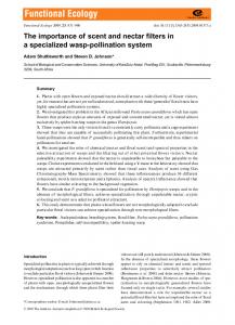
Platyzoa Fig. 1. Examples of protostomes that produce ommochromes. Ommochromes are found in the three main protostomian phyla: Ecdysozoa (insects and spiders), Lophozoa (cephalopods) and Platyzoa (flatworms). (A–C) Ommochromes generally function as eye pigments in insects, such as in Drosophila melanogaster (A; body length 3 mm), Bombyx mori (B; 20 mm) and Ephestia kuehniella (C; 10 mm). (D) Several Sympetrum species show sexual dimorphism, which results from different redox states of ommochromes: a yellow female and a red male of S. darwinianum are shown (body length 40 mm). (E) Some crab spiders, such as Thomisus onustus (body length 10 mm), can change their colour from white to yellow by producing and degrading ommochromes. Some individuals also show an irreversible purple-stripe pattern. (F) Cephalopods, such as Loligo vulgaris (body length 40 cm), can rapidly change their colour pattern by expanding or shrinking saccules of ommochromasomes, the ommochrome-containing organelles. The inset shows integumental chromatophores of different colours (from yellow to brown) corresponding to different ommochromes. (G) The flatworm Girardia dorotocephala (body length 5 mm) possesses epithelial chromatophores producing ommochromes. In presence of light, these chromatophores are lost leading to an unpigmented animal. Photograph credits (all CC BY SA): (A) Sanjay Acharya, (B) Ash Bowie, (C) Magne Fl˚aten, (D) Alpsdake, (E) Fritz Geller-Grimm, Paul-Henri Cahier and Hectonichus, (F) adapted from Hans Hillewaert, (G) adapted from Stubenhaus et al. (2016) https://doi.org/10.7554/eLife.14175.003.
in terms of structure and biogenesis (Lloyd, Ramaswami & Kr¨amer, 1998). Considering both ommochromes and ommochromasomes in their chemical and cellular contexts should help in teasing apart the complexity of their biological functions. Ommochromes are mainly found in insect ommatidia (from which their name is derived), in cephalopod eyes and in most protostomian integuments (Fig. 1) (Linzen, 1974; Needham, 1974). They are known to mediate colour patterning and colour changes, often in association with other chromes (Fuzeau-Braesch, 1985; Kayser, 1985; Oxford & Gillespie, 1998). Their colours range from pale yellow to dull brown, as well as bright red and deep purple. Other chromes, like melanins and pterins, and structural colours, such as in butterfly wing scales, can alter the chemical colours of ommochromes (Stavenga, Leertouwer & Wilts, 2014; Wilts et al., 2017). Colour patterns associated with ommochromes
and their precursors are involved in crypsis (Williams et al., 2016), mimicry (Reed, McMillan & Nagy, 2008; Ferguson & Jiggins, 2009; Bybee et al., 2012), colour changes (Insausti & Casas, 2008; Llandres et al., 2013; Umbers et al., 2014; Williams et al., 2016), sexual maturation and seasonal forms (Nijhout, 1997; Futahashi et al., 2012), as well as many other colour-based functions. Like most chromes, ommochromes not only produce colours, but also function in many metabolic and biological processes (Linzen, 1974; Needham, 1974). The chemical properties of chromophores often make them suitable for transporting electrons or reacting with oxidants, reducers and free radicals, as well as functioning in vision (Needham, 1974). All these functions have been proposed to be fulfilled by ommochromes in nature (Needham, 1974; Stavenga, 2002; Insausti, Le Gall & Lazzari, 2013; Romero & Martínez, 2015), often without direct proof. Furthermore, protostomes Biological Reviews (2018) 000–000 © 2018 Cambridge Philosophical Society
Florent Figon and J´erˆome Casas
4
(A)
'Ommochrome' in the text
Publications/decade
500
'Ommochrome' in the title
50
400
40
300
30
200
20
100
10
0 '40 s
'50s
'60 s
(B) 1940
1950
'70s
'80s
'90s
1960
0 2000s 2010s 1970
1980
1990
2000
2010
Photochemistry
Biochemistry
Genetics
1962 Butenandt & Schäffer 1961 Ziegler Ultrastructure
1967 Linzen
1974 Linzen Needham 1975 Dickinson & Sullivan
Physiology
Development
1982 Summers et al.
Ion content
Cell Biology
Lysosome-related organelles Puri fi cation
1972 Fuzeau-Braesch 1974 Needham
Quantum chemistry
1985 Kayser Fuzeau-Braesch
1998 Lloyd et al.
1987 Holl 1989 Stavenga
Ecology
2002 Stavenga
1998 Oxford & Gillespie
2010 Nijhout
2009 2014 Théry Umbers & Casas et al.
Omics
Fig. 2. History of ommochrome study. (A) The number of publications related to ommochromes since their first description. Results include papers found in Google Scholar with the term ‘ommochrome’ either in their whole content (blue line) or only in their title (red line). After a peak in the 1960s, the number of publications with ommochrome in the title decreased at a regular rate, reaching the lowest number in the last decade. By contrast, papers mentioning ommochromes in the main text reached a new peak during the last decade, after a low point in the 1990s. (B) Main periods in the history of ommochrome study. Major reviews for each topic are indicated. No review on ommochrome biochemistry and genetics has been published for almost 30 years. Interruption of lines implies almost no work on that topic.
generally lack the glutarate and nicotinamide biosynthesis pathway that catabolises the amino acid tryptophan (Linzen, 1974). In insects, tryptophan is toxic at high concentrations, such as those that occur during moulting (Linzen, 1974; Manoukas, 1981). Therefore ommochromes are also thought to be end-products of tryptophan detoxification (Linzen, 1974). Most difficulties in studying ommochromes arise from their chemical behaviour. Not all of them are soluble in the same solvents, some tend to form aggregates and they usually react with the extraction mixture (Linzen, 1974; Bolognese & Liberatore, 1988). However, they have led to many advances in biology. Their genetics paved the way for understanding the genome structure of eukaryotes and Biological Reviews (2018) 000–000 © 2018 Cambridge Philosophical Society
its inheritance (Morgan, 1910), the relationship between genes and enzymes (Butenandt, Weidel & Becker, 1940; Beadle & Ephrussi, 1936) and the importance of cell interactions during development (Beadle & Ephrussi, 1936). This was made possible by studies of eye-colour mutants of Drosophila melanogaster that were impaired in particular steps of the ommochrome pathway. During recent decades, ommochromes also helped advance understanding of general developmental mechanisms of colour patterning in animals, especially in butterfly wings (Reed & Nagy, 2005; Reed et al., 2008; Wittkopp & Beldade, 2009; Nijhout, 2010; Sekimura & Nijhout, 2017). Hence, ommochromes have been studied in many contexts since the most recent reviews, more than three decades ago (Fig. 2B) (Linzen, 1974; Kayser, 1985). It
Ommochromes in invertebrates is therefore now appropriate to provide an updated insight into the biochemistry and cell biology of ommochromes.
II. OMMOCHROME BIOCHEMISTRY: FROM THE INDOLE TO THE PHENOXAZONE CHROMOPHORE (1) Ommochromes, a phenoxazone-based chrome family restricted to invertebrates Ommochromes are a class of chromes (pigments and dyes) restricted to protostomes (referred to as invertebrates; Fig. 1). Ommochromes are composed of three main families of molecules: ommatins, ommidins and ommins (Fig. 3). They are based on either a phenoxazone (ommatins) or a phenothiazine (ommins and possibly ommidins) ring (Fig. 3) (Linzen, 1974; Needham, 1974). Ommochromes were historically classified according to their dialysis profile: ommatins are small molecules that can be dialysed, ommins are large (and often aggregated) molecules unable to be dialysed, while ommidins are in between (Linzen, 1974). From the detailed chemical studies of Becker, Butenandt and their colleagues in the mid-1920s, six ommatins were identified and one ommin structure was proposed (Table 1; Fig. 3B) (Linzen, 1974; Needham, 1974; Kayser, 1985). The most recently identified ommochromes are the decarboxylated forms of xanthommatin and H2 -xanthommatin, which were first found in in vitro oxidation of ommochrome precursors (Bolognese et al., 1988b; Vogliardi et al., 2004) but were subsequently described in extracts of crab spiders, dragonflies, silkworms and cephalopods (Riou & Christid`es, 2010; Futahashi et al., 2012; Osanai-Futahashi et al., 2016; Williams et al., 2016). Unfortunately, almost nothing is known about ommidins. Phenoxazone and phenothiazine rings act as the main chromophores of ommochromes. These chromophores provide an electronic delocalisation system based on a polycyclic and asymmetric aromatic ring, which is composed of heteroatoms (N and O or S) and is associated with strong polar side-chains (Fig. 3). These characteristics create a high dipole moment allowing the absorption of low-energy-carrying photons (Needham, 1974). Changes in side-chains (e.g. H2 -xanthommatin to ommatin D) or in redox states (e.g. xanthommatin to H2 -xanthommatin) modify the ability of these chromophores to absorb specific wavelengths and thus their colour (Table 1). The typical absorption spectra of ommochromes show three peaks: two in the UV and near-UV regions and one in the 430–520 nm range (Table 1) (Linzen, 1974; Riou & Christid`es, 2010). Therefore their colouration ranges from yellow (xanthommatin), to red (H2 -xanthommatin and its derivatives) to purple (ommins) (Table 1). Depending on the solvent used to measure ommochrome absorbance (5 N HCl, acidified methanol or phosphate buffer), there can be a shift of ∼10 nm in their absorbance peaks (Linzen, 1974; Riou & Christid`es, 2010). Interestingly, kynurenines
5 are fluorescent whereas ommochromes only fluoresce in their crystal state (Linzen, 1974; Insausti & Casas, 2008). Internal or solvent-based quenching mechanisms have been proposed to explain this absence of fluorescence in solution (Linzen, 1974; Needham, 1974). Some studies have used the difference in fluorescence between kynurenines and ommochromes to detect an accumulation of kynurenines in tissues of white crab spiders and eye-colour mutants of fruit flies (Insausti & Casas, 2008; Harris et al., 2011). (2) Extraction, analysis and identification of ommochromes The chemistry of ommochromes is still largely unknown for one main reason: they are hard to extract and solubilise in conventional solvents (Linzen, 1974; Needham, 1974). Because ommidins and ommins tend to form aggregates and are thus hard to purify, these ommochromes are less characterised than ommatins. However, new analytical techniques, particularly liquid chromatography coupled with mass spectrometry, are now available to extract, identify and quantify ommochrome diversity more easily. The most common solvent used to extract ommochromes is methanol acidified with 0.5–5% hydrochloric acid (MeOH–HCl) (Butenandt & Sch¨afer, 1962; Linzen, 1974; Kayser, 1985; Riou & Christid`es, 2010; Williams et al., 2016). It allows the extraction of ommochrome precursors, most ommatins and to some extent ommins. Only ommatin D and rhodommatin can be extracted with neutral aqueous solvents due to the presence of sulphate and glucose, respectively. Despite the convenience of MeOH–HCl extraction, photochemical studies (described in Section II.6), demonstrated that, upon visible light radiation and at room temperature, ommochromes can undergo reversible transformation (photoreduction and methanol addition), as well as non-reversible reactions (phenoxazone opening, hydroxylation and methylation; Fig. 4) (Bolognese & Liberatore, 1988; Bolognese et al., 1988a, 1988b). This implies that not all compounds found in MeOH–HCl extracts may be biologically relevant. It is also well known that ommatin D and rhodommatin spontaneously degrade into xanthommatin (Linzen, 1974; Nijhout, 1997). Thus, ommochrome extractions should be performed and stored in darkness at low temperature to avoid the production of chemical artefacts. Furthermore, it has been reported that the aspartic amino acid chain is susceptible to deamination (forming fumaric acid) either in an acidic environment or by the action of aspartases (Fig. 4B) (Bolognese et al., 1988a). Finally, acidic solvents can also lead to the decarboxylation of the pyrido[a] ring of ommatins (Fig. 4B) (Bolognese & Liberatore, 1988). Hence, extraction steps should be performed as rapidly as possible to ensure the identification of unaltered ommochromes. The in vivo oxidized/reduced state of ommochromes depends directly on the redox state of their biological environment. This is of relevance regarding the role of red H2 -xanthommatin in filtering stray light within compound eyes and in the colour-changing ability of some dragonflies Biological Reviews (2018) 000–000 © 2018 Cambridge Philosophical Society
Florent Figon and J´erˆome Casas
6
(A) H N
H N
NH2
O
OH
o-aminophenol
Indole
N
O
Phenoxazine
(B) Tyrosine Alanine
NH
O
O
H N
S
Pyrido[a]phenoxazone
Phenoxazone
Anthranilic acid
Papiliochromes
O
N
Kynurenic acid
Phenothiazine
Xanthurenic acid HKT
KAT OH
OH
OH
O
OH
O
O NH2
O NH2
NH2
TDO
NH2
KFase?
KMO O
O NH
O NH2
NH O
NH2
OH
Tryptophan
Formylkynurenine
OH
OH
OH
3-hydroxykynurenine
O NH2
NH2
NH2
PHS? Cardinal?
PHS? Cardinal?
OH
Cardinal?
O
O
O
Kynurenine
NH2
OH O NH2
OH
HO
HO O
O
O H N
O
H N
S
Cardinal?
O
Ommin A (?)
O
O N
N
O
O
N
N
O
O
?
Decarboxylated xanthommatin
Xanthommatin
Cardinal? OH
Methionine Cysteine
Xanthommatin reductase?
O NH2
OH
Ox .
HO O
Re d .
Ox . OH
OH
H N
N O
O O O
O
NH2
S
NH2
OH
OH HO
HO
OH
Re d .
O
Ommatin D
O
O
?
O NH2
H N
N
?
HO O H N
N
O
O
H N
N
O
OH
?
OH
O
O
O
O
Dihydroxanthommatin
OH
Decarboxylated dihydroxanthommatin
Glucose
Rhodommatin
Fig. 3. Legend on next page.
(Stavenga, 2002; Futahashi et al., 2012). The redox conditions of an extraction solvent, which is in contact with the oxidising atmosphere, are likely to differ greatly from the buffered cytoplasm of a cell, which might also differ from the ommochromasome interior. Hence, reporting a ratio of xanthommatin/H2 -xanthommatin measured in a particular Biological Reviews (2018) 000–000 © 2018 Cambridge Philosophical Society
extraction solvent might not provide valid information regarding the real in vivo ratio of these two ommochromes (Futahashi et al., 2012). Using standard samples (e.g. synthesised xanthommatin and H2 -xanthommatin) should provide information about the importance of redox reactions happening within the extract. The best approach would be
Ommochromes in invertebrates
7
Table 1. Chemical characteristics of ommochromes. Theoretical MS [M + H]+ m/zb
Ommochrome
Formula
Colour
Main absorbance peaks (nm)a
Ommatins Xanthommatin
C20 H13 N3 O8
Yellow
230; 450
424.07808
C19 H13 N3 O6
Yellow
230; 440
380.08826
C20 H15 N3 O8
Red
230; 480; 495
426.09373
424.0761; 407.0512; 389.0410; 361.0458; 351.0614 380.0865; 363.0616; 345.0511; 317.0561; 307.0713 n/a
C19 H15 N3 O6
Red
n/a
382.10391
n/a
C20 H15 N3 O11 S
Red
230; 370; 490
506.05055
n/a
C26 H25 N3 O13
Red
230; 380; 500
588.14656
n/a
C30 H27 N5 O10 S n/a
Purple Yellow
520 230; 320; 430
650.15568 n/a
n/a n/a
Decarboxylated xanthommatin (Dc-xanthommatin) Dihydroxanthommatin (H2 -xanthommatin) Decarboxylated dihydroxanthommatin (Dc-H2 -xanthommatin) Ommatin D (H2 -xanthommatin O-sulphate) Rhodommatin (O-glucosyl-H2 -xanthommatin) Ommins Ommin A Ommidins
MS [M + H]+ and MS2 fragment m/zc
a There
can be shifts depending on the solvent (Linzen, 1974; F. Figon, personal observations). with MassBank (http://www.massbank.jp/MassCalc.html). MS, mass spectrometry; [M + H]+ m/z, mass-to-charge ratio of the H adduct ion deriving from the considered molecule (M) after ionization. c From postitive-mode electrospray source ionisation-based mass spectrometry coupled to high pressure liquid chromatography (HPLC-ESI+ -MS) and tandem MS (MS2 ) data in Williams et al. (2016). b Calculated
to measure the redox potential of ommochrome-containing organelles as has been done for mitochondria (Go & Jones, 2008). Historically, the identification of isolated ommochromes was performed with chromatography and spectrophotometry using, whenever possible, standard compounds (Linzen, 1974). It is important to note that no commercial ommochromes are available to date, meaning that ommochromes either have to be synthesised in the laboratory (Butenandt, Schiedt & Biekert, 1954; Butenandt et al., 1960, 1963) or purified from, for example, Calliphora erythrocephala eyes (xanthommatin), Sepia officinalis eyes (ommin A) and
Vanessa cardui secretions (ommatin D and rhodommatin) (Butenandt & Sch¨afer, 1962). Migration profiles and absorption spectra of extracts can be compared to these standards, leading to a relatively precise identification of the compound of interest. This method is rather long, complex and error-prone. Today, with more sensitive detectors available, the absorption spectra of compounds even at very low concentrations can be measured using high-pressure liquid chromatography (HPLC) (Riou & Christid`es, 2010; Llandres et al., 2013). The increasing availability of mass spectrometry (MS) further enhances the identification power for both known and unknown ommochromes (Table 1)
Fig. 3. The ommochrome biosynthetic pathway. (A) The cyclised chemical structure of ommochromes and their precursors. (B) Biochemical steps involved in ommochrome formation from tryptophan and their relationships with other kynurenine derivatives. Compounds that can be used as chromes are shown with a coloured background. Ommochrome formation starts with the breakdown of tryptophan via the oxidised opening of the indole ring, which is catalysed by TDO. Formylkynurenine is then transformed into kynurenine either through a spontaneous reaction or by the enzyme KFase. Kynurenine can be transformed into either anthranilic acid or kynurenic acid by HKT and KAT, respectively. It can also lead to the formation of papiliochromes by reaction with other amino acids. During ommochrome biosynthesis, kynurenine is processed into 3-hydroxykynurenine by the mitochondrial enzyme KMO. Both kynurenine and 3-hydroxykynurenine can be turned by HKT into xanthurenic acid, a yellow zoochrome. The condensation of two 3-hydroxykynurenines into the ommochrome xanthommatin may be either spontaneous or catalysed by a putative PHS. Both lead to the production of a phenoxazone ring, a ketone-derivative of phenoxazine. Redox reactions can lead to an equilibrium with the reduced state dihydroxanthommatin. The latter can be either O-sulphated or O-glycosylated to form ommatin D and rhodommatin, respectively. Both reduced and oxidised states of xanthommatin may be decarboxylated in vivo through an unknown mechanism. Ommin A formation involves two other amino acids, methionine and cysteine, which provide the sulphur atom for the phenothiazine system. Cardinal is thought to be the enzyme catalysing the late steps of ommin formation by using either 3-hydroxykynurenine or xanthommatin as substrates. HKT, 3-hydroxykynurenine transaminase; KAT, kynurenine aminotransferase; KFase, kynurenine formamidase; KMO, kynurenine 3-monooxygenase; Ox., oxidation; PHS, phenoxazone synthase; Red., reduction; TDO, tryptophan 2,3-dioxygenase. Biological Reviews (2018) 000–000 © 2018 Cambridge Philosophical Society
8 (Vogliardi et al., 2004; Daniels & Reed, 2012; Futahashi et al., 2012; Williams et al., 2016). The ommochrome field would benefit from the creation of a community-based MS library (Dunn et al., 2012) containing precursor and fragment ions from a large range of ommochromes and phenoxazone-based compounds (Table 1). As for plant metabolomics (Heiling et al., 2016), such a library would help in the identification of new compounds and thereby in unravelling the diversity of ommochromes arising both in vivo and in vitro (Vogliardi et al., 2004). Other analytical techniques [IR spectrometry, nuclear magnetic resonance (NMR), electron paramagnetic resonance (EPR)] have been used successfully to identify ommochrome-related compounds, which differ to ommochromes in methylation and side chains; unfortunately, natural ommochromes are largely unsuitable for these methods due to their low concentrations and poor solubility in neutral organic solvents, particularly chloroform (Bolognese et al., 1988b). (3) Ommochrome biogenesis: the tryptophan oxidation pathway The steps leading to ommatin formation from tryptophan (Trp) were first studied using the eye-colour mutants vermilion and cinnabar of Drosophila melanogaster (Summers, Howells & Pyliotis, 1982). Ephestia kuehniella (Caspari, 1949), Bombyx mori (Uda, 1932; Kikkawa, 1953; Tanaka, 1953) and Apis mellifera (Dustmann, 1987) are also important genetic models that proved the generality of this ommochrome biosynthetic pathway. Supplementation of eye-colour mutants with tryptophan derivatives [N -formylkynurenine (FKyn), kynurenine (Kyn) and 3-hydroxykynurenine (3OHKyn)] allowed clarification of the so-called Tryptophan→Ommochrome pathway (Fig. 3B) (Linzen, 1974). Xanthommatin biogenesis involves two spontaneous steps (loss of the formyl group, and the final cyclisation of the aspartyl chain), while the other steps rely exclusively on enzymatic activities (Fig. 3B). It is still debated whether the condensation of two 3-hydroxykynurenines to form the phenoxazone ring takes place spontaneously in vivo (Phillips & Forrest, 1970; Yamamoto, Howells & Ryall, 1976; Bolognese et al., 1990; Li, Beerntsen & James, 1999). It should be noted that a labile ommochrome precursor, thought to be non-cyclised xanthommatin, can be extracted in cold solvents from cephalopods, crustaceans and insects; this ommochrome spontaneously forms xanthommatin at room temperature (Bolognese & Scherillo, 1974). In this manner, non-cyclised xanthommatin might be stabilised within the cell. The final steps leading to the reduction of xanthommatin to H2 -xanthommatin and the addition of sulphate (ommatins D) or glucose (rhodommatin) are completely unknown (Fig. 3B). Compared to xanthommatin, the biogenesis of ommins is poorly understood. Early studies on radioactive incorporation of methionine and cysteine demonstrated that these two amino acids provide sulphur for the phenothiazine ring (Fig. 3B) (Linzen, 1974), but at which step and how this sulphur addition occurs is not known Biological Reviews (2018) 000–000 © 2018 Cambridge Philosophical Society
Florent Figon and J´erˆome Casas (Linzen, 1974; Osanai-Futahashi et al., 2012). Ommidins, which also seem to contain sulphur, might use the same pathway as ommins (Linzen, 1974). Recent studies on the silkworm B. mori and its eggs suggested that cardinal, a heme-peroxidase-encoding gene, is involved in the final steps of both ommatin and ommin synthesis (Fig. 3B) (Harris et al., 2011; Osanai-Futahashi et al., 2016; Zhang et al., 2017b). Since sulphur-containing ommochromes are found in a variety of animals and tissues (Linzen, 1974; Evans, Acosta & Bolstad, 2015), further studies of their synthesis will be essential to a better understanding of the biological role of these animal chromes. Several by-products of the tryptophan oxidation pathway have been described and might be of importance when considering the tryptophan-detoxification hypothesis. On the one hand, kynureninases can form anthranilic (or 3-hydroxyanthranilic) acid from kynurenine (or 3-hydroxykynurenine, respectively) (Fig. 3B). Anthranilic acids could be used as end-catabolites, rather than intermediates, of tryptophan catabolism in the absence of the glutarate/nicotinamide biosynthesis pathway (Linzen, 1974). In B. mori, the kynureninase mutant rb accumulates 3-hydroxykynurenine and has a red body, suggesting that diverting the tryptophan flux towards either the 3-hydroxykynurenine or the anthranilic acid pathway can alter ommochrome production (Meng et al., 2009). On the other hand, kynurenine and 3-hydroxykynurenine can be transaminated to form kynurenic acid and xanthurenic acid, respectively (Fig. 3B) (Li & Li, 1997). In the crab spider Thomisus onustus and the butterfly Junonia coenia, the coloured intermediates 3-hydroxykynurenine and xanthurenic acid, respectively, may be directly responsible for integument colour patterns without involving ommochrome production (Daniels & Reed, 2012; Llandres et al., 2013). (4) Enzymes of the tryptophan→Ommochrome pathway Enzymes of the tryptophan catabolism pathway have been known for several decades, mainly from the availability of two D. melanogaster mutants: vermilion and cinnabar (Linzen, 1974; Summers et al., 1982). These enzymes and their genes have since been characterised in many different insects (Table 2), and also in planarians (Stubenhaus et al., 2016; He et al., 2017), implying their conservation in protostomes (Fig. 1). Recent years have seen the publication of crystal structures for these enzymes, including from D. melanogaster. Below, we detail the involvement of each enzyme in the formation of ommochromes from tryptophan. The precise description of these enzymatic steps, together with crystallographic data, should help in the design of new specific inhibitors and, therefore, in clarifying the roles of these enzymes, particularly in non-model organisms. The first step of tryptophan catabolism is catalysed by the vermilion-encoded enzyme, tryptophan 2,3-dioxygenase (TDO or tryptophan pyrrolase; EC 1.13.11.11; Table 2). TDO accelerates the opening of the indole ring, the rate-limiting step of tryptophan catabolism (Fig. 3B).
Ommochromes in invertebrates
9
(A) a
OH
b HO
CH3
R2
O
O R1 N
N
O
O
Light MeOH H+
Darkness
N
O
O
Xanthommatin Light H2O H+
Darkness
CH3
CH3
O
O
OH
OH
HO
HO
R2
R1
R1
N
H N
N
N
O
OH
O
O
N
N
O
OH HO
O H N
O
OH HO
N
OH
OH
O
Photoreduction
R2
N
CH3
OH
Photo-induced opening
Photoaddition R1 = COCH 2 CH(NHCOCH 3 )COOCH 3
R2 = COCH 2 CH(NH 2 )COOH
(B)
(C) a
OH
OH
NH2
NH2
2
O N
O
O
3
3
OH
NH2
OH O
O O
O N
•OR
O
1
OH HO
OH
•OR
O
O
O
O
O
N
NH
H N
O
O
O
2
NH
1 O
O
Xanthommatin Xanthommatin (Xan) MeOH H+ O
OH O
CH3
O H3 C
NH2
Ommatin D (OmmD)
S
OH
O
HO•
CH3O•
HO2•
NO2•
Xan
++
+
+
-
Xan•(-H)
OmmD
+++
++
+
+
OmmD•(-H)
H2O
CH3OH
H2O2
NO2H
O HO
O
O
O
O
N
N
H N
N
O
O
O
O
b
RO• Low enthalpy of N-H bond dissociation
Decarboxylated xanthommatin
H N
Low entropy of electron delocalization
CH3
O
O
O
Electron donating group CH3
CH3
3,7-dimethoxyphenoxazine
Fig. 4. The reactivity of ommochromes and related compounds. (A) The photoreactivity of xanthommatin and one of its derivatives in acidified solvents. Photo-induced modifications are highlighted in yellow. (a) Ommochrome-related compounds can undergo reversible reduction and solvent addition upon visible light irradiation. These transformations are not exclusive. In darkness, the initial compound is restored through spontaneous oxidation and demethylation. (b) In an acidified aqueous environment, where xanthommatin relatively insoluble, an irreversible opening of the phenoxazone ring can take place upon visible light irradiation. This is driven by the hydrolysis of O bonds and can be accompanied by photoreduction. Hence, subsequent closure of the phenoxazone ring can lead to the irreversible formation of new ommochrome compounds in xanthommatin mixtures (not shown). (B) Photo-independent reactions between xanthommatin and acidified methanol can either lead to pyrido[a] decarboxylation, side-chain methylation or aspartyl-to-fumaryl transformation. (C) Antiradical properties of ommochromes and phenoxazone-based compounds. Ommochromes are able to quench free radicals, RO· , by a H-donor mechanism (curved arrow). (a) The three best H-donor sites (1–3), as calculated by computational chemistry, of xanthommatin and ommatin D are highlighted in brown. (b) Three chemical properties can explain the radical-trapping efficiency of phenoxazine-based compounds by decreasing the energy barrier of N–H bond dissociation. Biological Reviews (2018) 000–000 © 2018 Cambridge Philosophical Society
Florent Figon and J´erˆome Casas
10
Table 2. Enzymes involved in ommochrome biosynthesis and their related mutants in insects. Enzyme
Cofactor
Mutants
Crystallographic data
Inhibitors
References
Tryptophan 2,3-dioxygenase (TDO)
Heme
For D. melanogaster
LM10 (competitive inhibitor)
Summers et al. (1982); Lorenzen et al. (2002); Huang et al. (2013)
Kynurenine formamidase (KFase)
n/a
vermilion (Drosophila melanogaster), green (Musca domestica), yellowish (Lucilia cuprina), ivory (Sarcophaga barbata), a (Ephestia kuehniella), snow (Apis mellifera) and vermilionwhite (Tribolium castaneum) n/aa
For D. melanogaster
Diazoxon, PMSFb and diazinon
Kynurenine 3-monooxygenase (KMO)
FADc
No data for ommochromeproducing animals
See Smith, Jamie & Guillemin (2016)
Phenoxazone synthase (PHS)?
n/a
cinnabar (D. melanogaster), ocra (M. domestica), yellow (L. cuprina), w-1 (Bombyx mori), khw ( Aedes aegypti) and ivory (A. mellifera) chartreuse? (A. mellifera) and alb? (E. kuehniella)
Moore & Sullivan (1978); Summers et al. (1982); Han, Robinson & Li (2012) Summers et al. (1982); Cornel et al. (1997); Smith et al. (2016)
n/a
n/a
Heme peroxidased
Heme
cardinal (D. melanogaster, B. mori and T. castaneum)
n/a
n/a
Xanthommatin reductase?
n/a
n/a
n/a
n/a
Phillips & Forrest (1970); Phillips, Forrest & Kulkarni (1973); Yamamoto et al. (1976); Rasgon & Scott (2004) Howells, Summers & Ryall (1977); Harris et al. (2011); OsanaiFutahashi et al. (2016) Santoro & Parisi (1987)
a Reduced
activity of kynurenine formamidase in vermilion. fluoride. c Flavin adenine dinucleotide. d Proposed to act as a PHS to form xanthommatin and ommins. b Phenylmethylsulfonyl
Although this enzyme was first identified in the fruit fly several decades ago, its crystal structure has only been reported recently (Huang et al., 2013). TDO is a tetrameric complex containing a heme that is important for tryptophan oxidation (Capece et al., 2010). Three highly conserved loops are required for binding the heminic cofactor and for the induced-fit mechanism involved in binding tryptophan (Huang et al., 2013; Michels et al., 2016). Information on conserved sequences, structures and mechanisms are of great importance in the synthesis of new inhibitors of this enzymatic step (Huang et al., 2013; Michels et al., 2016). Hence, crystallographic data are needed if one intends to inhibit enzymes in both non-model organisms and model organisms for which no tryptophan pathway mutants are available. The second step leading to the formation of kynurenine can be either spontaneous or catalysed by kynurenine formamidase (KFase; EC 3.5.1.9; Table 2; Fig. 3B). The absence of isolated mutants for this step in insects, and particularly in D. melanogaster (Moore & Sullivan, 1978), Biological Reviews (2018) 000–000 © 2018 Cambridge Philosophical Society
suggests that KFase is either essential or expendable. Interestingly, N -formylkynurenine is unstable and thus rapidly converted into kynurenine in vitro. Hence, KFase might not be essential, but could be necessary in specific contexts for fine-tuning of the tryptophan pathway or to produce kynurenine derivatives at a higher rate. The KFase structure of D. melanogaster was also described recently, meaning that its in vivo functions can now be investigated using purpose-designed inhibitors (Han et al., 2012). The third step involves the hydroxylation of kynurenine to 3-hydroxykynurenine by kynurenine 3-monooxygenase (KMO; EC 1.14.13.9; Table 2; Fig. 3B), which is encoded by cinnabar in D. melanogaster. KMO reduces its cofactor flavin adenine dinucleotide (FAD) to FADH2 by oxidising dihydronicotinamide adenine dinucleotide phosphate (NADPH) (or NADH) to NADP+ (or NAD+ , respectively). FADH2 and kynurenine are then both oxidised by O2 , leading to the recovery of FAD and the formation of 3-hydroxykynurenine (Smith et al., 2016). Interestingly, KMO localises at the outer mitochondrial membrane,
Ommochromes in invertebrates which links this enzyme and the ommochrome pathway to the oxidative metabolism. To date, no crystal structure of any insect KMO exists; only yeast and Pseudomonas KMO have been successfully purified and crystallised (Amaral et al., 2013; Smith et al., 2016; Gao et al., 2018). New KMO inhibitors based on crystallographic data could provide tools to study the ommochrome pathway in model and non-model organisms. However, further studies on interspecific differences among KMOs are required because KMO inhibitors produce different effects in different species (Smith et al., 2016). There remains strong debate about the involvement of a phenoxazone synthase (PHS; EC 1.10.3.4; Table 2) in the ommochrome pathway. If present, PHS would catalyse the condensation of two 3-hydroxykynurenines into xanthommatin (Fig. 3B). Some reports showed that ommochrome-containing organelles possess both enzymatic and non-enzymatic activities leading to xanthommatin formation (Phillips & Forrest, 1970; Phillips et al., 1973; Yamamoto et al., 1976; Rasgon & Scott, 2004). However, recent studies on mosquitoes suggested that PHS is unlikely to function in this pathway (Li et al., 1999). The situation is even more complex because other enzymes (laccase, catalase or tyrosinase) are also able to form the phenoxazone ring from o-aminophenol compounds, such as xanthurenic acid (Le Roes-Hill et al., 2009). Several studies on D. melanogaster, B. mori and T. castaneum have linked the cardinal mutation to a loss of ommochromes and an accumulation of 3-hydroxykynurenine in eyes (Howells et al., 1977; Harris et al., 2011; Osanai-Futahashi et al., 2016). They all suggest that Cardinal, a heme peroxydase, is the PHS catalysing the final formation of ommatins and ommins (Table 2; Fig. 3B). Interestingly, in D. melanogaster S9 cells, an overexpressed and tagged version of Cardinal was located to intracellular vesicles, which is coherent with ommochrome formation occurring in a specific cell compartment, the ommochromasome (Harris et al., 2011). However, Cardinal acting as a PHS has been recently refuted in the hemipteran Nilaparvata lugens (Liu et al., 2017). We still lack sufficient biochemical data on the Cardinal enzyme to clarify its exact role in ommochrome biosynthesis. We hypothesise that Cardinal, as a redox enzyme, might be involved in the oxidative condensation of 3-hydroxykynurenine to xanthommatin. However, this catalysed step might complement other ways to produce xanthommatin (e.g. spontaneous oxidation, tyrosinase reaction, etc.) and might not act through the PHS reaction mechanism. Only a few studies have investigated the formation of H2 -xanthommatin by reduction of xanthommatin. Xanthommatin was first proposed to be a cofactor of a soluble cytochrome c reductase that oxidised NADH (Harano & Chino, 1971). Reducing xanthommatin to H2 -xanthommatin would thus lead to the oxidation of NADH to NAD+ . A mitochondrial process oxidising H2 -xanthommatin with O2 was proposed to regenerate xanthommatin. Similarly, xanthommatin was proposed to be a cofactor of xanthine dehydrogenase in the biogenesis of
11 pterins in fruit fly eyes (Parisi, Carfagna & D’Amora, 1976a, 1976b). Finally, Santoro & Parisi (1987) suggested that H2 -xanthommatin was formed by the action of a specific enzyme, called xanthommatin reductase, that used NADH as a cofactor; in this case, pterins were involved as potent inhibitors of this redox reaction. To date, xanthommatin reductase has not been purified nor its gene identified. Its involvement in ommochrome formation remains therefore highly speculative. Enzymes of the ommochrome pathway differ in their cellular and tissue localisation (Sullivan, Grillo & Kitos, 1974). TDO and KFase are both present in the soluble fraction of cells, so they may localise to the cytosol. On the contrary, KMO is anchored to the outer membrane of mitochondria. If both PHS and xanthommatin reductase exist, they should be associated with ommochrome organelles. Thus, ommochrome biogenesis is a process that involves enzymes in various cell compartments, implying the involvement of transporters to deliver their substrates through membranes. Furthermore, not all steps of the ommochrome pathway occur in pigmented cells; it is known that precursors such as kynurenine and 3-hydroxykynurenine are taken up by ommochrome-producing cells from the haemolymph in insects (Linzen, 1974; Reed et al., 2008). These precursors are produced by other organs containing TDO, KFase or KMO, such as the fat body, and are subsequently transported to target tissues. A comprehensive understanding of ommochrome biosynthesis thus requires a broad vision of cell and tissue processes. (5) Putative cross-talk between ommochromes and other chromes Since it was discovered that the white mutant of D. melanogaster lacked two different eye zoochromes, ommochromes and pterins, it has been hypothesised that biochemical cross-talk exists between chromes (Summers et al., 1982). At that time, when the function of the white gene was unknown, it was believed that either ommochromes or pterins were needed for the synthesis of the other class of chromes. Even though the hypothesis that both chromes shared a biochemical relationship was later ruled out by white encoding a transmembrane transporter and not a common enzyme, some studies have continued to focus on chemical interrelationships between ommochromes and three other insect chromes: pterins, melanins and papiliochromes. In the following, we do not discuss the gene regulation and developmental patterns that can affect these chromogenic pathways because such signalling cross-talk is not directly related to the biochemistry of chromes (but see Nijhout, 2010; Fujiwara & Nishikawa, 2016; Nadeau, 2016; Sekimura & Nijhout, 2017; Zhang, Mazo-Vargas & Reed, 2017c). The red pterins are associated with ommochromes in D. melanogaster eyes (Shoup, 1966). Thus, mutants lacking pterins display brown eyes that only contain xanthommatin. Both pterin and ommochrome pathways can occur within the same cells; since they involve redox steps, ommochromes and their precursors were proposed to act as electron Biological Reviews (2018) 000–000 © 2018 Cambridge Philosophical Society
12 donors/acceptors for pterin biogenesis, and vice versa (Ziegler, 1961; Ziegler & Harmsen, 1970). To date, no metabolic connections between those pathways have been demonstrated in vivo. Changes in both chrome levels in some mutants are rather interpreted as a common defect in cell trafficking (Summers et al., 1982; Reaume, Knecht & Chovnick, 1991; Lloyd et al., 1998). The melanin pathway can be linked to the last step of xanthommatin biogenesis. As previously mentioned, tyrosinase, a key enzyme in the biosynthesis of melanins, is able to produce in vitro the phenoxazone ring, as well as xanthommatin, from o-aminophenols (Vogliardi et al., 2004; Le Roes-Hill et al., 2009). In the same way, dopaquinone, the product of tyrosinase, can convert two 3-hydroxykynurenine into xanthommatin; dopaquinone could then be converted back to 3,4-dihydroxyphenylalanine (DOPA) by tyrosinase and would thus act as a catalyst in the biogenesis of xanthommatin, at least in vitro (Needham, 1974). As for pterins, this chemical interrelationship between melanins and ommochromes has never been properly investigated in vivo. Papiliochromes are a class of chromes only found in the wing of papilionid butterflies (Umebachi, 1985). Papiliochromes are formed from three amino acids: alanine, tyrosine and tryptophan (Fig. 3B) (Koch, Behnecke & ffrench-Constant, 2000; Nishikawa et al., 2013). These chromogenic amino acids tightly link papiliochromes to other chromes, including ommochromes and melanins. The enzyme catalysing the formation of papiliochrome II from kynurenine and N -β-alanyldopamine was purified some years ago (Yago, 1989). However, to date, no butterfly has been described to possess both papiliochromes and ommochromes. Thus, a direct and in vivo relationship between these chromogenic pathways seems hard to address. Nonetheless, these observations indicate that producing ommochromes is not the only route to catabolise free tryptophan in insects; papiliochromes might serve as tryptophan detoxifiers in insects that do not have ommochromes. Why papilionid butterflies favour papiliochromes over ommochromes is not known and deserves to be studied in more detail. (6) Ommochrome reactivity Once produced, ommochromes are not chemically inert, they can be further modified by interaction with radiation (visible light in particular) and through redox reactions. The reduction of xanthommatin to H2 -xanthommatin has been demonstrated to be of biological importance in dragonflies since it provides the chemical basis of colour change during sexual maturation (Futahashi et al., 2012). It is noteworthy that kynurenines and their derivatives are also very reactive upon light exposure, in redox reactions and during radical scavenging. This field has already been extensively reviewed elsewhere (Giles et al., 2003; Tsentalovich, Snytnikova & Sagdeev, 2008; Colín-Gonz´alez, Maldonado & Santamaría, 2013; Avila, Friguet & Silva, 2015). Biological Reviews (2018) 000–000 © 2018 Cambridge Philosophical Society
Florent Figon and J´erˆome Casas The photochemistry of ommochromes and of some of their synthesised derivatives was extensively studied in the 1980s (e.g. Bolognese & Liberatore, 1988). These photochemical studies provided much of what we know about how ommochromes react to visible light. Since ommochromes are weakly soluble and can be extracted in only small amounts, Bolognese and coworkers examined the photostability of related compounds (Fig. 4A, B). They showed that, in acidified methanol (a common extraction solvent for ommochromes), a reversible photoreduction process can lead to the formation of H2 -xanthommatin from xanthommatin (without any reducing agents and in the presence of O2 ) (Bolognese et al., 1988a; Bolognese, Liberatore & Scherillo, 1988c). Photo-induced methylation (from the solvent) of the phenoxazone ring took place in parallel with this photoreduction (Fig. 4A) (Bolognese et al., 1988c). Methylations and acetylations are common modifications of biomolecules within the cell (Su, Wellen & Rabinowitz, 2016) and they might therefore also affect ommochromes after their photoactivation. These reactions might explain the diversity of ommochrome-like spectra reported in some studies (Riou & Christid`es, 2010; Llandres et al., 2013). Another unexpected result is the ease with which the phenoxazone ring could be opened upon irradiation, either by methylation in pure methanol or by water addition in acidified solvents (Fig. 4B) (Bolognese & Liberatore, 1988; Bolognese et al., 1988c; Bolognese, Liberatore & Scherillo, 1988d). Compared to photoreduction and photoaddition, the photo-induced opening of the phenoxazone system was irreversible and led to new and stable ommochrome-related compounds. Interestingly, some ommochrome-like compounds could not undergo phenoxazone opening in methanol upon irradiation; it was only possible for molecules either with a pyrido[a] ring or with a methylated ketone group (Bolognese et al., 1988d). This result means that rhodommatin and ommatin D might well fall into this class of openable ommochromes in methanol (Fig. 3), whereas xanthommatin and H2 -xantommatin should not suffer from phenoxazone opening and subsequent conformational change in this solvent. However, in an acidified aqueous solvent, this opening occurred by photoaddition of water even with a complete ketone group (Bolognese & Liberatore, 1988). The open phenoxazone was highly unstable and could be hydroxylated (Fig. 4B) before closing by methylation, acetylation or by reacting with other ommochromes in a redox reaction. Since ommochromes are deposited in an aqueous environment and are irradiated by sunlight, it is not improbable that natural ommochromes undergo multiple opening and closing episodes after conformational change, which would lead to a high diversity of ommochromes within the same cell. The large number of unidentified ommochrome-related compounds might be the result of these processes (Vogliardi et al., 2004; Riou & Christid`es, 2010). Ommochromes are not only photosensitive, they can also perform redox reactions both in vivo and in vitro,
Ommochromes in invertebrates as demonstrated by the colour shift of xanthommatin reduction to H2 -xanthommatin in dragonflies (Futahashi et al., 2012). This bathochromic change (from short wavelengths to longer ones) upon reduction is uncommon in zoochromes (Needham, 1974). It occurs because the electronic delocalisation of the phenoxazine chromophore is increased in H2 -xanthommatin, leading to greater stabilisation by resonance of the oxonium ion (O+ ) (Sch¨afer & Geyer, 1972). Recent studies suggested that these redox and related antiradical behaviours allow phenoxazine-based compounds to buffer oxidative stress (Romero & Martínez, 2015; Farmer et al., 2017; Shah, Margison & Pratt, 2017). The theoretical basis of this putative biological role was modelled using computational and quantum chemistry, as well as in biological assays. Engineers working in the field of organic matter preservation acknowledge that phenoxazine-based compounds are one of the best radical-trapping antioxidants (Farmer et al., 2017). In vivo, phenoxazines and phenothiazines were shown to be the most potent inhibitors of autoxidation and ferroptosis (iron-dependent oxidative stress) by trapping lipid radicals, thus breaking the propagation mechanism (Shah et al., 2017). Interestingly, this antiradical property was directly linked to the N–H bond of phenoxazine/phenothiazine rings (Fig. 4C) (Farmer et al., 2017), meaning that only reduced ommochromes could act as potent antiradicals and antioxidants in cells. This result is consistent with in silico measurements of hydrogen-donor capacities of ommochromes (Fig. 4C) (Romero & Martínez, 2015; Zhuravlev et al., 2016). For xanthommatin, the three best H-donor sites localise to the aspartyl chain while the two best ones of ommatin D are within the pyrido[a]phenoxazone system (Fig. 4C). This probably arises from the reduced state of ommatin D, which extends its electronic delocalisation. Interestingly, the antiradical power of the phenoxazone N–H (Fig. 4C) may be partially inhibited in ommatin D by hydrogen bonds with the two carbonyl groups, making it less effective than the pyrido[a] N–H (Fig. 4C). Using ommochrome-related compounds, another study proposed a chemical explanation for this antiradical property of phenoxazone N–H (Farmer et al., 2017). First, stabilisation by resonance of the phenoxazine system decreases the N–H bond dissociation enthalpy. Then, a low increase in entropy during electronic delocalisation in this same phenoxazine system further decreases the energy barrier of N–H bond dissociation. Finally, electron-donating side-chains could reinforce electronic delocalisation (Fig. 4C). Those three properties are maximised in ommatin D compared to xanthommatin, which explains why it is a better antiradical ommochrome. However, for ommatin D, hydrogen bonds increase N–H dissociation enthalpy; hence, it is not the best H-donor site in this molecule (Fig. 4C). To assess whether ommochromes could act as antiradical compounds in vivo, free energies of reactions involving ommochromes and four relevant radicals were calculated in silico (Romero & Martínez, 2015). Ommatin D could react spontaneously and liberate more energy with the four different radicals
13 compared to xanthommatin (Fig. 4C) (Romero & Martínez, 2015). These results are in agreement with the differences in H-donor capacities of these two ommochromes. Thus, reduced ommochromes, which also are the reddest ones, may act as better antiradical compounds in vivo than their oxidised forms (Romero & Martínez, 2015; Zhuravlev et al., 2016). To date, the ratio of these two redox forms within ommochrome-containing organelles is not known.
III. OMMOCHROMES WITHIN THE CELL: THE OMMOCHROMASOME LIFE CYCLE Their chemical properties and low solubility in neutral aqueous solvents mean that ommochromes are deposited within intracellular membrane-bound organelles (Linzen, 1974). These organelles were first called ‘ommochrome granules’. In an attempt to unify a science of chromes (chromatology), Needham (1974) proposed the name ‘ommochromasome’ in analogy with melanosomes, pterinosomes, and more generally chromasomes. As we will discuss in Sections III.3–4, the biogenesis of ommochrome-containing organelles is too complex to be summarised by the term ‘ommochrome granule’, which implies a rather static view of these organelles. In the following, we therefore adopt the term ommochromasomes. Furthermore, ‘granule’ is typically used to designate a membrane-less aggregate of molecules [e.g. melanin aggregates of cephalopod inks (Sun et al., 2017)] whereas known ommochrome organelles are membrane-bound. It is not known whether ommochromes are aggregated or fully soluble in this environment. Thus, to avoid confusion, we only use the term ‘ommochromasome’. Throughout this section, we compare ommochromasomes of invertebrates with the related and well-characterised melanosomes of vertebrates. Melanosomes are the melanin-containing organelles that mediate hair, skin and eye pigmentation, among others (Borovansky & Riley, 2011). Melanosomes have been widely studied for their role in skin colouration (Jablonski & Chaplin, 2017), as well as in melanomas (Dobry & Fisher, 2018) and various human pigmentation disorders (Yamaguchi & Hearing, 2014). The ultrastructure, biochemistry, cell biology and physiology of melanosomes are now relatively well understood (Borovansky & Riley, 2011), which makes these organelles an interesting subject for comparison with ommochromasomes. The genetics of ommochromasomes enlightened in the past the biology of melanosomes and related organelles (Lloyd et al., 1998), in return, the melanosome field can provide important insights in understanding ommochromasomes. (1) Methods for studying ommochromasomes Ommochromasomes were first identified in D. melanogaster and its eye-colour mutants (Ziegler-G¨under & Jaenicke, 1959; Ziegler, 1960). Subsequently, ommochromasomes were described in all known ommochrome-producing groups, Biological Reviews (2018) 000–000 © 2018 Cambridge Philosophical Society
14 i.e. insects, spiders, crustaceans and cephalopods (Mirow, 1972; Needham, 1974; Insausti & Casas, 2008). However, relative to ommochromes, ommochromasomes have been poorly studied, despite this being noted by Linzen more than 40 years ago (Linzen, 1974). While several attempts to describe their ultrastructure, content and functions have been performed (Fig. 2B), our knowledge of these specialised organelles does not match yet those of melanosomes (Raposo & Marks, 2007). Ommochromasomes range from 200 nm to more than 1 μm in some species (Shoup, 1966; Linzen, 1974; Kayser, 1985; Stark & Sapp, 1988) and thus their structure is best studied using electron microscopy (EM). Ultrastructural studies allowing direct observation of ommochromasomes in tissues have been mainly performed using conventional EM approaches on chemically fixed and resin-embedded specimens. This in situ technique allowed the description, at least in part, of both ommochromasome anabolism and catabolism during morphological colour changes in crab spiders (Insausti & Casas, 2008, 2009). Recently, scanning EM allowed visualisation of the filamentous network that tethers ommochromasomes together in squid chromatophores, as well as their shrinkage upon ommochrome extraction (Williams et al., 2016). Unfortunately, preparation techniques for EM often cause artifacts due to the fixation and cutting processes. It has been reported several times that ommochromes can be readily extracted and lost during fixation (Kolb, 1977; C¨olln, Hedemann & Ojijo, 1981; Stark & Sapp, 1988; Insausti & Casas, 2008). Furthermore, ommochromasome rigidity can lead to ‘holes’ in their content during the cutting process (Insausti & Casas, 2008). Modified EM protocols can be used to achieve better results, such as using faster fixation techniques (Prum, Cole & Torres, 2004), reduced ethanol concentration (Mackenzie et al., 2000) and avoiding glutaraldehyde that readily extracts ommochromes (Kolb, 1977). Interestingly, new EM methods like cryo-EM and particularly the development of high-pressure freezing (HPF), have not yet been applied to the study of ommochromasomes. These techniques allow a far better conservation of organelle ultrastructure (Studer, Humbel & Chiquet, 2008), which led to major advances in the understanding of melanosomes when coupled with electron tomography or correlative light and electron microscopy (CLEM) (Hurbain et al., 2008, 2017; Delevoye et al., 2016). We anticipate that applying such techniques to ommochromasomes should also lead to major insights in their functional characterisation. Purifying organelles is necessary when one seeks to understand how they work and what their specific components are. Early studies on ommochromasome-associated enzymes suggested that mitochondrial, peroxisomal and lysosomal enzymes were present within ommochromasomes (Ziegler-G¨under & Jaenicke, 1959). These discoveries were later refuted on the grounds that preparations were not devoid of other organelle types (Linzen, 1974). A specific purification method for ommochromasomes was thus needed to assess their exact composition. This was achieved for Biological Reviews (2018) 000–000 © 2018 Cambridge Philosophical Society
Florent Figon and J´erˆome Casas insect ommochromasomes more than 30 years ago (C¨olln et al., 1981). However, interest in ommochromes declined in the 1980s (Fig. 2), meaning that this purification protocol has remained unused. Yet, this technique can be highly efficient; it represents a finely tuned protocol that minimises ommochrome extraction, ommochromasome aggregation and membrane contamination. By taking advantage of the high relative density of ommochromasomes (ranging from 1.32 to 1.38), ultracentrifugation steps on gradients of sucrose and metrizamide (now replaced by iohexol, also known as histodenz or nycodenz) allows removal of other membrane-bound organelles (C¨olln et al., 1981; Kayser, 1985). This purification protocol produces ommochromasome fractions that are suitable for performing cryo-EM, thus enabling characterisation of ommochromasome ultrastructure in detail. Other ultracentrifugation methods used to extract ommochromasomes from other tissues and organisms tend to be based on sucrose-only gradients or slow centrifugation, and might either lead to contaminated preparations (Sawada et al., 1990; Sawada, 1994) and/or be only suitable for ommochromasomes of lower relative density, for example in squid (DiBona et al., 2016). Purifying ommochromasomes is not the only way to analyse their contents and their intraluminal environment. In situ spectroscopic studies using X-ray microprobes on ommatidia and single ommochromasomes were successfully carried out in mayflies, house crickets, migratory locusts, fruit flies and honeybees (Lhonor´e, Anglo & Marcaillou, 1973; Burovina et al., 1978; Bouthier & Lhonor´e, 1984; Gribakin et al., 1987; Ukhanov, 1991). These studies demonstrated that ommochromasomes sequester K, Ca, Mg and possibly S, and could hence act as cationic reservoirs in the cell. Ca2+ is known to be required in ommochromasome extracts to stabilise them (C¨olln et al., 1981). It is not known whether ommochromes can sequester cations themselves; however, pirenoxine, a xanthommatin-derived drug used to treat cataracts, was shown to bind calcium (Liao et al., 2011). Because ommochromes and pirenoxine share similar structures and chemical functions, ommochromes may also bind calcium, which could explain ommochromasome stabilisation by Ca2+ . X-ray fluorescence studies using Synchrotron radiation have recently been applied to various organelles in order to map trace elements at the sub-100 nm scale (Gorniak et al., 2014; Kashiv et al., 2016; Zhu et al., 2017). These studies revealed populations of melanosomes with different patterns of elemental heterogeneity (Gorniak et al., 2014). Such techniques should also be suitable for ommochromasomes and would allow us to unravel their internal chemistry. In particular, their chemistry might change during ommochrome production as they may require calcium and other ions for ommochrome stabilisation and function. Finally, to our knowledge, only two studies have succeeded in performing immuno-EM on ommochromasomes. Using this method, two proteins were localised to the ommochromasome membrane of the fruit fly: the transporter White, which is involved in ommochrome biosynthesis (Mackenzie et al., 2000), and the small
Ommochromes in invertebrates GTPase Ras-associated binding (RAB)-related protein 1 (RAB-RP1)/Lightoid, which is implied in ommochromasome biogenesis (Fujikawa et al., 2002). Expanding immuno-EM to other ommochrome-related proteins, especially putative PHS, should help to decipher their role in the ommochrome pathway by clarifying their localisation to ommochromasomes. (2) Ommochromasome structure and composition Ommochromasomes are sub-micrometric spherical organelles delimited by a single membrane (Langer, 1975). They can be found in the cytoplasm of primary and secondary pigment cells of ommatidia, in arthropod and cephalopod epidermis, and in butterfly brain and eggs (Shoup, 1966; Elofsson & Hallberg, 1973; Veron, O’Farrell & Dixon, 1974; Langer, 1975; Sawada et al., 1990, 2000; Insausti & Casas, 2008). Although ommochromes, especially ommatin D and rhodommatin, are present in butterfly wings, the existence of ommochromasomes in those tissues has never been investigated. Unravelling the subcellular localisation of these water-soluble ommochromes in butterfly scales will be of importance for a better understanding of colour patterning (Nijhout, 2010). Compared to pterinosomes (pterin organelles also present in pigment cells of insect eyes) and maturing melanosomes, the internal structure of ommochromasomes is often undistinguishable because of the electron-dense and osmiophilic material they contain. This material is thought to be made of precursors and/or ommochromes that are associated with ommochrome-binding proteins (OBPs). In crab spiders, during colour change different stages of ommochromasomes can be identified on the basis of their osmiophilic properties and the presence of intraluminal vacuoles of varying sizes and electron-density (Insausti & Casas, 2008, 2009). It is not known whether these vacuoles are membrane-bound vesicles. If this is the case, they would be similar to the intraluminal vesicles of pre-melanosomes in stage I and II upon which melanin-nucleating fibrils form (Hurbain et al., 2008). Interestingly, similar fibrils have been observed in pterinosomes (Shoup, 1966; Fuge, 1967; Hearl & Bruce Jacobson, 1984). The most recent studies on ommochromasome ultrastructure were performed in squid (e.g. Williams et al., 2016). Ommochromasomes are important in these species because they provide one of the colouration systems facilitating their extraordinary and rapid colour-changing ability (Van Den Branden & Decleir, 1976; Messenger, 2001; Sutherland et al., 2008). The shape, ommochrome content and protein composition, particularly in reflectins, of ommochromasomes controlled the refractive index of squid ommochromasomes (Deravi et al., 2014; Dinneen et al., 2016; Williams et al., 2016). Ommochromasomes are thus not only light-absorbing granules but also very efficient and complex nanostructures that participate in modifying the optical properties of squid epidermis. These light scattering and absorption functions are based on three main characteristics. First, the tethering
15 of ommochromasomes within saccules by a fibrous network allows expansion and contraction of the colouration of a single chromatophore (Deravi et al., 2014). Second, the high refractive index of reflectins and, to some extent, of ommochromes efficiently scatters light (Dinneen et al., 2016). Finally, the apparent contribution of both ommochromes and proteins in maintaining the spherical shape and diameter of ommochromasomes helps them to act as photonic nanostructures (Williams et al., 2016). To date, it is not known whether the same conclusions can be drawn for other ommochromasome-producing animals because reflectins and neurologically controlled ommochromasomes saccules have only been described in cephalopods (Messenger, 2001; Guan et al., 2017). (3) The origin of the ommochromasome, a lysosome-related organelle As for melanosomes (Seiji et al., 1963), the question of ommochromasome origins was a puzzling problem. Mitochondrial enzymatic activities of ommochromasome fractions (now known to be artefacts) led researchers to suggest that ommochromasomes originated from mitochondria (Ziegler-G¨under & Jaenicke, 1959); an hypothesis that was also proposed for melanosomes (du Buy, Showacre & Hesselbach, 1963). Ultrastructural studies, however, suggested that ommochromes were first deposited within the endoplasmic reticulum (ER) (Fuge, 1967) or within the Golgi apparatus (Shoup, 1966); again, similar hypotheses were suggested for melanosomes (Wellings & Siegel, 1959). In the case of ommochromasomes, these conclusions were mainly drawn from the presence within the ER and Golgi apparatus of highly electron-dense granule-like deposits. However, in absence of biochemical evidence, it is impossible to determine the chemical nature of a deposit based solely on its electron-density. Thus, the origin of ommochromasomes remained speculative until a few decades ago. Fruit fly mutants provided the answer. Some eye-colour mutants of D. melanogaster are part of the so-called ‘granule group’ (Table 3) because they lack both ommochromasomes and pterinosomes. Sequence homologies and functional analogies demonstrated that granule group genes encoded orthologues of intracellular trafficking proteins (Table 3), which are involved in vacuole formation (the equivalent of lysosomes in yeast) and in the biogenesis of lysosome-related organelles (LROs, comprising lysosomes, melanosomes, lytic granules, etc.) in vertebrates (Raposo & Marks, 2007). Thus, ommochromasomes were proposed to be part of this LRO family, a subset of organelles with very different shapes and structures, but which share features with lysosomes such as an acidic lumen and lysosomal proteins (Schraermeyer & Dohms, 1993; Lloyd et al., 1998). However, quantification of the ommochromasome pH has never been performed, nor has the detection of LRO markers such as the integral membrane protein lysosome-associated membrane protein 1 (LAMP-1) (Zhou et al., 1993). All LROs receive material from endosomes and most share a direct endosomal origin (Marks, Heijnen & Raposo, 2013). Hence, ommochromasomes could Biological Reviews (2018) 000–000 © 2018 Cambridge Philosophical Society
Florent Figon and J´erˆome Casas
16
Table 3. Orthologues and functions of proteins encoded by ommochrome-related genes of the granule group in Drosophila melanogaster. Fruit fly mutant
Orthologue proteina
References
Commentsb
carmine garnet orange ruby
μ3 δ3 σ3 β3
Mullins et al. (1999) Lloyd et al. (1999) Mullins, Hartnell & Bonifacino (2000) Ooi et al. (1997); Kretzschmar et al. (2000)
Subunits of the AP-3 complex that controls a vesicular sorting pathway
carnation deep orange light maroon
VPS33a VPS18p VPS41p VPS16a
Sevrioukov et al. (1999); Suzuki et al. (2003) Shestopal et al. (1997); Sevrioukov et al. (1999) Warner et al. (1998) Grant et al. (2016)
Subunits of the HOPS and CORVET complexes that control an endosomal and a Golgi-derived trafficking pathway to lysosomes and LROs
lightoid
RAB32/38 (RAB-RP1)
Fujikawa et al. (2002); Ma et al. (2004)
claret blos1 pink
RAB32 GEF? BLOC1S1 HPS5
Cheli et al. (2010) Falc´on-P´erez et al. (2007)
dHPS4 mauve
HPS4 CHS1/LYST
Harris et al. (2011) Rahman et al. (2012)
purpleoid
n/a
Localises at the limiting membrane of lysosomes and ommochromasomes, and regulates an AP-3-independent vesicular pathway Putative activator of RAB32/38 Subunit of BLOC-1 Subunit of BLOC-2 that regulates eye pigmentation Subunit of BLOC-3 Prevents homotypic fusion of lysosomes and LROs n/a
a BLOC1S1, biogenesis of lysosome-related organelle complex subunit 1; CHS1, Chediak–Higashi syndrome; GEF, guanine-exchange factor; HPS, Hermansky–Pudlak syndrome; LYST, lysosomal trafficking regulator; RAB, Ras-associated binding protein; RAB-RP1, RAB-related protein 1; VPS, vacuolar protein sorting-associated protein. b AP, adaptor protein. BLOC, biogenesis of lysosome-related organelle complex; CORVET, class C core vacuole/endosome tethering; HOPS, homotypic fusion and protein sorting; LRO, lysosome-related organelle; RAB, Ras-associated binding protein.
primarily arise from endosomes and subsequently receive material from the ER and the Golgi apparatus (Fig. 5). This is further supported by the many orthologues of known LRO biogenesis-related proteins that were recently assigned to fruit fly eye-colour mutants (see Table 3 and references therein). Altogether, these results on the relationship between ommochromasomes, melanosomes and LROs lead us to propose for the first time a LRO-based model of ommochromasome biogenesis (Fig. 5). This model is neither definitive nor complete and it should be tested in coming years. In the following, we detail this model, emphasising its limitations and how they could be overcome. (4) Ommochromasome biogenesis As a LRO, ommochromasome precursors (preommochromasomes) should be derived from an endosomal compartment (Fig. 5) (Klumperman & Raposo, 2014) although no ultrastructural study has yet shown this lineage. HPF-EM, which allows very good preservation of membrane-bound organelles (Hawes et al., 2007; Hurbain et al., 2017), might be the technique of choice to assess this endosomal origin. Furthermore, this cellular process would be easier to study in cell cultures that are able to synthesise ommochromasomes (Li & Meinertzhagen, 1997) rather than in complex organs, such as insect eyes. Maturing pre-ommochromasomes may then receive materials (membrane, lipids, proteins, etc.) mainly from Biological Reviews (2018) 000–000 © 2018 Cambridge Philosophical Society
endosomal compartments, which sort proteins (or cargoes) to the plasma membrane, late endosomes, lysosomes and LROs (Fig. 5) (Klumperman & Raposo, 2014). The Golgi apparatus, the ER and the plasma membrane could also provide materials to maturing ommochromasomes (Fig. 5). These pathways would be controlled by a set of molecular actors, such as adaptor proteins (APs), homotypic fusion and protein sorting proteins (HOPS), class-C core vaculole/endosome tethering (CORVET), Ras-associated binding (RAB) proteins and biogenesis of lysosome-related organelles complexes (BLOCs) (Table 3; Fig. 5). During the last decade, several of these molecular actors have been specifically studied in D. melanogaster to understand how they regulate the endolysosomal system, and, more precisely, ommochromasome biogenesis. The RAB32/38 orthologue Lightoid/RAB-RP1, an important small GTPase involved in LRO biogenesis, localises at the ommochromasome membrane (Table 3; Fig. 5) (Fujikawa et al., 2002). When a negative dominant version of RAB-RP1 was overexpressed in fruit fly eyes, RAB-RP1-positive autophagosomes and multivesicular endosomes accumulated in pigment cells, suggesting that RAB-RP1 is involved in an endolysosomal pathway that possibly controls ommochromasome biogenesis (Fujikawa et al., 2002). RAB-RP1 activation might be under the control of the guanine-nucleotide exchange factor (GEF) Claret, another protein of the granule group (Table 3; Fig. 5) (Ma et al., 2004). The latter authors also suggested that this RAB-RP1/Claret
Ommochromes in invertebrates
17
7 Plasma membrane
3
10 RAB-RP1
Lysosome
?
Claret?
Mauve?
-
11
+
Preommochromasome
9 BLOC-1/2/3? AP-1?
?
4
5 ?
Golgi Apparatus
8
White
2
MVB
1
AP-3 HOPS? CORVET?
6
Endosome
RER
Fig. 5. Putative scheme of ommochromasome biogenesis. Ommochromasomes, the organelles that produce and store ommochromes, are part of the lysosome-related organelle (LRO) family. LROs are derived from endosomes (1), which give also rise to multivesicular bodies (MVBs) and thence lysosomes (2). Endosomes receive material from the plasma membrane (endocytic pathway, 3) and the Golgi apparatus (link not shown). The latter can also directly target carriers to the plasma membrane (secretory pathway, 4) or to LROs (5). The rough endoplasmic reticulum (RER) is often associated with maturing ommochromasomes, suggesting that it can also participate in their biogenesis (6). The plasma membrane might also be directly involved in ommochromasome formation (7). The ommochrome precursor transporter White is the only protein known to travel from endosomes to ommochromasomes in an AP-3-dependent pathway (8). This endosomal trafficking route might also involve the HOPS and CORVET complexes (8). An AP-3-independent pathway, which is regulated by BLOCs, AP-1 and RAB-RP1, might bring other cargoes from endosomes to ommochromasomes (9). RAB-RP1/Lightoid associates with the ommochromasome membrane and is necessary for their proper maturation. The GEF Claret potentially activates this RAB GTPase promoting its association with ommochromasomes (10). Finally, Mauve could prevent the homotypic fusion of ommochromasomes, and hence could regulate their size (11). AP, adaptor protein; BLOC, biogenesis of lysosome-related organelle complex; CORVET, class-C core vacuole/endosome tethering; GEF, guanine-nucleotide exchange factor; HOPS, homotypic fusion and protein sorting; RAB-RP1, Ras-associated binding-related protein 1.
pathway is independent of AP-3, a complex first described in fruit fly eye-colour mutants and known to regulate the sorting of specific cargoes from early endosomes to LROs (Table 3). During melanosome biogenesis, BLOC-1/2/3, AP-1 and, more interestingly, RAB32/38 define an AP-3-independent endosomal pathway, which sorts specific cargoes from endosomes to melanosomes (Sitaram & Marks, 2012). To our knowledge, no D. melanogaster eye-colour mutant of AP-1 has been reported, however, orthologues of AP-1 subunits exist in the fruit fly genome (Gramates et al., 2017). Interestingly, AP-1 works in the endosomal trafficking pathway of developmental genes and in the formation of mucin-containing secretory granules of D. melanogaster (Benhra et al., 2011; Burgess et al., 2011; Kametaka et al., 2012). Subunits of BLOC-1, the BLOC1S1 orthologue BLOS1, of BLOC-2, the HPS5 orthologue Pink, and of BLOC-3, the HPS4 orthologue dHPS4, are directly involved in eye-colour mutant phenotypes (Table 3) (Falc´on-P´erez et al., 2007; Cheli et al., 2010; Harris et al., 2011). Other subunit genes of these three complexes were found in
the fruit fly genome without involvement in any natural or artificial eye-colour mutant (Falc´on-P´erez et al., 2007). Thus, the parallel pathways controlling LRO biogenesis, i.e. the AP-3- and the BLOCs-AP-1-RAB32/38-dependent trafficking routes, might function in a similar way during ommochromasome biosynthesis (Fig. 5). It is noteworthy that the production of uric acid-containing organelles in the silkworm relies on orthologues of BLOC-1, BLOC-2 and the BLOC-3-interacting Vps9-domain ankyrin repeat protein (VARP) (Ito et al., 2009; Fujii et al., 2010, 2012; Wang et al., 2013a; Zhang et al., 2017a). This further supports the idea that LROs and their related molecular complexes play a major role in colour patterning in animals. In the endolysosomal system, trafficking pathways also define, partly at least, to which organelles specific cargoes are targeted (Luzio et al., 2014). This is of great importance because lysosomes and LROs, whose respective functions are very different, often coexist within the same cell (Raposo & Marks, 2007). This is even truer in D. melanogaster eyes Biological Reviews (2018) 000–000 © 2018 Cambridge Philosophical Society
18 since lysosomes, ommochromasomes and pterinosomes were described in the same pigment cells (Shoup, 1966). Thus, studying ommochromasome biogenesis in the context of the fruit fly eye should help to unravel the cell trafficking pathways that ensure the concomitant biogenesis of three different LROs. LROs are not only known to fuse with carriers of the endocytic pathway, but also with vesicles budding from the Golgi apparatus through a co-opted secretory pathway (Patwardhan et al., 2017). In the D. melanogaster eye, this would explain why the Golgi apparatus was suggested to be a precursor of ommochromasomes (Shoup, 1966). The close proximity of ER to ommochromasomes in several species also indicates that both organelles might directly exchange materials (Insausti & Casas, 2008); an hypothesis that still requires proper testing. Unlike melanosomes, no specific cargo of ommochromasomes has been clearly demonstrated to be transported via endosomal pathways. It is a problem of growing concern for the complete understanding of ommochromasome biogenesis. Earlier studies on ommochrome precursor transport suggested two putative cargoes: White and Scarlet, two intramembrane transporters from the ATP binding cassette (ABC) family (Lloyd et al., 1998). It is now well established that White specifically localises at the ommochromasome membrane (Fig. 5) (Mackenzie et al., 2000). The putative function of these two ABC transporters would be to incorporate ommochrome precursors (especially 3-hydroxykynurenine) from the cytosol to the ommochromasome lumen by an ATP-driven mechanism (Fig. 6) (Mackenzie et al., 2000). The incorporation of White and Scarlet (as well as Brown and Ok in the case of pterinosomes and uric-acid-containing organelles, respectively) within ommochromasome membranes should rely on a fusion process between a tubulo-vesicular carrier and the ommochromasome itself (Fig. 5). This hypothesis is supported by a cryptic allele of the white gene, enhancer of garnet (AP-3) (Lloyd et al., 2002). When this allele was mutated in D. melanogaster, White was not dysfunctional but rather mis-localised, suggesting that White is transported through an AP-3-dependent route to ommochromasomes (Lloyd et al., 2002). Whether White travels directly from Golgi or endosomes to ommochromasomes remains to be determined. Evidence for an endosomal route comes from the fact that White tagged with a green fluorescent protein (GFP) localised to the membrane of vesicular compartments resembling endosomes in Malpighian tubules of fruit flies (Evans et al., 2008). Thus, overexpressing a tagged version of White in the eye of wild-type flies, AP-3 mutants and enhancer of garnet mutants should provide sufficient evidence for White being sorted in an AP-3-dependent way from endosomes to ommochromasomes. Interestingly, White was also proposed to take an alternative route by travelling first to the plasma membrane and then undergoing endocytosis to reach ommochromasomes (Lloyd et al., 2002); a behaviour that has been described for the premelanosome protein (PMEL) (Raposo & Marks, 2007). Biological Reviews (2018) 000–000 © 2018 Cambridge Philosophical Society
Florent Figon and J´erˆome Casas (5) Ommochrome biosynthesis in relation to ommochromasome maturation In all eukaryotic cells, compartmentalisation allows specific metabolic pathways to proceed in a controlled environment (Martin, 2010). The best-known examples of cellular compartmentalisation are gene expression (nuclear transcription and cytosolic translation) and aerobic respiration (cytosolic glycolysis and mitochondrial oxidation). In all these processes, compartmentalisation allows fine-tuning of the rates of enzymatic reactions in response to the cell environment (Barbier-Brygoo et al., 1997). In the same way, the ommochrome pathway is compartmentalised between the cytosol, mitochondria and ommochromasomes. Its first step, the production of formylkynurenine from tryptophan, occurs in the cytosol (Fig. 6), which is in accordance with TDO being in the soluble fraction of centrifuged cells. By contrast, KMO localises to the outer membrane of mitochondria (Fig. 6) meaning that 3-hydroxykynurenine should occur at the highest intracellular concentration near these organelles. Finally, the only known site of ommochrome production is within ommochromasomes (Fig. 6), which is correlated to the specific presence of ommochrome precursor transporters in their delimiting membrane (Mackenzie et al., 2000). Interestingly, mitochondria are often found in close proximity to ommochromasomes (Insausti & Casas, 2008, 2009), which suggests that these two organelles might cooperate tightly in producing ommochromes (Fig. 6). Physical contact between mitochondria and melanosomes has recently been described to be involved in melanogenesis (Daniele et al., 2014). Further investigations are needed to assess whether ommochromasome biogenesis also requires such inter-organelle contacts. ABC transporters play a key role in the biosynthesis of insect chromes, as has been demonstrated in the genetic models D. melanogaster, B. mori, Helicoverpa armigera and Tribolium castaneum (Ewart et al., 1994; Tatematsu et al., 2011; Grubbs et al., 2015; Khan et al., 2017). These model species, especially the lepidopteran models, are highly useful in terms of their suitability for CRISPR/Cas9-based genome editing (Zhang & Reed, 2017). This method is of great interest now that there are candidate genes to test, which are already known to work in the ommochrome pathway or in ommochromasome biogenesis in other species. ABC transporters are likely to drive the influx of 3-hydroxykynurenine, pterins and uric acid by an ATP-driven mechanism (Mackenzie et al., 2000; Tatematsu et al., 2011; Grubbs et al., 2015; Wang et al., 2013b). Their specificity to one of these molecules is based on the heterodimerization of White with either Scarlet, Brown or Ok, respectively (Fig. 6). Since ommochromasomes and pterinosomes only accumulate ommochromes and pterins, respectively, this means that specific intracellular routes are taken by Scarlet and Brown to reach their organelles (Fig. 6). Whether only these ABC transporters define the identity of chromasomes within pigment cells or other molecular patterns are involved is not known.
Ommochromes in invertebrates
19 Plasma membrane
Met Cys
Trp
Ommochromasome
FKyn NAD(P)H
Ommin Car?
KFase? Kyn
Ommatin PHS? Car?
3OHKyn ATP NAD(P)
+
1. Maturation
OBP
ADP Pi 2. Autolysis
4. Recycling
Glycogen 3. Degradation
Autocatalytic Ommochromasome
Autophagosome Enzymes TDO
ABC Transporters KMO
White
MFS Transporter
Scarlet
Brown
Re
Fig. 6. Putative model for the formation and recycling of ommochromasomes and ommochromes. Following or concomitantly with ommochromasome formation (Fig. 5), ommochrome biosynthesis begins with the uptake of tryptophan into the pigment cell. Tryptophan is further processed into kynurenines by cytosolic and mitochondrial enzymes, TDO, KFase and KMO. Kynurenines can also be taken up from the extracellular space (not shown here). Kynurenines can be translocated to ommochromasomes by dimers of the ABC transporters, White and Scarlet. By hydrolysing ATP into ADP and Pi, they transport ommochromasome precursors within ommochromasomes. In their lumen, PHS might catalyse the condensation of two 3-hydroxykynurenines into ommatins. Another enzyme, Cardinal, might form ommins from ommatins and methionine/cysteine; those amino acids could be transported by another class of transporters, the MFS transporter Re. Ommochrome-binding proteins could stabilise ommochromes and form a proteinaceous matrix within the ommochromasome. During their maturation, ommochromasomes might be able to fuse with White/Scarlet/Re-transporting vesicles, but not with White/Brown-transporting vesicles that are specific to pterinosomes. In some organisms, ommochromasomes might be recycled through a two-step process. First, ommochromasomes become autocatalytic and promote glycogen formation in the cytosol. Intraluminal vesicles can be seen. Layers of endoplasmic reticulum forming an autophagosome then surround such autocatalytic ommochromasomes. These autophagosomes do not fuse with lysosomes; degraded ommochromasomes are rather thought to be recycled and then refilled with ommochromes. 3OHkyn, 3-hydroxykynurenine; ABC, ATP-binding cassette; Car, Cardinal; Cys, cysteine; FKyn, formylkynurenine; KFase, kynurenine formamidase; KMO, kynurenine 3-monooxygenase; Kyn, kynurenine; Met, methionine; MFS, major facilitator superfamily; OBP, ommochrome-binding protein; PHS, phenoxazone synthase; TDO, tryptophan 2,3-dioxygenase; Trp, tryptophan.
Interestingly, during colour change in crab spiders, different stages of ommochromasome maturation were described based on the content of ommochromasomes. The initial stages appeared to contain kynurenine, then 3-hydroxykynurenine and lastly ommochromes themselves (Insausti & Casas, 2008). These results imply that kynurenine, along with 3-hydroxykynurenine, can be incorporated within ommochromasomes, probably through the same ABC heterodimer. We suggest that measuring the levels of ommochrome precursors in purified ommochromasomes should provide an answer to the question of which of these acts as an ABC substrate. If kynurenine is incorporated within ommochromasomes, it is not known whether it then leaves ommochromasomes to be hydroxylated or is further processed within the organelles.
Another transporter, from the major facilitator superfamily (MFS), was shown to mediate the synthesis of ommochromes in both B. mori and T. castaneum (Osanai-Futahashi et al., 2012). This transporter, called Re because its mutation led to the red egg phenotype in silkworms, is thought to incorporate sulphur-containing precursors, such as cysteine and methionine, within ommochromasomes (Osanai-Futahashi et al., 2012; Zhang et al., 2017b). Hence, ommin synthesis, which requires sulphur, may rely on the presence and activity of Re at the ommochromasome membrane (Fig. 6). To date, this MFS transporter has not been described in the fruit fly, which would explain why this insect does not produce ommins in its eyes. Interestingly, another MFS transporter, BmmucK, was shown to be involved in the formation of melanin in Biological Reviews (2018) 000–000 © 2018 Cambridge Philosophical Society
20 silkworms (Ito et al., 2012; Zhao et al., 2012). Thus, studying ommochromasome transporters might provide information on chromogenesis that is also relevant for melanosomes and other LROs. How ommochromes are deposited within ommochromasomes is poorly understood. Studies on the development of these organelles during colour changes in crab spiders suggested that ommochromes formed around membrane-bound intraluminal vesicles; ommochromes then completely filled the lumen with only a highly electron-dense material visualisable by EM (Insausti & Casas, 2008). Early work on ommochromes proposed that they were bound to proteins within the cell (Fig. 6) (Shoup, 1966; Fuge, 1967; Riddiford & Ajami, 1971). Such OBPs were purified and identified in butterflies (Riddiford & Ajami, 1971; Yepiz-Plascencia et al., 1993; Sawada, Iino & Tsuu´e, 1997; Sawada et al., 2002, 2007). These glycosylated proteins are rather large (13–30 kDa), and have an affinity for phenoxazone-based molecules. They might play a role in stabilising the reduction state of H2 -xanthommatin or the uncyclised form of ommatins. In the same way, if 3-hydroxykynurenine does serve as a chrome (Llandres et al., 2013), the presence of specific proteins within ommochromasomes might preserve it from oxidation and condensation (Reed et al., 2008). Interestingly, a glycosylated OBP of 31 kDa has been found in the haemolymph of M. sexta, with an increase in concentration at pupation onset (Martel & Law, 1991, 1992). This OBP could be bound to ommatin D, and thus would define the colour change of caterpillars during their growth. This finding suggests that ommochromasome contents could be released, possibly by an exocytosis and secretion mechanism, within the body circulation. Since ommatin D is normally found in butterfly wings and excreta, it could also mean that ommochromes undergo chemical transformations, such as O-glycosylations that make them water soluble, prior to their secretion (Martel & Law, 1991). (6) Ommochromasome recycling If our knowledge of ommochromasome biogenesis is still incomplete, our understanding of their catabolism is nearly non-existent. This also applies to other LROs, including melanosomes (Borovansk´y & Elleder, 2003). However, crab spiders, which are able to change their colour within a few days depending on the flower in which they forage (Morse, 2007; Llandres et al., 2013), concomitantly produce and degrade ommochromasomes within the same cells (Insausti & Casas, 2008, 2009). Thus, crab spiders might well be a suitable model to study how LROs are degraded and/or recycled to produce new LROs. However, since crab spiders are difficult to breed and given that very little is known about their genetics, future research should also focus on finding new model organisms or cell cultures with which to address this question. In their ultrastructural studies using EM on crab spiders, Insausti & Casas (2009) suggested that a single ommochromasome could undergo several cycles of ommochrome production and degradation. They describe Biological Reviews (2018) 000–000 © 2018 Cambridge Philosophical Society
Florent Figon and J´erˆome Casas the catabolic process as taking place in two steps (Fig. 6). First, part of the ommochromasome content is degraded by an autocatalytic mechanism, which might give rise to the production of glycogen, a possible source of energy for ommochrome reformation (Insausti & Casas, 2009). Interestingly, all LROs share characteristics with lysosomes, such as an acidic pH and lysosomal enzymes (Klumperman & Raposo, 2014). Thus, if ommochromasomes are indeed LROs, lysosomal properties might be acting during the putative autocatalytic process of ommochromasomes. In whitening crab spiders, degraded ommochromasomes lost their electron-dense content and accumulated as electron-lucent vacuoles at the base of the cell (Insausti & Casas, 2009). Before the ommochromasome content is completely degraded, an intermediate step exists in which vacuoles of different electron-dense properties are present within the ommochromasome lumen (Fig. 6). Subsequently, onion-shaped ER layers sequester these electron-lucent vacuoles, as well as the surrounding cytosol; these structures are reminiscent of autophagosomes (Fig. 6) (Insausti & Casas, 2009), which indicates that an active degradative process is occurring. Lysosomes were often seen close to these autophagocytic structures, without any evidence of fusion events leading to the formation of autolysosomes (Insausti & Casas, 2009), in which autophagosome content would be entirely degraded by the lysosomal content. Thus, the vacuolar structure of electron-lucent ommochromasomes was preserved; on this basis, the authors suggested that catabolic ommochromasomes could be reused for another round of ommochrome production and degradation during colour change (Insausti & Casas, 2009). To date, this is the only evidence for a cyclic turnover of chromes and their related organelles within the same cell (Fig. 6) and it might be related to the colour-changing ability of this group. This hypothesis still needs further experimental confirmation both in crab spiders and in other ommochrome-producing organisms. Interestingly, these crab spiders can develop an irreversible purple colour after moulting (F. Figon, personal observations) perhaps suggesting that not all ommochromes can undergo this cyclic turnover, and highlighting the complexity and the plasticity of LRO metabolism. Autophagy has also been linked to ommochromasome homeostasis in the fruit fly. This organism has been used to study autophagy in relation to endolysosomal pathways (L˝orincz et al., 2016). Recently, loss-of-function of the autophagy-related gene Atg2 was shown to ameliorate the eye-colour phenotype of AP-3, BLOC-1 and RAB-RP1 mutants (Rodriguez-Fernandez & Dell’Angelica, 2015). Thus, autophagy might also be required during pigmentation to control the quantity of ommochromes that are produced by ommochromasomes. Such a link between autophagy and pigmentation has also been shown for melanosomes, which can be differentially degraded after being transferred to skin keratinocytes (Ho & Ganesan, 2011; Murase et al., 2013). Hence, studying both melanosomes and ommochromasomes could help in unravelling how LROs are catabolised.
Ommochromes in invertebrates IV. OMMOCHROMES IN CONTEXT: THE INSECT COMPOUND EYE Ommochromes are best known in the context of light capture in ommatidia, which are the optical units of compound eyes in insects and some crustaceans (Nilsson & Kelber, 2007). Each ommatidium perceives light coming from a single point leading to the formation of a mosaic image (Cronin, 2014). Ommatidia are separated from each other by pigment cells that produce screening chromes, such as ommochromes and pterins (Stavenga, 1989). Screening chromes perform two functions in fruit fly ommatidia: absorbing stray light, hence limiting light from entering adjacent ommatidia (Fig. 7A), and promoting the photogeneration of metarhodopsins back to rhodopsins, the light-sensor molecules of rhabdomeres (Stavenga, 2002). In the following, we take an integrative approach to draw links between ommochrome and ommochromasome characteristics with their biological functions in compound eyes. In D. melanogaster, primary pigment cells (PPCs) produce both chromasomes and pterinosomes, while secondary pigment cells (SPCs) only form ommochromasomes (Fig. 7A) (Shoup, 1966). This indicates that a single cell can produce at least three different LROs, namely ommochromasomes, pterinosomes and lysosomes in the fruit fly, without confusing their biogenesis (Fig. 7B). Thus, mechanisms to sort LRO type-specific materials within the cell must be present. Similarly, melanosomes and lysosomes coexist in melanocytes (Raposo & Marks, 2007), thus SPCs might a good model to study how these sorting mechanisms work. In dark-adapted eyes, ommochromasomes migrate on microtubules within pigment cells (Fig. 7A), which allows the leaking of stray light to adjacent ommatidia, thus enhancing eye sensitivity (Stavenga, 1989, 2002). Actin filaments and microtubules are two important players in the formation and proper function of LROs such as melanosomes (Hume & Seabra, 2011). It is not known whether cytoskeletons participate in the maturation of ommochromasomes, but they certainly regulate ommochromasome functions by modifying their intracellular localisation (Fig. 7B). In light-adapted eyes, ommochromasomes and ommochromes are known to be involved in the maintenance of photoreceptors. In the blowfly Lucilia sp., degraded rhabdomere membranes seem to be transported to pigment cells (Schraermeyer, Rack & Stieve, 1993). Residual bodies originating from this degradation might then fuse with ommochromasomes (Schraermeyer et al., 1993). The function of this membrane exchange between photoreceptors and pigment cells is not known. We can hypothesise that the possible lysosomal characteristics of ommochromasomes, i.e. an acidic pH and the presence of hydrolases, might help in degrading photoreceptor materials. Antioxidant and antiradical properties of ommochromes might also serve to quench harmful products arising from the degradation of membrane lipids and photo-activated rhodopsins. In red-eye mutants of triatomine bugs lacking ommochromes, UV exposure led
21 to severely damaged ommatidia, especially when bugs were blood-fed, and thus experienced high oxidative stress (Insausti et al., 2013). Furthermore, in wild-type bugs, light exposure induced a major increase in ommochrome synthesis. Thus, these results link ommochromes to the protection of the eye structure against both light and oxidation. Similarly to crab spiders, degradative ommochromasomes associated with glycogen occur naturally in ommatidia of horseshoe crabs, crickets and mantis shrimps (Fahrenbach, 1969; Wachmann, 1969; Perrelet, Orci & Baumann, 1971). However, in these species, ommochromasomes do not seem to be recycled, but are localised to autophagic vacuoles and lysosomal compartments with acid phosphatase activity, indicating that these ommochromasomes are intended for degradation. Thus, depending on the cellular context, mature ommochromasomes could be diverted toward either a fully degradative or a recycling process (Fig. 7B). The choice between these two routes might rely on a last fusion step of ommochromasome-containing autophagosomes with lysosomes (Fig. 7B). Overall, the study of compound eyes demonstrates the importance of taking an integrative approach when one seeks to unravel the biological role of ommochromes. Their functions are directly linked to their chromophores and to ommochromasomes. Thus, as noted over 40 years ago (Needham, 1974), there is a strong requirement to understand the biochemistry, as well as the cell biology, of ommochromes in order to link them to their biological function.
V. CONCLUSIONS (1) Since the nineteenth century, animal colouration has been an ever-growing field of research with important implications for both basic and applied sciences. Thus, understanding the mechanisms underlying colouration and its related functions is of great importance. In invertebrates, chemical colours are often produced by coloured molecules of the ommochrome family. These chromes and the related ommochromasomes have been implied in a broad range of biological functions, including camouflage, vision and cell homeostasis, as well as in many general biological processes, such as colour pattern development and lysosome-related family biogenesis. Furthermore, the phenoxazine-derived chromophore of ommochromes has been used in a wide array of classic and new technologies, from histological dyes to photovoltaic sensitizers. Hence, future studies on the biochemistry of ommochromes will shed light on their biological and technological use. (2) Ommochromes are widespread coloured compounds in invertebrates and they have been studied for almost 80 years. With their unique phenoxazone-based chromophore, ommochromes perform different functions from vision and colour patterning to amino acid detoxification. Ommochromes are products of tryptophan Biological Reviews (2018) 000–000 © 2018 Cambridge Philosophical Society
Florent Figon and J´erˆome Casas
22
(A)
(B) Cornea Colour patterning
Pseudocone Primary pigment cell (PPC)
Antioxidant Antiradical
Screening (vision) UV protection
Membrane degradation
Cone cell
Tryptophan detoxification Nitrogen storage
Biological functions of ommochromes and ommochromasomes
Secondary pigment cell (SPC)
3
Photoreceptor cell
4 5
Xan
Rhabdomere
3OHKyn KMO Kyn
2
Trp
TDO
6a 6b 1c
1a 1b
Nucleus
Ommochromatocyte
Light
Ommochromasome
Golgi apparatus
Stray light
Pterinosome
RER
Preommochromasome
Autocatalytic ommochromasome Phagosome
Glycogen
Microtubule
Endosome Mitochondria
Lysosome
Fig. 7. Integrated biology of ommochromes and ommochromasomes. (A) View of an insect ommatidium showing the different parts, cells and organelles involved in vision. Direct light is perceived by the rhabdomeres of photoreceptor cells. Stray light, either out of the ommatidial axis or coming from another ommatidium, is absorbed by ommochromasomes, as well as pterinosomes, of primary and secondary pigment cells. Both organelles can be moved along the cytoskeleton of these cells (black dashed double-headed arrow). Thus, an ommatidium receives light from only one direction. (B) Biological functions of ommochromes and ommochromasomes in relation to their biochemistry and cell biology in ommochrome-producing cells (ommochromatocytes). (1) Endosomes can ommochrome pathway leads become either (a) preommochromasomes, (b) lysosomes or (c) pterinosomes. (2) The tryptophan to the formation of 3-hydroxykynurenine in coordination with mitochondria. This ommochrome precursor is loaded into maturing ommochromasomes and then condenses into xanthommatin to form mature ommochromasomes. (3) Ommochromasomes can be moved within the cells via microtubule tracks. (4) Prior to their degradation or recycling, ommochromasomes enter into an autocatalytic process, which is associated with depigmentation and glycogen formation. (5) Autocatalytic ommochromasomes then transform into autophagosomes that either (6a) lead to recycled ommochromasomes or (6b) fuse with lysosomes that degrade their content. Note that not all these steps might be present in all ommochromatocytes. 3OHKyn, 3-hydroxykynurenine; KMO, kynurenine 3-monooxygenase; Kyn, kynurenine; RER, rough endoplasmic reticulum; TDO, tryptophan 2,3-dioxygenase; Trp, tryptophan; Xan, xanthommatin.
Biological Reviews (2018) 000–000 © 2018 Cambridge Philosophical Society
Ommochromes in invertebrates metabolism, which involves the formation and the condensation of ommochrome precursors, called kynurenines, in four biochemical steps. These steps are either spontaneous or catalysed by specific enzymes, for some of which crystallographic structures are now available. The Tryptophan→Ommochrome pathway might be biochemically linked to other chromogenic pathways either through common precursors or through redox reactions. (3) Ommochromes are classified into three compound families: ommatins, ommins and ommidins. Ommatin structure and chemistry are now well understood, but almost nothing is known about the two other groups. Following recent studies on silkworms, future research should focus on unravelling the chemical structure of ommochromes and how they are produced, especially for ommidins that are abundant in several model species for both basic and applied biology. (4) Ommochromes are photosensitive molecules and can undergo redox reactions, two characteristics shared by chromes in general. Hence, ommochromes can be modified by light exposure and chemical reactions. Thus, their stability both in vivo and in vitro is questionable and further studies should address the way ommochromes are spontaneously modified in cells, as well as during extraction and identification. Moreover, the putative biological role of ommochromes in buffering oxidative stress may rely on their light and redox reactivity. (5) Even though the formation of ommochrome precursors has been well described, we still lack considerable understanding of the last steps leading to ommochrome biosynthesis. In particular, whether a phenoxazone synthase is involved in the in vivo condensation of 3-hydroxykynurenine to xanthommatin is still highly debated. Future studies should also address the mechanisms by which H2 -xanthommatin derivatives, such as rhodommatin and ommatin D, are produced. These ommochromes differ from other ommochromes by being water soluble. Thus, rhodommatin and ommatin D might perform different biological functions from other ommochromes. (6) Ommochrome precursor loading and ommochrome deposition both take place in specialised membrane-bound organelles termed ommochromasomes. Ommochromasomes also contain cations and proteins, especially ommochrome-binding proteins. Ommochromasomes are thought to belong to the lysosome-related organelle (LRO) family and therefore could originate from endosomes. Ommochromasome biogenesis and maturation rely on the functioning of highly conserved traffic-regulating complexes: APs, BLOCs, RABs, HOPS and CORVET. Many other organelles and membrane systems might also play a role in the proper formation of ommochromasomes. Mitochondria have a significant role by hosting in their outer membrane a key enzyme of the Tryptophan→Ommochrome pathway. This highlights the importance of cell compartmentalisation in ommochrome production. (7) Ommochromasome structure and content has only been deciphered in the context of organisms and tissues. Purifying them and identifying appropriate cell models
23 will also be required to fully appreciate their intrinsic characteristics. Hence, future studies should perform morphological, biochemical and analytical studies, as successfully done for melanosomes, on highly purified fractions of ommochromasomes. (8) How the different lysosome-related organelles (LROs), i.e. ommochromasomes, pterinosomes and lysosomes, can coexist within the same cell is not known. Even though endosomal complexes are known to control ommochromasome formation, how they do this has only been described through the study of other LROs, in particular the melanosomes of vertebrates. It is now necessary to unravel the specific details of their involvement in the ommochrome context by precisely describing the endosomal routes taken by ommochromasome-specific proteins. (9) The fate of LROs is a key question in cell biology. Even for melanosomes, whether they are degraded or recycled and how these processes happen remain unknown. Mature ommochromasomes may be recycled and re-loaded with ommochromes under specific conditions. This part of the ommochromasome life cycle might involve an autocatalytic process of ommochromasome content degradation, followed by the formation of autophagosomes. In other contexts, ommochromasome-containing autophagosomes might fuse with lysosomes leading to the complete degradation of ommochromasomes. (10) An integrated approach is needed to understand the biological role of ommochromes. Their functions depend directly on their chemistry and their organelles. Thus, future ecological and physiological studies on ommochromes should take into account recent advances in understanding their biochemistry and cell biology to clarify their involvement in biological processes.
VI. ACKNOWLEDGEMENTS We are grateful to C´edric Delevoye, Teresita Insausti and two anonymous reviewers for their comments on the manuscript. This study formed part of the doctoral dissertation of F.F. under the supervision of J.C. The ENS de Lyon is thanked for its financial support.
VII. REFERENCES Amaral, M., Levy, C., Heyes, D. J., Lafite, P., Outeiro, T. F., Giorgini, F., Leys, D. & Scrutton, N. S. (2013). Structural basis of kynurenine 3-monooxygenase inhibition. Nature 496, 382–385. Avila, F., Friguet, B. & Silva, E. (2015). Photosensitizing activity of endogenous eye lens chromophores: an attempt to unravel their contributions to photo-aging and cataract disease. Photochemistry and Photobiology 91, 767–779. Barbier-Brygoo, H., Joyard, J., Pugin, A. & Ranjeva, R. (1997). Intracellular compartmentation and plant cell signalling. Trends in Plant Science 2, 214–222. Bates, H. W. (1862). XXXII. Contributions to an insect Fauna of the Amazon Valley. Lepidoptera: Heliconidae. Transactions of the Linnean Society of London 23, 495–566. Beadle, G. W. & Ephrussi, B. (1936). The differentiation of eye pigments in Drosophila as studied by transplantation. Genetics 21, 225.
Biological Reviews (2018) 000–000 © 2018 Cambridge Philosophical Society
24 Becker, E. (1939). Uber die Natur des Augenpigments von Ephestia kuhniella und seinen Vergleich mit den Augenpigmenten anderer Insekten. Biologisches Zentralblatt 59, 597–627. ¨ Becker, E. (1942). Uber Eigenschaften, Verbreitung und die genetischentwicklungsphysiologische Bedeutung der Pigmente der Ommatin-und Ommingruppe (Ommochrome) bei den Arthropoden. Molecular and General Genetics MGG 80, 157–204. Benhra, N., Lallet, S., Cotton, M., Le Bras, S., Dussert, A. & Le Borgne, R. (2011). AP-1 controls the trafficking of Notch and Sanpodo toward E-cadherin junctions in sensory organ precursors. Current Biology 21, 87–95. Bolognese, A., Bonomo, R. P., Chillemi, R. & Sciuto, S. (1990). Oxidation of 3-hydroxykynurenine. An EPR investigation. Journal of Heterocyclic Chemistry 27, 2207–2208. Bolognese, A. & Liberatore, R. (1988). Photochemistry of ommochrome pigments. Journal of Heterocyclic Chemistry 25, 1243–1246. Bolognese, A., Liberatore, R., Piscitelli, C. & Scherillo, G. (1988a). A light-sensitive yellow ommochrome pigment from the house fly. Pigment Cell Research 1, 375–378. Bolognese, A., Liberatore, R., Riente, G. & Scherillo, G. (1988b). Oxidation of 3-hydroxykynurenine. A reexamination. Journal of Heterocyclic Chemistry 25, 1247–1250. Bolognese, A., Liberatore, R. & Scherillo, G. (1988c). Photochemistry of ommochromes and related compounds. Journal of Heterocyclic Chemistry 25, 979–983. Bolognese, A., Liberatore, R. & Scherillo, G. (1988d ). Photochemistry of ommochromes and related compounds. Part II. Journal of Heterocyclic Chemistry 25, 1251–1254. Bolognese, A. & Scherillo, G. (1974). Occurrence and characterization of a labile xanthommatin precursor in some invertebrates. Experientia 30, 225–226. Borovansk´y, J. & Elleder, M. (2003). Melanosome degradation: fact or fiction. Pigment Cell Research 16, 280–286. Borovansky, J. & Riley, P. A. (2011). Melanins and Melanosomes: Biosynthesis, Structure, Physiological and Pathological Functions. John Wiley & Sons, Weinheim. Bouthier, A. & Lhonor´e, J. (1984). Developmental changes in the amount of pigments, inorganic material and uric acid in Locusta migratoria cinerascens Fabr. (Orthoptera, Acrididae) epidermis, during the last larval instar and the imaginal life. Journal of Comparative Physiology B 154, 549–560. Burgess, J., Jauregui, M., Tan, J., Rollins, J., Lallet, S., Leventis, P. A., Boulianne, G. L., Chang, H. C., Le Borgne, R., Kramer, H. & Brill, J. A. (2011). AP-1 and clathrin are essential for secretory granule biogenesis in Drosophila. Molecular Biology of the Cell 22, 2094–2105. Burovina, I. V., Gribakin, F. G., Petrosyan, A. M., Pivovarova, N. B., Pogorelov, A. G. & Polyanovsky, A. D. (1978). Ultrastructural localization of potassium and calcium in an insect ommatidium as demonstrated by X-ray microanalysis. Journal of Comparative Physiology A 127, 245–253. ¨ Butenandt, A., Biekert, E., Koga, N. & Traub, P. (1960). Uber Ommochrome, XXI. Konstitution und Synthese des Ommatins D. Hoppe-Seyler’s Zeitschrift f¨ur physiologische Chemie 321, 258–275. ¨ ¨ Butenandt, A., Biekert, E., Kubler, H., Linzen, B. & Traub, P. (1963). Uber Ommochrome, XXII. Konstitution und Synthese des Rhodommatins. Hoppe-Seyler’s Zeitschrift f¨ur physiologische Chemie 334, 71–83. Butenandt, A. & Sch¨afer, W. (1962). Ommochromes. In Recent Progress in the Chemistry of Natural and Synthetic Colouring Matters and Related Fields, pp. 13–33. Academic Press, New York, NY. ¨ Butenandt, A., Schiedt, U. & Biekert, E. (1954). Uber Ommochrome, III. Mitteilung:. Synthese des Xanthommatins. Justus Liebigs Annalen der Chemie 588, 106–116. Butenandt, A., Weidel, W. & Becker, E. (1940). Kynurenin als Augenpigmentbildung ausl¨osendes Agens bei Insekten. Die Naturwissenschaften 28, 63–64. du Buy, H. G., Showacre, J. L. & Hesselbach, M. L. (1963). Enzymic and other similarities of melanoma granules and mitochondria. Annals of the New York Academy of Sciences 100, 569–583. Bybee, S. M., Yuan, F., Ramstetter, M. D., Llorente-Bousquets, J., Reed, R. D., Osorio, D. & Briscoe, A. D. (2012). UV photoreceptors and UV-yellow wing pigments in Heliconius butterflies allow a color signal to serve both mimicry and intraspecific communication. The American Naturalist 179, 38–51. Capece, L., Lewis-Ballester, A., Batabyal, D., Di Russo, N., Yeh, S.-R., Estrin, D. A. & Marti, M. A. (2010). The first step of the dioxygenation reaction carried out by tryptophan dioxygenase and indoleamine 2,3-dioxygenase as revealed by quantum mechanical/molecular mechanical studies. JBIC Journal of Biological Inorganic Chemistry 15, 811–823. Caro, T. (2017). Wallace on coloration: contemporary perspective and unresolved insights. Trends in Ecology & Evolution 32, 23–30. Caro, T., Stoddard, M. C. & Stuart-Fox, D. (2017). Animal coloration research: why it matters. Philosophical Transactions of the Royal Society B: Biological Sciences 372, 20160333. Caspari, E. (1949). Physiological action of eye color mutants in the moths Ephestia k¨uhniella and Ptychopoda seriata. The Quarterly Review of Biology 24, 185–199.
Biological Reviews (2018) 000–000 © 2018 Cambridge Philosophical Society
Florent Figon and J´erˆome Casas Chauhan, P., Hansson, B., Kraaijeveld, K., de Knijff, P., Svensson, E. I. & Wellenreuther, M. (2014). De novo transcriptome of Ischnura elegans provides insights into sensory biology, colour and vision genes. BMC Genomics 15, 808. Cheli, V. T., Daniels, R. W., Godoy, R., Hoyle, D. J., Kandachar, V., Starcevic, M., Martinez-Agosto, J. A., Poole, S., DiAntonio, A., Lloyd, V. K., Chang, H. C., Krantz, D. E. & Dell’Angelica, E. C. (2010). Genetic modifiers of abnormal organelle biogenesis in a Drosophila model of BLOC-1 deficiency. Human Molecular Genetics 19, 861–878. Colín-Gonz´alez, A. L., Maldonado, P. D. & Santamaría, A. (2013). 3-Hydroxykynurenine: an intriguing molecule exerting dual actions in the central nervous system. Neurotoxicology 34, 189–204. ¨ Colln, K., Hedemann, R. & Ojijo, E. (1981). A method for the isolation of ommochrome-containing granules from insect eyes. Experientia 37, 44–46. Connahs, H., Rhen, T. & Simmons, R. B. (2016). Transcriptome analysis of the painted lady butterfly, Vanessa cardui during wing color pattern development. BMC Genomics 17, 270. Cornel, A. J., Benedict, M. Q., Rafferty, C. S., Howells, A. J. & Collins, F. (1997). Transient expression of the Drosophila melanogaster cinnabar gene rescues eye color in the white eye (WE) strain of Aedes aegypti. Insect Biochemistry and Molecular Biology 27, 993–997. Cronin, T. W. (2014). Visual Ecology. Princeton University Press, Princeton. Croucher, P. J., Brewer, M. S., Winchell, C. J., Oxford, G. S. & Gillespie, R. G. (2013). De novo characterization of the gene-rich transcriptomes of two color-polymorphic spiders, Theridion grallator and T. californicum (Araneae: Theridiidae), with special reference to pigment genes. BMC Genomics 14, 862. Cuthill, I. C., Allen, W. L., Arbuckle, K., Caspers, B., Chaplin, G., Hauber, M. E., Hill, G. E., Jablonski, N. G., Jiggins, C. D., Kelber, A., Mappes, J., Marshall, J., Merrill, R., Osorio, D., Prum, R., Roberts, N. W., Roulin, A., Rowland, H. M., Sherratt, T. N., Skelhorn, J., Speed, M. P., Stevens, M., Stoddard, M. C., Stuart-Fox, D., Talas, L., Tibbetts, E. & Caro, T. (2017). The biology of color. Science 357, eaan0221. Daniele, T., Hurbain, I., Vago, R., Casari, G., Raposo, G., Tacchetti, C. & Schiaffino, M. V. (2014). Mitochondria and melanosomes establish physical contacts modulated by Mfn2 and involved in organelle biogenesis. Current Biology 24, 393–403. Daniels, E. V. & Reed, R. D. (2012). Xanthurenic acid is a pigment in Junonia coenia butterfly wings. Biochemical Systematics and Ecology 44, 161–163. Delevoye, C., Heiligenstein, X., Ripoll, L., Gilles-Marsens, F., Dennis, M. K., Linares, R. A., Derman, L., Gokhale, A., Morel, E., Faundez, V., Marks, M. S. & Raposo, G. (2016). BLOC-1 brings together the actin and microtubule cytoskeletons to generate recycling endosomes. Current Biology 26, 1–13. Deravi, L. F., Magyar, A. P., Sheehy, S. P., Bell, G. R. R., Mathger, L. M., Senft, S. L., Wardill, T. J., Lane, W. S., Kuzirian, A. M., Hanlon, R. T., Hu, E. L. & Parker, K. K. (2014). The structure-function relationships of a natural nanoscale photonic device in cuttlefish chromatophores. Journal of the Royal Society Interface 11, 20130942–20130942. DiBona, C. W., Williams, T. L., Dinneen, S. R., Jones Labadie, S. F. & Deravi, L. F. (2016). A method for extracting pigments from squid Doryteuthis pealeii. Journal of Visualized Experiments 117, e54803. Dickinson, W. J. & Sullivan, D. T. (1975). Eye color mutants and their enzymes. In Gene-Enzyme Systems in Drosophila (eds W. Beerman, J. Reinert and H. Ursprung), pp. 32–48. Springer, Berlin, Heidelberg, New York. Dinneen, S. R., Osgood, R. M., Greenslade, M. E. & Deravi, L. F. (2016). Color richness in cephalopod chromatophores originating from high refractive index biomolecules. The Journal of Physical Chemistry Letters 8, 313–317. Dobry, A. S. & Fisher, D. E. (2018). The biology of pigmentation. In Melanoma (eds D. E. Fisher and B. C. Bastian), pp. 1–30. Springer New York, New York. Dunn, W. B., Erban, A., Weber, R. J. M., Creek, D. J., Brown, M., Breitling, R., Hankemeier, T., Goodacre, R., Neumann, S., Kopka, J. & Viant, M. R. (2012). Mass appeal: metabolite identification in mass spectrometry-focused untargeted metabolomics. Metabolomics 9, 44–66. Dustmann, J. H. (1987). Eye-colour mutants of the honeybee. Bee World 68, 124–128. Elofsson, R. & Hallberg, E. (1973). Correlation of ultrastructure and chemical composition of crustacean chromatophore pigment. Journal of Ultrastructure Research 44, 421–429. Endler, J. A. & Mappes, J. (2017). The current and future state of animal coloration research. Philosophical Transactions of the Royal Society B: Biological Sciences 372, 20160352. Evans, A. B., Acosta, M. L. & Bolstad, K. S. (2015). Retinal development and ommin pigment in the cranchiid squid Teuthowenia pellucida (Cephalopoda: Oegopsida). PLoS One 10, e0123453. Evans, J. M., Day, J. P., Cabrero, P., Dow, J. A. T. & Davies, S.-A. (2008). A new role for a classical gene: white transports cyclic GMP. Journal of Experimental Biology 211, 890–899. Ewart, G. D., Cannell, D., Cox, G. B. & Howells, A. J. (1994). Mutational analysis of the traffic ATPase (ABC) transporters involved in uptake of eye pigment precursors in Drosophila melanogaster. Implications for structure-function relationships. The Journal of Biological Chemistry 269, 10370–10377.
Ommochromes in invertebrates Fahrenbach, W. H. (1969). The morphology of the eyes of Limulus. II. Ommatidia of the compound eye. Zeitschrift Fur Zellforschung Und Mikroskopische Anatomie 1948(93), 451–483. ´ ´ R., Brooks, E. S., Krantz, D. E. Falcon-P´ erez, J. M., Romero-Calderon, & Dell’Angelica, E. C. (2007). The Drosophila pigmentation gene pink (p) encodes a homologue of human Hermansky-Pudlak syndrome 5 (HPS5). Traffic 8, 154–168. Farmer, L. A., Haidasz, E. A., Griesser, M. & Pratt, D. A. (2017). Phenoxazine: a privileged scaffold for radical-trapping antioxidants. The Journal of Organic Chemistry 82, 10523–10536. Ferguson, L. C. & Jiggins, C. D. (2009). Shared and divergent expression domains on mimetic Heliconius wings. Evolution & Development 11, 498–512. Fox, D. L. (1944). Biochromes. Science 100, 470–471. Fuge, H. (1967). Die Pigmentbildung im Auge von Drosophila melanogaster und ihre Beeinflussung durch den white +−Locus. Zeitschrift f¨ur Zellforschung und Mikroskopische Anatomie 83, 468–507. Fujii, T., Banno, Y., Abe, H., Katsuma, S. & Shimada, T. (2012). A homolog of the human Hermansky–Pudluck syndrome-5 (HPS5) gene is responsible for the oa larval translucent mutants in the silkworm, Bombyx mori. Genetica 140, 463–468. Fujii, T., Daimon, T., Uchino, K., Banno, Y., Katsuma, S., Sezutsu, H., Tamura, T. & Shimada, T. (2010). Transgenic analysis of the BmBLOS2 gene that governs the translucency of the larval integument of the silkworm, bombyx mori: transgenic analysis of silkworm mutant distinct oily. Insect Molecular Biology 19, 659–667. Fujikawa, K., Satoh, A. K., Kawamura, S. & Ozaki, K. (2002). Molecular and functional characterization of a unique Rab protein, RABRP1, containing the WDIAGQE sequence in a GTPase motif. Zoological Science 19, 981–993. Fujiwara, H. & Nishikawa, H. (2016). Functional analysis of genes involved in color pattern formation in Lepidoptera. Current Opinion in Insect Science 17, 16–23. Futahashi, R., Kurita, R., Mano, H. & Fukatsu, T. (2012). Redox alters yellow dragonflies into red. Proceedings of the National Academy of Sciences 109, 12626–12631. Fuzeau-Braesch, S. (1972). Pigments and color changes. Annual Review of Entomology 17, 403–424. Fuzeau-Braesch, S. (1985). Colour changes. In Comprehensive Insect Physiology, Biochemistry and Pharmacology (eds G. A. Kerkut and L. I. Gilbert), pp. 549–590. Pergamon Press, Oxford. Gao, J., Yao, L., Xia, T., Liao, X., Zhu, D. & Xiang, Y. (2018). Biochemistry and structural studies of kynurenine 3-monooxygenase reveal allosteric inhibition by Ro 61-8048. The FASEB Journal 32, 2036–2045. Giles, G. I., Collins, C. A., Stone, T. W. & Jacob, C. (2003). Electrochemical and in vitro evaluation of the redox-properties of kynurenine species. Biochemical and Biophysical Research Communications 300, 719–724. Go, Y.-M. & Jones, D. P. (2008). Redox compartmentalization in eukaryotic cells. Biochimica et Biophysica Acta (BBA) - General Subjects 1780, 1273–1290. Gorniak, T., Haraszti, T., Suhonen, H., Yang, Y., Hedberg-Buenz, A., Koehn, D., Heine, R., Grunze, M., Rosenhahn, A. & Anderson, M. G. (2014). Support and challenges to the melanosomal casing model based on nanoscale distribution of metals within iris melanosomes detected by X-ray fluorescence analysis. Pigment Cell & Melanoma Research 27, 831–834. Gramates, L. S., Marygold, S. J., dos Santos, G., Urbano, J.-M., Antonazzo, G., Matthews, B. B., Rey, A. J., Tabone, C. J., Crosby, M. A., Emmert, D. B., Falls, K., Goodman, J. L., Hu, Y., Ponting, L., Schroeder, A. J., et al. (2017). FlyBase at 25: looking to the future. Nucleic Acids Research 45, D663–D671. Grant, P., Maga, T., Loshakov, A., Singhal, R., Wali, A., Nwankwo, J., Baron, K. & Johnson, D. (2016). An eye on trafficking genes: identification of four eye color mutations in Drosophila. G3 Genes—Genomes—Genetics 6, 3185–3196. Gribakin, F. G., Burovina, I. V., Chesnokova, Y. G., Natochin, Y. V., Shakmatova, Y. I., Ukhanov, K. Y. & Woyke, E. (1987). Reduced magnesium content in non-pigmented eyes of the honey bee (Apis mellifera L.). Comparative Biochemistry and Physiology Part A: Physiology 86, 689–692. Grubbs, N., Haas, S., Beeman, R. W. & Lorenzen, M. D. (2015). The ABCs of eye color in Tribolium castaneum: orthologs of the Drosophila white, scarlet, and brown genes. Genetics 199, 749–759. Guan, Z., Cai, T., Liu, Z., Dou, Y., Hu, X., Zhang, P., Sun, X., Li, H., Kuang, Y., Zhai, Q., Ruan, H., Li, X., Li, Z., Zhu, Q., Mai, J., Wang, Q., Lai, L., Ji, J., Liu, H., Xia, B., Jiang, T., Luo, S. J., Wang, H. W. & Xie, C. (2017). Origin of the reflectin gene and hierarchical assembly of its protein. Current Biology 27, 2833–2842. Han, Q., Robinson, H. & Li, J. (2012). Biochemical identification and crystal structure of kynurenine formamidase from Drosophila melanogaster. Biochemical Journal 446, 253–260. Harano, T. & Chino, H. (1971). A new diaphorase from Bombyx silkworm eggs—cytochrome c reductase activity mediated with xanthommatin. Archives of Biochemistry and Biophysics 146, 467–476. Harris, D. A., Kim, K., Nakahara, K., V´asquez-Doorman, C. & Carthew, R. W. (2011). Cargo sorting to lysosome-related organelles regulates siRNA-mediated gene silencing. The Journal of Cell Biology 194, 77–87.
25 Hawes, P., Netherton, C. L., Mueller, M., Wileman, T. & Monaghan, P. (2007). Rapid freeze-substitution preserves membranes in high-pressure frozen tissue culture cells. Journal of Microscopy 226, 182–189. He, X., Lindsay-Mosher, N., Li, Y., Molinaro, A. M., Pellettieri, J. & Pearson, B. J. (2017). FOX and ETS family transcription factors regulate the pigment cell lineage in planarians. Development 144, 4540–4551. Hearl, W. G. & Bruce Jacobson, K. (1984). Eye pigment granules of Drosophila melanogaster. Insect Biochemistry 14, 329–335. Heiling, S., Khanal, S., Barsch, A., Zurek, G., Baldwin, I. T. & Gaquerel, E. (2016). Using the knowns to discover the unknowns: MS-based dereplication uncovers structural diversity in 17-hydroxygeranyllinalool diterpene glycoside production in the Solanaceae. The Plant Journal 85, 561–577. Ho, H. & Ganesan, A. K. (2011). The pleiotropic roles of autophagy regulators in melanogenesis: autophagy and melanogenesis. Pigment Cell & Melanoma Research 24, 595–604. Holl, A. (1987). Coloration and chromes. In Ecophysiology of Spiders (ed N. Wolfgang), pp. 16–25. Springer, Berlin, Heidelberg. Howells, A. J., Summers, K. M. & Ryall, R. L. (1977). Developmental patterns of 3-hydroxykynurenine accumulation in white and various other eye color mutants of Drosophila melanogaster. Biochemical Genetics 15, 1049–1059. Huang, W., Gong, Z., Li, J. & Ding, J. (2013). Crystal structure of Drosophila melanogaster tryptophan 2,3-dioxygenase reveals insights into substrate recognition and catalytic mechanism. Journal of Structural Biology 181, 291–299. Hume, A. N. & Seabra, M. C. (2011). Melanosomes on the move: a model to understand organelle dynamics. Biochemical Society Transactions 39, 1191–1196. Hurbain, I., Geerts, W. J. C., Boudier, T., Marco, S., Verkleij, A. J., Marks, M. S. & Raposo, G. (2008). Electron tomography of early melanosomes: implications for melanogenesis and the generation of fibrillar amyloid sheets. Proceedings of the National Academy of Sciences 105, 19726–19731. Hurbain, I., Romao, M., Bergam, P., Heiligenstein, X. & Raposo, G. (2017). Analyzing lysosome-related organelles by electron microscopy. In Lysosomes (eds K. ¨ Ollinger and H. Appelqvist), pp. 43–71. Springer, New York. Insausti, T. C. & Casas, J. (2008). The functional morphology of color changing in a spider: development of ommochrome pigment granules. Journal of Experimental Biology 211, 780–789. Insausti, T. C. & Casas, J. (2009). Turnover of pigment granules: cyclic catabolism and anabolism of ommochromes within epidermal cells. Tissue and Cell 41, 421–429. Insausti, T. C., Le Gall, M. & Lazzari, C. R. (2013). Oxidative stress, photodamage and the role of screening pigments in insect eyes. Journal of Experimental Biology 216, 3200–3207. Ito, K., Katsuma, S., Yamamoto, K., Kadono-Okuda, K., Mita, K. & Shimada, T. (2009). A 25bp-long insertional mutation in the BmVarp gene causes the waxy translucent skin of the silkworm, Bombyx mori. Insect Biochemistry and Molecular Biology 39, 287–293. Ito, K., Kidokoro, K., Katsuma, S., Shimada, T., Yamamoto, K., Mita, K., Kadono-Okuda, K. & Bell, J. (2012). Positional cloning of a gene responsible for the cts mutation of the silkworm, Bombyx mori. Genome 55, 493–504. Jablonski, N. G. & Chaplin, G. (2017). The colours of humanity: the evolution of pigmentation in the human lineage. Philosophical Transactions of the Royal Society B: Biological Sciences 372, 20160349. Kametaka, S., Kametaka, A., Yonekura, S., Haruta, M., Takenoshita, S., Goto, S. & Waguri, S. (2012). AP-1 clathrin adaptor and CG8538/Aftiphilin are involved in notch signaling during eye development in Drosophila melanogaster. Journal of Cell Science 125, 634–648. Kashiv, Y., Austin, J. R., Lai, B., Rose, V., Vogt, S. & El-Muayed, M. (2016). Imaging trace element distributions in single organelles and subcellular features. Scientific Reports 6, 21437. Kayser, H. (1985). Pigments. In Biochemistry (eds G. A. Kerkut and L. I. Gilbert), pp. 367–415. Elsevier, Pergamon, Amsterdam. Khan, S. A., Reichelt, M. & Heckel, D. G. (2017). Functional analysis of the ABCs of eye color in Helicoverpa armigera with CRISPR/Cas9-induced mutations. Scientific Reports 7, 40025. Kikkawa, H. (1953). Biochemical genetics of Bombyx mori (silkworm). In Advances in Genetics (ed. M. Demerec), pp. 107–140. Elsevier, Berlin, Heidelberg, New York and Tokyo. Klumperman, J. & Raposo, G. (2014). The complex ultrastructure of the endolysosomal system. Cold Spring Harbor Perspectives in Biology 6, a016857–a016857. Koch, P. B., Behnecke, B. & ffrench-Constant, R. H. (2000). The molecular basis of melanism and mimicry in a swallowtail butterfly. Current Biology 10, 591–594. Kolb, G. (1977). The structure of the eye of Pieris brassicae L. (Lepidoptera). Zoomorphologie 87, 123–146. Kretzschmar, D., Poeck, B., Roth, H., Ernst, R., Keller, A., Porsch, M., Strauss, R. & Pflugfelder, G. O. (2000). Defective pigment granule biogenesis and aberrant behavior caused by mutations in the Drosophila AP-3β adaptin gene ruby. Genetics 155, 213–223. Langer, H. (1975). Properties and functions of screening pigments in insect eyes. In Photoreceptor Optics, pp. 429–455. Springer, Berlin, Heidelberg.
Biological Reviews (2018) 000–000 © 2018 Cambridge Philosophical Society
26 Le Roes-Hill, M., Goodwin, C. & Burton, S. (2009). Phenoxazinone synthase: what’s in a name? Trends in Biotechnology 27, 248–258. Lhonor´e, D., Anglo, A. & Marcaillou, C. (1973). Donn´ees histophysiologiques sur le d´eveloppement post-embryonnaire d’un insecte Trichopt`ere (Phryganea varia Fab.). Annales de Limnologie 9, 157–176. Li, C. & Meinertzhagen, I. A. (1997). The effects of 20-hydroxyecdysone on the differentiation in vitro of cells from the eye imaginal disc from Drosophila melanogaster. Invertebrate Neuroscience 3, 57–69. Li, J., Beerntsen, B. T. & James, A. A. (1999). Oxidation of 3-hydroxykynurenine to produce xanthommatin for eye pigmentation: a major branch pathway of tryptophan catabolism during pupal development in the yellow fever mosquito, Aedes aegypti. Insect Biochemistry and Molecular Biology 29, 329–338. Li, J. & Li, G. (1997). Transamination of 3-hydroxykynurenine to produce xanthurenic acid: a major branch pathway of tryptophan metabolism in the mosquito, Aedes aegypti, during larval development. Insect Biochemistry and Molecular Biology 27, 859–867. Li, P., Cui, Y., Song, C. & Zhang, H. (2017). A systematic study of phenoxazine-based organic sensitizers for solar cells. Dyes and Pigments 137, 12–23. Liao, J.-H., Chen, C.-S., Hu, C.-C., Chen, W.-T., Wang, S.-P., Lin, I.-L., Huang, Y.-H., Tsai, M.-H., Wu, T.-H., Huang, F.-Y. & Wu, S.-H. (2011). Ditopic complexation of selenite anions or calcium cations by pirenoxine: an implication for anti-cataractogenesis. Inorganic Chemistry 50, 365–377. Linzen, B. (1967). Zur biochemie der ommochrome. Naturwissenschaften 54, 259–267. Linzen, B. (1974). The Tryptophan → Ommochrome pathway in insects. In Advances in Insect Physiology (eds J. E. Treherne, M. J. Berridge and V. B. Wigglesworth), pp. 117–246. Elsevier. Liu, S.-H., Luo, J., Yang, B.-J., Wang, A.-Y. & Tang, J. (2017). karmoisin and cardinal ortholog genes participate in the ommochrome synthesis of Nilaparvata lugens (Hemiptera: Delphacidae). Insect Science. https://doi.org/10.1111/1744-7917 .12501. Llandres, A. L., Figon, F., Christides, J.-P., Mandon, N. & Casas, J. (2013). Environmental and hormonal factors controlling reversible colour change in crab spiders. Journal of Experimental Biology 216, 3886–3895. Lloyd, V., Ramaswami, M. & Kr¨amer, H. (1998). Not just pretty eyes: Drosophila eye-colour mutations and lysosomal delivery. Trends in Cell Biology 8, 257–259. Lloyd, V. K., Sinclair, D. A., Alperyn, M. & Grigliatti, T. A. (2002). Enhancer of garnet /δ AP-3 is a cryptic allele of the white gene and identifies the intracellular transport system for the white protein. Genome 45, 296–312. Lloyd, V. K., Sinclair, D. A., Wennberg, R., Warner, T. S., Honda, B. M. & Grigliatti, T. A. (1999). A genetic and molecular characterization of the garnet gene of Drosophila melanogaster. Genome 42, 1183–1193. Lorenzen, M. D., Brown, S. J., Denell, R. E. & Beeman, R. W. (2002). Cloning and characterization of the Tribolium castaneum eye-color genes encoding tryptophan oxygenase and kynurenine 3-monooxygenase. Genetics 160, 225–234. L˝ orincz, P., Tak´ats, S., K´arp´ati, M. & Juh´asz, G. (2016). iFly: the eye of the fruit fly as a model to study autophagy and related trafficking pathways. Experimental Eye Research 144, 90–98. Luzio, J. P., Hackmann, Y., Dieckmann, N. M. G. & Griffiths, G. M. (2014). The biogenesis of lysosomes and lysosome-related organelles. Cold Spring Harbor Perspectives in Biology 6, a016840–a016840. Ma, J., Plesken, H., Treisman, J. E., Edelman-Novemsky, I. & Ren, M. (2004). Lightoid and Claret: a rab GTPase and its putative guanine nucleotide exchange factor in biogenesis of Drosophila eye pigment granules. Proceedings of the National Academy of Sciences 101, 11652–11657. Mackenzie, S. M., Howells, A. J., Cox, G. B. & Ewart, G. D. (2000). Sub-cellular localisation of the white/scarlet ABC transporter to pigment granule membranes within the compound eye of Drosophila melanogaster. Genetica 108, 239–252. Manoukas, A. G. (1981). Effect of excess levels of individual amino acids upon survival, growth and pupal yield of Dacus oleae (Gmel.) larvae. Journal of Applied Entomology 91, 309–315. Marks, M. S., Heijnen, H. F. & Raposo, G. (2013). Lysosome-related organelles: unusual compartments become mainstream. Current Opinion in Cell Biology 25, 495–505. Martel, R. R. & Law, J. H. (1991). Purification and properties of an ommochrome-binding protein from the hemolymph of the tobacco hornworm, Manduca sexta. The Journal of Biological Chemistry 266, 21392–21398. Martel, R. R. & Law, J. H. (1992). Hemolymph titers, chromophore association and immunological cross-reactivity of an ommochrome-binding protein from the hemolymph of the tobacco hornworm, Manduca sexta. Insect Biochemistry and Molecular Biology 22, 561–569. Martin, W. (2010). Evolutionary origins of metabolic compartmentalization in eukaryotes. Philosophical Transactions of the Royal Society B: Biological Sciences 365, 847–855. Meng, Y., Katsuma, S., Mita, K. & Shimada, T. (2009). Abnormal red body coloration of the silkworm, Bombyx mori, is caused by a mutation in a novel kynureninase. Genes to Cells 14, 129–140. Messenger, J. B. (2001). Cephalopod chromatophores: neurobiology and natural history. Biological Reviews 76, 473–528.
Biological Reviews (2018) 000–000 © 2018 Cambridge Philosophical Society
Florent Figon and J´erˆome Casas Michels, H., Seinstra, R. I., Uitdehaag, J. C. M., Koopman, M., van Faassen, M., Martineau, C. N., Kema, I. P., Buijsman, R. & Nollen, E. A. A. (2016). Identification of an evolutionary conserved structural loop that is required for the enzymatic and biological function of tryptophan 2,3-dioxygenase. Scientific Reports 6, 39199. Mirow, S. (1972). Skin color in the squids Loligo pealii and Loligo opalescens: I. Chromatophores. Zeitschrift f¨ur Zellforschung und Mikroskopische Anatomie 125, 143–175. Moore, G. P. & Sullivan, D. T. (1978). Biochemical and genetic characterization of kynurenine formamidase from Drosophila melanogaster. Biochemical Genetics 16, 619–634. Morgan, T. H. (1910). Sex limited inheritance in Drosophila. Science 32, 120–122. Morse, D. H. (2007). Predator upon a Flower: Life History and Fitness in a Crab Spider. Harvard University Press, Cambridge, London. ¨ Muller, F. (1879). Ituna and Thyridia: a remarkable case of mimicry in butterflies. Proceedings of the Entomological Society of London20–29. Mullins, C., Hartnell, L. M. & Bonifacino, J. S. (2000). Distinct requirements for the AP-3 adaptor complex in pigment granule and synaptic vesicle biogenesis in Drosophila melanogaster. MGG - Molecular and General Genetics 263, 1003–1014. Mullins, C., Hartnell, L. M., Wassarman, D. A. & Bonifacino, J. S. (1999). Defective expression of the μ3 subunit of the AP-3 adaptor complex in the Drosophila pigmentation mutant carmine. Molecular and General Genetics MGG 262, 401–412. Murase, D., Hachiya, A., Takano, K., Hicks, R., Visscher, M. O., Kitahara, T., Hase, T., Takema, Y. & Yoshimori, T. (2013). Autophagy has a significant role in determining skin color by regulating melanosome degradation in keratinocytes. Journal of Investigative Dermatology 133, 2416–2424. Nadeau, N. J. (2016). Genes controlling mimetic colour pattern variation in butterflies. Current Opinion in Insect Science 17, 24–31. Needham, A. E. (1974). The Significance of Zoochromes. Springer, Berlin, Heidelburg, New York. Nijhout, H. F. (1997). Ommochrome pigmentation of the linea and rosa seasonal forms of Precis coenia (Lepidoptera: Nymphalidae). Archives of Insect Biochemistry and Physiology 36, 215–222. Nijhout, H. F. (2010). Molecular and physiological basis of colour pattern formation. In Advances in Insect Physiology (eds J. Casas and S. J. Simpson), pp. 219–265. Elsevier. Nilsson, D.-E. & Kelber, A. (2007). A functional analysis of compound eye evolution. Arthropod Structure & Development 36, 373–385. Nishikawa, H., Iga, M., Yamaguchi, J., Saito, K., Kataoka, H., Suzuki, Y., Sugano, S. & Fujiwara, H. (2013). Molecular basis of wing coloration in a Batesian mimic butterfly, Papilio polytes. Scientific Reports 3, 3184. Ooi, C. E., Moreira, J. E., Dell’Angelica, E. C., Poy, G., Wassarman, D. A. & Bonifacino, J. S. (1997). Altered expression of a novel adaptin leads to defective pigment granule biogenesis in the Drosophila eye color mutant garnet. The EMBO Journal 16, 4508–4518. Osanai-Futahashi, M., Tatematsu, K., Futahashi, R., Narukawa, J., Takasu, Y., Kayukawa, T., Shinoda, T., Ishige, T., Yajima, S., Tamura, T., Yamamoto, K. & Sezutsu, H. (2016). Positional cloning of a Bombyx pink-eyed white egg locus reveals the major role of cardinal in ommochrome synthesis. Heredity 116, 135–145. Osanai-Futahashi, M., Tatematsu, K. -i., Yamamoto, K., Narukawa, J., Uchino, K., Kayukawa, T., Shinoda, T., Banno, Y., Tamura, T. & Sezutsu, H. (2012). Identification of the Bombyx red egg gene reveals involvement of a novel transporter family gene in late steps of the insect ommochrome biosynthesis pathway. Journal of Biological Chemistry 287, 17706–17714. Oxford, G. S. & Gillespie, R. G. (1998). Evolution and ecology of spider coloration. Annual Review of Entomology 43, 619–643. Parisi, G., Carfagna, M. & D’Amora, D. (1976a). A proposed biosynthesis pathway of drosopterins in Drosophila melanogaster. Journal of Insect Physiology 22, 415–423. Parisi, G., Carfagna, M. & D’Amora, D. (1976b). Biosynthesis of dihydroxanthommatin in Drosophila melanogaster: possible involvement of xanthine dehydrogenase. Insect Biochemistry 6, 567–570. Patwardhan, A., Bardin, S., Miserey-Lenkei, S., Larue, L., Goud, B., Raposo, G. & Delevoye, C. (2017). Routing of the RAB6 secretory pathway towards the lysosome related organelle of melanocytes. Nature Communications 8, 15835. Perrelet, A., Orci, L. & Baumann, F. (1971). Evidence for granulolysis in the retinula cells of a stomatopod crustacean, Squilla mantis. The Journal of Cell Biology 48, 684–688. Phillips, J. P. & Forrest, H. S. (1970). Terminal synthesis of xanthommatin in Drosophila melanogaster. II. Enzymatic formation of the phenoxazinone nucleus. Biochemical Genetics 4, 489–498. Phillips, J. P., Forrest, H. S. & Kulkarni, A. D. (1973). Terminal synthesis of xanthommatin in Drosophila melanogaster. III. Mutational pleiotropy and pigment granule association of phenoxazinone synthetase. Genetics 73, 45–56. Poulton, E. B. (1890). The Colours of Animals, their Meaning and Use, Especially Considered in the Case of Insects. D. Appleton and Company, New York. Prum, R. O., Cole, J. A. & Torres, R. H. (2004). Blue integumentary structural colours in dragonflies (Odonata) are not produced by incoherent Tyndall scattering. Journal of Experimental Biology 207, 3999–4009.
Ommochromes in invertebrates Qiu, Z., Liu, F., Lu, H. & Huang, Y. (2017). Characterization and analysis of a de novo transcriptome from the pygmy grasshopper Tetrix japonica. Molecular Ecology Resources 17, 381–392. Rahman, M., Haberman, A., Tracy, C., Ray, S. & Kr¨amer, H. (2012). Drosophila mauve mutants reveal a role of LYST homologs late in the maturation of phagosomes and autophagosomes: a Drosophila model for Chediak-Higashi Syndrome. Traffic 13, 1680–1692. Raposo, G. & Marks, M. S. (2007). Melanosomes — dark organelles enlighten endosomal membrane transport. Nature Reviews Molecular Cell Biology 8, 786–797. Rasgon, J. L. & Scott, T. W. (2004). Crimson: a novel sex-linked eye color mutant of Culex pipiens L. (Diptera: Culicidae). Journal of Medical Entomology 41, 385–391. Reaume, A. G., Knecht, D. A. & Chovnick, A. (1991). The rosy locus in Drosophila melanogaster: xanthine dehydrogenase and eye pigments. Genetics 129, 1099–1109. Reed, R. D., McMillan, W. O. & Nagy, L. M. (2008). Gene expression underlying adaptive variation in Heliconius wing patterns: non-modular regulation of overlapping cinnabar and vermilion prepatterns. Proceedings of the Royal Society B: Biological Sciences 275, 37–46. Reed, R. D. & Nagy, L. M. (2005). Evolutionary redeployment of a biosynthetic module: expression of eye pigment genes vermilion, cinnabar, and white in butterfly wing development. Evolution & Development 7, 301–311. Riddiford, L. M. & Ajami, A. M. (1971). Purification and characterization of an ommochrome-protein from the eyes of saturniid moths. Biochemistry 10, 1455–1460. Riou, M. & Christid`es, J.-P. (2010). Cryptic color change in a crab spider (Misumena vatia): identification and quantification of precursors and ommochrome pigments by HPLC. Journal of Chemical Ecology 36, 412–423. Rodriguez-Fernandez, I. A. & Dell’Angelica, E. C. (2015). Identification of Atg2 and ArfGAP1 as candidate genetic modifiers of the eye pigmentation phenotype of adaptor protein-3 (AP-3) mutants in Drosophila melanogaster. PLoS One 10, e0143026. Romero, Y. & Martínez, A. (2015). Antiradical capacity of ommochromes. Journal of Molecular Modeling 21, 220. Santoro, P. & Parisi, G. (1987). Biosynthesis of dihydroxanthommatin. Insect Biochemistry 17, 635–638. Sawada, H. (1994). Biochemical Studies on the Pigment Granules of Insects: Accumulation and Localization of Ommochrome in Epidermal Cells of the Silkworm, Bombyx mori. Sawada, H., Iino, T. & Tsuu´e, M. (1997). Properties of ommochrome-binding proteins from the pigment granules in epidermal cells of the silkworm, Bombyx mori. The Journal of Sericultural Science of Japan 66, 393–402. Sawada, H., Nakagoshi, M., Mase, K. & Yamamoto, T. (2000). Occurrence of ommochrome-containing pigment granules in the central nervous system of the silkworm, Bombyx mori. Comparative Biochemistry and Physiology Part B: Biochemistry and Molecular Biology 125, 421–428. Sawada, H., Nakagoshi, M., Yamamoto, T., Kato, T., Mase, K., Yamamoto, T. & Izumi, S. (2002). Purification and characterization of an ommin-binding protein from an acid-methanol extract of diapause eggs of the silkworm, Bombyx mori. Journal of Insect Biotechnology and Sericology 71, 103–108. Sawada, H., Tsusu´e, M., Yamamoto, T. & Sakurai, S. (1990). Occurrence of xanthommatin containing pigment granules in the epidermal cells of the silkworm, Bombyx mori. Insect Biochemistry 20, 785–792. Sawada, H., Yamahama, Y., Mase, K., Hirakawa, H. & Iino, T. (2007). Molecular properties and tissue distribution of 30K proteins as ommin-binding proteins from diapause eggs of the silkworm, Bombyx mori. Comparative Biochemistry and Physiology Part B: Biochemistry and Molecular Biology 146, 172–179. ¨ Sch¨afer, W. & Geyer, I. (1972). Uber das redoxverhalten der ommochrome UV/S-spektren von 3h-phenoxazinonen-(3) und phenoxazinen. Tetrahedron 28, 5261–5279. Schraermeyer, U. & Dohms, M. (1993). Atypical granules in the eyes of the white mutant of Drosophila melanogaster are lysosome-related organelles. Pigment Cell Research 6, 73–84. Schraermeyer, U., Rack, M. & Stieve, H. (1993). Residual bodies resulting from photosensory membrane degradation are taken up by pigment cells in the eyes of the fly Lucilia sp. Cell & Tissue Research 271, 519–528. Seiji, M., Fitzpatrick, T. B., Simpson, R. T. & Birbeck, M. S. C. (1963). Chemical composition and terminology of specialized organelles (melanosomes and melanin granules) in mammalian melanocytes. Nature 197, 1082–1084. Sekimura, T. & Nijhout, H. F. (eds) (2017). Diversity and Evolution of Butterfly Wing Patterns. Springer Singapore, Singapore. Sevrioukov, E. A., He, J.-P., Moghrabi, N., Sunio, A. & Kr¨amer, H. (1999). A role for the deep orange and carnation eye color genes in lysosomal delivery in Drosophila. Molecular Cell 4, 479–486. Shah, R., Margison, K. & Pratt, D. A. (2017). The potency of diarylamine radical-trapping antioxidants as inhibitors of ferroptosis underscores the role of autoxidation in the mechanism of cell death. ACS Chemical Biology 12, 2538–2545. Shestopal, S. A., Makunin, I. V., Belyaeva, E. S., Ashburner, M. & Zhimulev, I. F. (1997). Molecular characterisation of the deep orange (dor) gene of Drosophila melanogaster. Molecular and General Genetics MGG 253, 642–648. Shoup, J. R. (1966). The development of pigment granules in the eyes of wild type and mutant Drosophila melanogaster. The Journal of Cell Biology 29, 223–249.
27 Sitaram, A. & Marks, M. S. (2012). Mechanisms of protein delivery to melanosomes in pigment cells. Physiology 27, 85–99. Smith, J. R., Jamie, J. F. & Guillemin, G. J. (2016). Kynurenine-3-monooxygenase: a review of structure, mechanism, and inhibitors. Drug Discovery Today 21, 315–324. Stark, W. S. & Sapp, R. (1988). Eye color pigment granules in wild-type and mutant Drosophila melanogaster. Canadian Journal of Zoology 66, 1301–1308. Stavenga, D. G. (1989). Pigments in compound eyes. In Facets of Vision (eds D. G. Stavenga and R. C. Hardie), pp. 152–172. Springer, Berlin, Heidelberg. Stavenga, D. G. (2002). Colour in the eyes of insects. Journal of Comparative Physiology A 188, 337–348. Stavenga, D. G., Leertouwer, H. L. & Wilts, B. D. (2014). Coloration principles of nymphaline butterflies - thin films, melanin, ommochromes and wing scale stacking. Journal of Experimental Biology 217, 2171–2180. Stubenhaus, B. M., Dustin, J. P., Neverett, E. R., Beaudry, M. S., Nadeau, L. E., Burk-McCoy, E., He, X., Pearson, B. J. & Pellettieri, J. (2016). Light-induced depigmentation in planarians models the pathophysiology of acute porphyrias. eLife 5, e14175. Studer, D., Humbel, B. M. & Chiquet, M. (2008). Electron microscopy of high pressure frozen samples: bridging the gap between cellular ultrastructure and atomic resolution. Histochemistry and Cell Biology 130, 877–889. Su, X., Wellen, K. E. & Rabinowitz, J. D. (2016). Metabolic control of methylation and acetylation. Current Opinion in Chemical Biology 30, 52–60. Sullivan, D. T., Grillo, S. L. & Kitos, R. J. (1974). Subcellular localization of the first three enzymes of the ommochrome synthetic pathway in Drosophila melanogaster. Journal of Experimental Zoology 188, 225–233. Summers, K. M., Howells, A. J. & Pyliotis, N. A. (1982). Biology of eye pigmentation in insects. In Advances in Insect Physiology (eds M. J. Berridge, J. E. Treherne and V. B. Wigglesworth), pp. 119–166. Elsevier. Sun, Y., Tian, L., Wen, J., Zhao, J., Zhang, W., Xie, C., Zhou, M., Qiu, X. & Chen, D. (2017). Morphologies of eumelanins from the ink of six cephalopods species measured by atomic force microscopy. Journal of Ocean University of China 16, 461–467. Sutherland, R. L., M¨athger, L. M., Hanlon, R. T., Urbas, A. M. & Stone, M. O. (2008). Cephalopod coloration model. I Squid chromatophores and iridophores. Journal of the Optical Society of America A 25, 588. Suzuki, T., Oiso, N., Gautam, R., Novak, E. K., Panthier, J.-J., Suprabha, P. G., Vida, T., Swank, R. T. & Spritz, R. A. (2003). The mouse organellar biogenesis mutant buff results from a mutation in Vps33a, a homologue of yeast vps33 and Drosophila carnation. Proceedings of the National Academy of Sciences 100, 1146–1150. Takeuchi, K., Satou, Y., Yamamoto, H. & Satoh, N. (2005). A genome-wide survey of genes for enzymes involved in pigment synthesis in an ascidian, Ciona intestinalis. Zoological Science 22, 723–734. Tanaka, Y. (1953). Genetics of the silkworm, Bombyx mori. In Advances in Genetics (ed. M. Demerec), pp. 239–317. Elsevier. Tatematsu, K., Yamamoto, K., Uchino, K., Narukawa, J., Iizuka, T., Banno, Y., Katsuma, S., Shimada, T., Tamura, T., Sezutsu, H. & Daimon, T. (2011). Positional cloning of silkworm white egg 2 (w-2) locus shows functional conservation and diversification of ABC transporters for pigmentation in insects: silkworm white egg 2 mutants. Genes to Cells 16, 331–342. Thayer, A. H. & Thayer, G. H. (1909). Concealing-coloration in the animal kingdom; an exposition of the laws of disguise through color and pattern: being a summary of Abbott H. Thayer’s discoveries. The Macmillan Co., New York. Th´ery, M. & Casas, J. (2009). The multiple disguises of spiders: web colour and decorations, body colour and movement. Philosophical Transactions of the Royal Society B: Biological Sciences 364, 471–480. Tsentalovich, Y. P., Snytnikova, O. A. & Sagdeev, R. Z. (2008). Photochemical and thermal reactions of kynurenines. Russian Chemical Reviews 77, 789–797. Uda, H. (1932). On egg coloration of domestic silkworm Scientific papers dedicated to the tenth anniversary of the Miye College of Agriculture and Forestry, 21–42. Ukhanov, K. Y. (1991). Ommochrome pigment granules: a calcium reservoir in the dipteran eyes. Comparative Biochemistry and Physiology Part A: Physiology 98, 9–16. Umbers, K. D. L., Fabricant, S. A., Gawryszewski, F. M., Seago, A. E. & Herberstein, M. E. (2014). Reversible colour change in Arthropoda. Biological Reviews 89, 820–848. Umebachi, Y. (1985). Papiliochrome, a new pigment group of butterfly. Zoological Science 2, 163–174. Van Den Branden, C. & Decleir, W. (1976). A study of the chromotaphore pigments in the skin of the cephalopod Sepia officinalis. Biologische Jaarb 44, 345–352. Veron, J. E. N., O’Farrell, A. F. & Dixon, B. (1974). The fine structure of odonata chromatophores. Tissue and Cell 6, 613–626. Vogliardi, S., Bertazzo, A., Comai, S., Costa, C. V. L., Allegri, G., Seraglia, R. & Traldi, P. (2004). An investigation on the role of 3-hydroxykynurenine in pigment formation by matrix-assisted laser desorption/ionization mass spectrometry. Rapid Communications in Mass Spectrometry 18, 1413–1420. Wachmann, E. (1969). Multivesikul¨are und andere Einschlubk¨orper in den Retinulazellen der Sumpfgrille Pteronemobius heydeni (Fischer). Zeitschrift f¨ur Zellforschung und Mikroskopische Anatomie 99, 263–276.
Biological Reviews (2018) 000–000 © 2018 Cambridge Philosophical Society
28 Wallace, A. R. (1877). The colors of animals and plants. The American Naturalist 11, 641–662. Wang, L., Kiuchi, T., Fujii, T., Daimon, T., Li, M., Banno, Y., Katsuma, S., Shimada, T. & Bell, J. (2013a). Reduced expression of the dysbindin -like gene in the Bombyx mori ov mutant exhibiting mottled translucency of the larval skin. Genome 56, 101–108. Wang, L., Kiuchi, T., Fujii, T., Daimon, T., Li, M., Banno, Y., Kikuta, S., Kikawada, T., Katsuma, S. & Shimada, T. (2013b). Mutation of a novel ABC transporter gene is responsible for the failure to incorporate uric acid in the epidermis of ok mutants of the silkworm, Bombyx mori. Insect Biochemistry and Molecular Biology 43, 562–571. Wang, P., Qiu, Z., Xia, D., Tang, S., Shen, X. & Zhao, Q. (2017). Transcriptome analysis of the epidermis of the purple quail-like (q-lp) mutant of silkworm, Bombyx mori. PLoS One 12, e0175994. Warner, T. S., Sinclair, D., Fitzpatrick, K. A., Singh, M., Devlin, R. H. & Honda, B. M. (1998). The light gene of Drosophila melanogaster encodes a homologue of VPS41, a yeast gene involved in cellular-protein trafficking. Genome 41, 236–243. Wellings, S. R. & Siegel, B. V. (1959). Role of Golgi apparatus in the formation of melanin granules in human malignant melanoma. Journal of Ultrastructure Research 3, 147–154. Williams, T. L., DiBona, C. W., Dinneen, S. R., Jones Labadie, S. F., Chu, F. & Deravi, L. F. (2016). Contributions of phenoxazone-based pigments to the structure and function of nanostructured granules in squid chromatophores. Langmuir 32, 3754–3759. Wilts, B. D., Vey, A. J. M., Briscoe, A. D. & Stavenga, D. G. (2017). Longwing (Heliconius) butterflies combine a restricted set of pigmentary and structural coloration mechanisms. BMC Evolutionary Biology 17, 226. Wittkopp, P. J. & Beldade, P. (2009). Development and evolution of insect pigmentation: genetic mechanisms and the potential consequences of pleiotropy. Seminars in Cell & Developmental Biology 20, 65–71. Xue, W.-H., Xu, N., Yuan, X.-B., Chen, H.-H., Zhang, J.-L., Fu, S.-J., Zhang, C.-X. & Xu, H.-J. (2017). CRISPR/Cas9-mediated knockout of two eye pigmentation genes in the brown planthopper, Nilaparvata lugens (Hemiptera: Delphacidae). Insect Biochemistry and Molecular Biology 93, 19–26. Yago, M. (1989). Enzymic synthesis of papiliochrome II, a yellow pigment in the wings of papilionid butterflies. Insect Biochemistry 19, 673–678. Yamaguchi, Y. & Hearing, V. J. (2014). Melanocytes and their diseases. Cold Spring Harbor Perspectives in Medicine 4, a017046–a017046. Yamamoto, M., Howells, A. J. & Ryall, R. L. (1976). The ommochrome biosynthetic pathway in Drosophila melanogaster: the head particulate phenoxazinone
(Received 5 January 2018; revised 9 June 2018; accepted 12 June 2018 )
Biological Reviews (2018) 000–000 © 2018 Cambridge Philosophical Society
Florent Figon and J´erˆome Casas synthase and the developmental onset of xanthommatin synthesis. Biochemical Genetics 14, 1077–1090. Yepiz-Plascencia, G. M., Ho, C., Martel, R. R. & Law, J. H. (1993). Molecular cloning and sequence of a novel ommochrome-binding protein cDNA from an insect, Manduca sexta. The Journal of Biological Chemistry 268, 2337–2340. Zhang, H., Kiuchi, T., Wang, L., Kawamoto, M., Suzuki, Y., Sugano, S., Banno, Y., Katsuma, S. & Shimada, T. (2017a). Bm-muted, orthologous to mouse muted and encoding a subunit of the BLOC-1 complex, is responsible for the otm translucent mutation of the silkworm Bombyx mori. Gene 629, 92–100. Zhang, H., Lin, Y., Shen, G., Tan, X., Lei, C., Long, W., Liu, H., Zhang, Y., Xu, Y., Wu, J., Gu, J., Xia, Q. & Zhao, P. (2017b). Pigmentary analysis of eggs of the silkworm Bombyx mori. Journal of Insect Physiology 101, 142–150. Zhang, L., Mazo-Vargas, A. & Reed, R. D. (2017c). Single master regulatory gene coordinates the evolution and development of butterfly color and iridescence. Proceedings of the National Academy of Sciences 114, 10707–10712. Zhang, L. & Reed, R. D. (2017). A practical guide to CRISPR/Cas9 genome editing in Lepidoptera. In Diversity and Evolution of Butterfly Wing Patterns (eds T. Sekimura and H. F. Nijhout), pp. 155–172. Springer Singapore, Singapore. Zhao, Y., Zhang, H., Li, Z., Duan, J., Jiang, J., Wang, Y., Zhan, S., Akinkurolere, R. O., Xu, A., Qian, H., Miao, X., Tan, A. & Huang, Y. (2012). A major facilitator superfamily protein participates in the reddish brown pigmentation in Bombyx mori. Journal of Insect Physiology 58, 1397–1405. Zhou, B.-K., Boissy, R. E., Pifko-Hirst, S., Moran, D. J. & Orlow, S. J. (1993). Lysosome-associated membrane protein-1 (LAMP-1) is the melanocyte vesicular membrane glycoprotein band II. Journal of Investigative Dermatology 100, 110–114. Zhu, Y., Zhang, J., Li, A., Zhang, Y. & Fan, C. (2017). Synchrotron-based X-ray microscopy for sub-100 nm resolution cell imaging. Current Opinion in Chemical Biology 39, 11–16. Zhuravlev, A. V., Zakharov, G. A., Shchegolev, B. F. & Savvateeva-Popova, E. V. (2016). Antioxidant properties of kynurenines: density functional theory calculations. PLoS Computational Biology 12, e1005213. Ziegler, I. (1960). Zur Feinstruktur der Augengranula bei Drosophila melanogaster. Zeitschrift f¨ur Vererbungslehre 91, 206–209. Ziegler, I. (1961). Genetic aspects of ommochrome and pterin pigments. In Advances in Genetics (eds E. W. Caspari and J. M. Thoday), pp. 349–403. Elsevier. Ziegler, I. & Harmsen, R. (1970). The biology of pteridines in insects. In Advances in Insect Physiology (eds J. W. L. Beament, J. E. Treherne and V. B. Wigglesworth), pp. 139–203. Elsevier. ¨ Ziegler-Gunder, I. & Jaenicke, L. (1959). Zur Wirkungsweise des white-Allels bei Drosophila melanogaster. Zeitschrift f¨ur Vererbungslehre 90, 53–61.