Abstract. Non-Hodgkins' lymphomas are relatively common tumors in the head and neck and should always be considered in the differential diagnosis of any ...
Eur Arch Otorhinolaryngol (1997) 254:470-473
9 Springer-Verlag 1997
A. F. P. T e m m e l 9 C. C z e r n y 9 M. S u s a n i 9 M. K a u t z k y
Ophthalmoplegia as an unusual initial symptom of non-Hodgkins' lymphoma in the head and neck
Received: 30 January 1997 / Accepted: 20 March 1997
A b s t r a c t N o n - H o d g k i n s ' lymphomas are relatively comm o n tumors in the head and neck and should always be considered in the differential diagnosis of any mass lesion in this region, especially in cases with single enlarged l y m p h n o d e s of u n k n o w n origin and symptoms that can only be explained by metastasis. The diagnosis is usually established by multidisciplinary collaboration. We report our experience in m a n a g i n g a 48-year-old m a n who was f o u n d to have a n o n - H o d g k i n s ' l y m p h o m a in his neck that was complicated b y a metastasis to his brain causing incomplete ophthalmoplegia, a rare primary complication. The differential diagnosis and the e x a m i n a t i o n s leading to the diagnosis are discussed.
sinus [4]. In the present report an u n c o m m o n initial m a n ifestation of a Burkitt's l y m p h o m a of the head and neck is presented. S y m p t o m s and signs b e g a n as a painless mass in the neck that persisted and slowly grew for 2 months. Acute onset of ipsilateral ptosis and the occurrence of dizziness and diplopia due to a paralytic strabismus resulted in a diagnosis. The differential diagnosis in this case included extracranial lesions extending intracranially, a primary process in the base of the skull and intracranial lesions extending extracranially.
Key words Non-Hodgkin's lymphoma Ophthalmoplegia 9 Differential diagnosis
A 48-year-old man presented with a history of progressive dizziness and had experienced ptosis and headache over the preceding 3 days. There was an 20-year history of alcohol abuse and heavy smoking. A painless mass had been noted in his right neck for 8 weeks. The mass had appeared after a brief febrile illness that he related to flu. Clinical examination revealed a symmetric face with a slight ptosis of the right eye. No abnormalities o1"the ear, nose and throat were detectable. No sinus tenderness or nasal drainage was present. A 3 • 8 cm firm, indolent tumor mass was readily palpable in the right carotid triangle, extending to the mandibular ramus and preauricular (parotid) soft tissue. The mass was fixed and perifocal edema was present. No other lymphadenopathy was noted. Clinical examination was consistent with an inflammatory or neoplastic lesion. Ophthalmology was consulted and described eye findings as right-sided ptosis pupillary isocoria and diplopia. Fundi were normal and there was no sign of increased intracranial pressure. Visual fields were intact. Neurology evaluation also uncovered hypesthesia in the region of the ophthalmic division of the trigeminial nerve. A diagnosis of cavernous sinus thrombosis was then considered, as well as a possible basal meningitis, borreliosis and diabetic neuropathy. Ultrasound examination of the neck was performed and showed that the mass lesion was a poorly vascularized, heterogeneous structure (Fig. 1). The mass could be moved against the wall of the common carotid artery and did not seem to infiltrate the vessel. The differential diagnosis now included either a primary or a secondary neoplastic process and a granulomatous lymph node (such as tuberculosis). A magnetic resonance imaging (MRI) examination was then performed and revealed an 8-ram mass in the right parasellar region (Fig. 2). Before intravenous injection of contrast material, the mass was isotense to gray material. The internal carotid artery,
Introduction Enlarged l y m p h nodes in the head and neck region are relatively frequent clinical findings and can be of either inflammatory or neoplastic origin. Gross differentiation can usually be established b y history and clinical examination. N o n - H o d g k i n s ' l y m p h o m a s are k n o w n to occur primarily in l y m p h nodes [9], whereas primary l y m p h o m a s in the region of the base of the skull invariably arise in the n a s o p h a r y n x or nasal cavity. Their occurrence in the sphenoid sinus is rare [7], as is i n v o l v e m e n t of the cavernous
A. F. P. Temmel (Szq) 9M. Kautzky Department of Otolaryngology-Head and Neck Surgery, University of Vienna Medical School, W~ihringer Gtirtel 18-20, A-1090 Vienna, Austria C. Czerny Department of Radiology, University of Vienna Medical School, W~hringer Gfirtel 18-20, A-1090 Vienna, Austria M. Susani Department of Pathology, University of Vienna Medical School, Wghringer Gtirtel 18-20, A-1090 Vienna, Austria
Case report
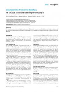
471 Fig. 1 Sonographic longitudinal section of the lateral neck demonstrating a well-demarcated mass (M) in the right carotid triangle. The mass is located anterior to the c o m m o n carotid artery (A) not infiltrating the wall of the vessel
Fig. 2 Coronal magnet resonance Tl-weighted spin-echo image (TR/TE 550 ms/25 ms) after contrast. A markedly enhancing, well-demarcated mass (arrow) is demonstrated within the parasellar region. The parasellar structures, internal carotid artery and cavernous sinus seem to be surrounded or infiltrated by the mass. The oculomotor nerve, the trochlear nerve and the abducens nerve cannot be depicted Fig.3 Histopathology, showing a uniform cellular infiltrate with densely packed lymphoblasts and some intermingled (arrow) phagocytosing reticulum cells ("starry sky cells")
cavernous sinus, oculomotor nerve, trochlear nerve and abducens nerve seemed to be infiltrated by the mass. After intravenous injection of gadolinium-DTPA as contrast agent, the mass showed a strong enhancement on T l - w e i g h t e d spin-echo sequences. The mass could be delineated against the hypophysis, but parasellar structures could not be demarcated. All other laboratory studies including blood count, chest X-ray, abdominal ultrasound and CT scans, were normal. One day later an incisional biopsy was carried out on the neck mass. Macroscopically, a tumor was found to be surrounded by a connective tissue capsule. Frozen section diagnosis was insufficient, but did show that the mass was not a reactive
lymph node. The final histologic diagnosis revealed a highly differentiated non-Hodgkins' lymphoma (Burkitt type, clg). The patient was then transferred to the Department of Hematology for further staging investigations and chemotherapeutic treatment. He has since received induction therapy with cyclophosphamide and prednisolone, a cycle with ifosfamide, vincristine, teniposide and cytoarabine intravenously, and methothrexate cytoarabine and dexomethasone intrathecally. A second chemotherapeutic cycle was given 4 weeks later, While tumor remission has been achieved to date, overall the prognosis remains poor.
472
Discussion A diagnosis of acute or chronic inflammatory lymphadenopathy had to be considered initially in our patient. According to his history of a brief fever, a viral genesis was not unlikely, although acute lymphadenopathy could be excluded by the duration of the mass for 2 months, the lack of pain and the absence of overt signs of inflammation in the neck. Chronic inflammatory lymphadenopathies frequently have a typical history, such as contact with animals when considering a diagnosis of brucellosis, toxoplasmosis, tularemia, cat scratch disease or actinomycosis, but the patient did not report any suitable history (see Table 1). Our patient's history of smoking and drinking was highly suggestive of a squamous cell carcinoma of the head and neck region. Lymph-node metastases to the neck usually derive from a neoplasm of the oral cavity, pharynx, larynx or skin. Although there was no clinical expression of a local carcinoma, final confirmation could only be gained by histological examination of a biopsy. With regard to extracranial lesions extending intracranially, primary malignant tumors of the sphenoid sinus or nasopharynx are the most likely group of neoplasms to explain our patient's clinical findings. The sphenoid sinus is most often implicated when a cranial neuropathy is present, particularly when involving the abducens nerve, although its actual incidence is less than 1% of malignant sinus tumors [14]. Granulomatous disease of the sphenoid (e.g., aspergillus granuloma) can present with surprisingly little systemic evidence of toxicity or abnormal laboratory findings. Cavernous sinus involvement with an associated lymphangitis has been reported to mimic central nervous system neoplasm or has led to the formation of a mycotic aneurysm with thrombosis, fistula formation or actual vessel rupture [2]. Among primary processes in the base of the skull, chondrosarcomas and various metastatic bone tumors need consideration. A case report of a sphenoid chondrosarcoma involved occlusion of the carotid arteries [8]. The clinical presentation with headache, visual and/or oculomotor symptoms or facial pain is typical but not specific. A tumor metastasis to either the sphenoid bone or cavernous sinus is rare, with only 26 cases of sphenoid metastases encountered in a recent 35-year review by Mickel and Zimmermann [10]. Typical symptoms in over
40% of cases include headaches, facial pain, visual abnormalities and especially oculomotor symptoms. Peripheral cranial third nerve palsy - as in our case can be distinguished as basilar, cavernous or orbital lesions. A complete palsy includes ptosis due to the weakness of the levator palpebrae, limitation in elevation or depression of the globe due to weakness of the superior rectus muscle and weakness of accommodation. In such cases abduction and the intorsion of the globe remain normal. Usually a peripheral third nerve palsy does not occur as an isolated lesion, because the 3rd, 4th and 6th cranial nerves run together in the lateral wall of the cavernous sinus. Possible etiologies include vascular causes due to diabetes, hypertension, or atherosclerosis, although other causes involve head trauma, aneurysm (mainly in women over the age of 50 years) and neoplasms, The most common tumors affecting the three cranial nerves are meningiomas [3], neurinomas [6], metastases, acoustic neurinomas, nasopharyngeal carcinomas, pontine/midbrain tumors, chordomas and pituitary tumor [1]. Intracranial vascular abnormalities include subclinical or partially thrombosed carotid aneurysms, hemangiomas and cavernous sinus thrombosis, but are rare. MRI loss of signal void will be noted in areas of thrombosis [15]. Additionally, sixth nerve symptoms are insidious and intermittent in the case of aneurysms. Inflammatory causes of ophthalmoplegia are mainly due to infections like otitis media with petrositis, basal meningitis, syphilis, and cavernous sinus thrombosis. Other causes include vasculitis, Tolosa Hunt syndrome, sarcoidosis and Guillain-Barr6 syndrome. Another cause is a carotid cavernous fistula, of which up to 25% are idiopathic. Frequently the first nerve to be affected is the abducens nerve, b e c a u s e it is the thinnest in diameter. Involvement of the nerves per se may indicate the site of a lesion (see Table 1). Non-Hodgkins' lymphoma is a well-known neoplastic disorder that occasionally presents in a cervical location as a primary site. Burkitt's lymphoma is a small, non-cleaved B-cell lymphoma, which is part of a continuum of B-cell lymphoproliferative disorders and was first described in 1958 as a mandibular tumor in African children [5]. Extranodal disease of the soft tissues and bones of the face occurs in less than 10% of patients. Common symptoms and signs are drenching night sweats, unexplained fever and weight loss > 10% and cytopenia, but a diagnosis may not be considered if the course of the disease is atypical. Our review of the literature revealed that there are very few single case reports of complicating ophthalmoplegias
Table 1 Differential diagnosis of ophthalmoplegia Syndrome
III palsy
IV palsy
VI palsy
V/1 palsy
Others
Cavernous sinus syndrome Syndrome of the orbital fissure Orbital apex syndrome Syndrome of the edge of the clivus
Yes Yes Yes Yes
Yes Sometimes Yes No
Yes Sometimes Yes No
Yes Sometimes Yes No
No
No I palsy (visual field) occasional mydriasis
473 in patients with n o n - H o d g k i n s ' l y m p h o m a [13]. M o s t o f the cases have b e e n r e p o r t e d in various retrospective reviews, with either a b r i e f or no detailed d e s c r i p t i o n of the intracranial lesion involved. R o s e b e r g et al. [12] f o u n d that only 1.3% o f cases s h o w e d any i n v o l v e m e n t o f the o c u l a r system, including orbital manifestation. In general, o p h t h a l m o p l e g i a creates an urgent n e e d to define the site o f a lesion, w h i c h can either be in muscle, nerve or central nucleus. R a d i o l o g i c a l i m a g i n g b y M R I p r o b a b l y gives the best r e s o l u t i o n o f the i n v o l v e d areas in order to define the e x a c t l o c a t i o n o f the lesion. H o w e v e r , in our case a b i o p s y p r o v e d to be the m o s t direct route to the diagnosis. C o n s i d e r i n g that m o s t therapeutic considerations are b a s e d on the grade o f malignancy, final confirm a t i o n o f disease requires h i s t o p a t h o l o g i c a l and i m m u n o logical e x a m i n a t i o n o f the b i o p s y specimens. The present consensus in m a n a g e m e n t o f n o n - H o d g k i n s ' l y m p h o m a (Burkitt t y p e cIg) is a g g r e s s i v e irradiation and c h e m o t h e r apy [11].
References 1.Ahmadi J, North CM, Segall HD, Fee CS, Weiss MH (1986) Cavernousous sinus invasion by piturity edemas. AJR Am J Roentgenol 146 : 257-262 2. Barat JL, Marchal JC, Paquis P, Angue J, Rache JL, Lepaire J (1990) Kyste 6pidermoide intracavemous extradural: ~ propos de deux observations. Neurochirurgic 36 : 242-245 3. Cioffi FA, Bemini FP, Punzo A, Natale M, Muras I (1987) Cavernous sinus meningeomas. Neurochirurgia (Stuttg) 30: 4047
4. Delpassand ES, Kirkpatrick JB (1988) Cavernous sinus syndrome as the presentation of malignant lymphoma: case report and review of the literature. Neurosurgery 23:501-504 5. Fukui K, Takeda S, Sadamoto K, et al (1985) A case of Burkitt's lymphoma with total ophthalmoplegia. Medline Abstract from No-Shinkei-Geka 13:1183-1189 6. Hansman ML, Haarer ED, Peyster RG (1986) Sixth nerve neurinoma in the cavernous sinus: CT features. J Comput Assist Tomogr 10:1030-1032 7.Inaki S, Okamura H, Chimameri Y (1988) Adult T-cell lymphoma. Riginating in the paranasal sinus. Arch Otolaryngol Head Neck Surg 114 : 1471-1473 8. Ishida M, Ashida K, Matsunga T, Wasaka K, Sakurai M (1986) Chondrosarcoma of the ethmoidal and sphenoidal sinuses: a case of chondrosarcoma arising from postparanasal sinuses. ORL J Otolaryngol Relat Spec 48 : 174-179 9. Lennert K, Mohri N, Stein H, Kaiserling E, M~ller-Hermelink HK (1978) Malignant lymphomas other than Hodgkins disease (Handbuch der speziellen pathologischen Anatomie und Histologie, vol 1, part 3B). Springer, Berlin Heidelberg New York, pp 3-15 10. Mickel RA, Zimmermann MC (1990) The sphenoid sinus - a site for metastasis. Otolaryngol Head Neck Surg 102:709-712 11. O'Reilly SE, Connors JM (1992) Non-Hodgkins' lymphoma. L Characterisation and treatment. BMJ 304:1682 12. Rosenberg SA, Diamond HD, Jaslowitz B, Caren LF (1961) Lymphosarcoma: a review of 1269 cases. Medicine 40 : 31 13. Russ SA, Yumen OH (1994) Burkitt's lymphoma presenting as ophthalmoplegia in an elderly male (letter). Am J Hematol 45 : 97-98 14. Sissen G, Snyderman NL, Becket S (1989) Cancer of the nasal cavity and paranasal sinuses. In: Suen JY, Myers EN (eds) Cancer of the head and neck, 2nd edn. Churchill Livingston, New York, pp 311-336 15. Weber AL, Dareis KR, Ojemann RG, Negri M (1982) X-ray study of the mouth; intracavemous internal carotid artery aneurysm. Ann Otol Rhinol Laryngol 91:543-545