JOURNAL OF OPTOELECTRONICS AND ADVANCED MATERIALS Vol. 8, No. 3, June 2006, p. 909 - 913
Optical properties of low-dimensional PbI2 particles embedded in polyacrylamide matrix N. PREDA*, L. MIHUT, I. BALTOG, T. VELULA, V. TEODORESCU National Institute R &D of Materials Physics, Lab. of Optics and Spectroscopy, P. O. Box MG-7, RO-77125, Bucharest, Romania
Small particles of PbI2 embedded in transparent polymer matrix have been studied by Raman scattering, UV-VIS absorption spectroscopy, low temperature photoluminescence (PL) and Scanning Electron Microscopy (SEM). The PbI2 crystallites were prepared by two different methods: i) by cooling to the room temperature a boiling saturated aqueous PbI2 solution containing polyacrylamide and ii) by chemical reaction of KI and Pb(NO3)2 in aqueous polyacrylamide solution. In both cases, beside the hexagonal platelets of PbI2 were identified rods whose luminescence and Raman signature is quite different than of PbI2 platelets. Regardless the rods nature, as distinct KPbI3 particles or lead iodide low-dimensional particles embedded in the polymer matrix, their photoluminescence is featured by an intense green emission (550 nm) appearing at low temperature at excitation wavelengths less then 350 nm. (Received March 15, 2006; accepted May 18, 2006) Keywords: Lead iodide crystallites, Polyacrylamide matrix, Raman scattering, Photoluminescence
1. Introduction Low-dimensional semiconducting particles embedded in polymer matrix form an interesting class of inorganic/organic hybrid materials for the fundamental physics as their applications in optical and optoelectronic devices [1-12]. Thermally stable, easy in processing and transparent to light, the polymers are an ideal host matrix for the inorganic particles. They can exert a direct influence on the semiconducting crystallite size, morphology and orientation [13]. Among the semiconductor compounds, a challenging material is lead iodide, that is a direct band gap semiconductor, having as structural unit a layer containing ions of lead, hexagonally packed, sandwiched between two layers of iodine atoms. The bulk PbI2 crystal is featured by a strong intralayer chemical bonding and a weak interlayer van der Waals interactions [14]. Low-dimensional lead iodide particles have been prepared in colloidal solutions [15-19] and embedded in transparent solid media (SiO2 [20-22], zeolite [23-25] and polymers [5,10,26-28]). Recently we have demonstrated by correlated studies of Scanning Electron Microscopy (SEM), Raman light scattering and absorption/emission spectroscopy that the reaction between Pb(NO3)2 and KI, carried out in different liquids media as water, methanol, ethanol and acetonitrile leads to hexagonal platelets of lead iodide and KPbI3 prismatic rods, of micro and nanometric size [29]. We have put in evidence that changing both, the liquid used as host medium and the stoichiometric ratio of compounds implied in the chemical reaction of preparation, the number of resulted platelets and rods is changed too. In this paper we show that the cooling down slowly a PbI2/polyacrylamide aqueous solution or the chemical synthesis of PbI2 from Pb(NO3)2 and KI carried out in acetonitrile, lead to the formation of the same type of particles, i.e hexagonal platelets and prismatic rods. The SEM pictures, as well as the Raman spectra are the most
convincing, they show a great resemblance with the particles formed by the reaction between Pb(NO3)2 and KI carried out in solvents as water, methanol, ethanol and acetonitrile, varying the stoichiometric ratio of the two implied compounds. The platelets are undoubtedly PbI2 crystalline particles. When the former preparation method was used the appearance of rods is amazing, all the more that no compound as KI or NaI were involved. In this case, the appearance of rod-like particles is the result of the intercalation during the crystallization process of PbI2 by polyacrylamide that leads to rhombohedral crystaline structures. As common feature of rods like particles resulted both from the reaction between Pb(NO3)2 and KI and the intercalation of PbI2 by polyacrylamide is a strong luminescence emission band at 2.23 eV (cca. 550 nm) that appears when the excitation is done at 334 nm. These data, compared with those concerning the intercalation of PbI2 by NH3 and alkyl amines [30] provide important evidence that polytypic phase transition from 1T to 3T on intercalation PbI2 ends in rhombohedral crystalline structures that display Raman and photoluminescence (PL) spectra quite similar with those measured on the rod-like particles, presumed as KPbI3 micro-crystals, resulted from the chemical synthesis of PbI2 from Pb(NO3)2 in acetonitrile. 2. Experimental The low-dimensional PbI2 particles were prepared by procedures described in [5, 10, 15] with some modifications. A method consists in the slow cooling to the room temperature of a boiling saturated aqueous PbI2 solution in which was added prior to the cooling 0.01 % aqueous polyacrylamide. Another method is based on the chemical reaction between KI and Pb(NO3)2 in a 0.01 % aqueous polyacrylamide solution. Into 100 cm3 of polymer solution, under vigorously ultrasonic homogenizing, was added 5 cm3 of 0.01 M aqueous solution of lead nitrate and then 5 cm3 of 0.05 M aqueous solution of potassium
910
N. Preda, L. Mihut, I. Baltog, T. Velula, V. Teodorescu
iodide. In both synthesis methods, the sudden appearance of the yellow colour is the first evidence of the formation of PbI2 crystallites. All materials used were of analytical grade quality. Considering the aqueous solution of PbI2/polyacrylamide it is worth notice that by crystallization were obtained the same type of platelets and rods using as starting PbI2 either bought powder of analytical grade quality or powder prepared grinding a crystalline ingot of PbI2 growth from the melt that was previously purified by fusion zone. The shape of the particles were determined by scanning electron microscopy (SEM, JEOL 200 CX). The Raman spectra were recorded under 676.4 nm excitation light at room temperature in backscattering geometry with a Jobin Yvon T64000 spectrophotometer equipped with a microprobe allowing the laser spot to be focused on the sample within a micrometer scale. This facility has permitted an individual Raman inspection of particles of different size and shape. A Perkin Elmer Lambda 2S spectrophotometer was used to measure the absorption spectra in UV-VIS at the room temperature. The luminescence spectra at liquid nitrogen temperature (LNT) were recorded in reflection at righta
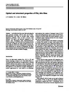
angle geometry under continuous excitation using a Coherent Innova 90 argon ion laser or a Hg lamp as the excitation light sources. The emission spectrometer was a SPEX double monochromator, equipped with a cooled EMI photomultiplier and a photon counting system. 3. Results and discussion Fig. 1a and Fig. 1b reveals the SEM images of the particles obtained slowly cooling to the room temperature of a hot saturated aqueous PbI2 solution in the absence and in the presence of the polymer, respectively. The lowdimensional PbI2 particles were prepared by procedures described in [5,10,15] with some modifications. The Fig. 1b is the thought provoking. It shows clearly that by the crystallisation of PbI2 in the presence of polyacrylamide are formed many rods. It is interesting notice that hexagonal platelets of PbI2 and rod-like particles reported into previous paper, were produced by the chemical reaction between lead nitrate and potassium iodide carried out in different liquid as water, methanol, ethanol and acetonitrile [29].
c
1µ
b
d
1µ
Fig. 1. SEM pictures of the particles produced by cooling to the room temperature of a hot saturated aqueous PbI2 solution in the absence (a) or in the presence of the polyacrylamide (b) and by chemical reaction between Pb(NO3)2 and KI carried out in water (c) and ethanol (d).
For comparison in Fig. 1c and Fig. 1d are presented the SEM pictures of such particles synthesised in water and ethanol, respectively. Only platelets or only rods were observed, when the reaction taken place in water or acetonitrile, respectively and the rods were attributed to the KPbI3, as auxiliary resulting compound of the reaction between Pb(NO3)2 and KI [29]. In the present case, when a saturated solution of PbI2 with polyacrylamide added was cooled down slowly, the appearance of rods as result of crystallization process is surprising because a compound
like KPbI3, cannot be invoked. Further, the insets from the Fig. 1b and Fig. 1d show another significant detail that the rods formed in the two cases have the same geometrical form. It has to mention that the same type of particles, i.e. platelets and rods, were observed as ending products of the reaction between lead nitrate and potassium iodide lead carried out in aqueous water polyacrylamide solution. In the previous work [29], we have demonstrated that the hexagonal platelets and the rods are featured by quite
911
Optical properties of low-dimensional PbI2 particles embedded in polyacrylamide matrix
different Raman spectra. The former show the well-known Raman spectrum of lead iodide, which is identical with that recorded on a crystalline sample cleaved from a melt grown crystal. The latter display a Raman spectrum strongly dependent on the rod orientation against the direction of incident radiation, incident polarization, observed polarization and direction of observation. Due to the great similarity of the Raman spectra of rods like particles and CsPbCl3 in the Pmma orthorhombic structure [29] we presumed that the rods could be KPbI3 crystallites that belong to the same crystalline system. a
Raman intensity (arb.units)
0
50
100
150
300
400
50
100
150
300
400
50
100
150
300
400
b
0
c
0
presumption is true, then the Raman spectra should differ because the Raman bands are associated to vibrations involving different atomic and molecular entities. In this scenario, considering the (PbI3)- ion as basic entity in the formation of rod like crystalline structure is hard to imagine that the bounding of these ions with different entities as K+ ions or polyacrylamide molecules is done without important changes in the phonon spectrum. Another explanation appeals to the intercalation of lead iodide with different atomic or molecular species [31]. Reasoning in this sense, the coincidence of the Raman spectra of rods like particles obtained by the two different synthesis methods as well as SEM micrographs of these particles conclude that by the intercalation of lead iodide are formed micro-crystals of the same geometrical form. As it is well known, in the PbI2 crystals the lowest conduction band is composed mainly of 6p lead atomic orbitals. This band is extremely flat [32,33] so that it will not be affected by the c-axis expansion after intercalation. The top of valence band of PbI2 crystal normally contains an admixture of Pb2+ s and p and I- p states [34]. For this layered material, by the intercalation of different atomic or molecular entities between the iodine atomic planes, one expects that the main change in the band structure relate the perturbed iodine orbitals. The modifications appear in the valence band where the contribution of the I- p sates increase as result of the compression produced by the foreign atoms, ions or molecules intercalated between the iodine atomic planes. The expansion in the direction of c - axis transforms the crystalline structure of PbI2 from a hexagonally packed one, with weak inter-layers van der Waals forces, in an orthorhombic structure that is featured by a quite different Raman spectrum. Such a result was already reported for the PbI2 intercalated with ammonia, methylamine, ethylamine and butylamine [30]. Besides, in [30] was signalled that the Raman spectra of the particles resulted from the reaction between Pb(NO3)2 and KI in acetonitrile are very similar to that of ammoniaintercalated PbI2. This similarity is shown also in Fig. 2, which presents the Raman spectra of the PbI2 intercalated with polyacrylamide and presumed KPbI3 rods like particles.
-1
Wavenumber (cm )
In this work we report another surprising result: the rods formed by the cooling down slowly the PbI2/polyacrylamide aqueous solution display the same Raman spectra as the presumed KPbI3 rods [29]. Such a result, naturally arises the question if these rods produced by different routes, by the reaction between Pb(NO3)2 and KI in a non-aqueous solvent (methanol, ethanol or acetonitrile that is the most convenient) or by the crystallization of PbI2 in an aqueous polyacrylamide solution, belong really to various materials or represent different products in the same morphic state. If the former
c
0.10
2.45
Absorbance
0.15
Fig. 2. Raman spectra at 676.4 nm excitation wavelengths of PbI2 platelets (a), rods like particles resulted from the reaction between Pb(NO3)2 and KI carried out in acetonitrile (b) and rods produced by the recrystalization of PbI2 crystaline powder in aqueous polyacrylamide solution (c).
d
0.05 b a
0.00 3.0
2.8
2.6
2.4
2.2
2.0
1.8
Energy (eV)
Fig. 3. The optical absorption spectra of polyacrylamide (a), particles obtained by cooling procedure in the absence (b) or in the presence of polymer (c) and crystallites synthesized by chemical reaction in aqueous polymer solution (d).
912
N. Preda, L. Mihut, I. Baltog, T. Velula, V. Teodorescu
2.48
a2
2.00
a1
2,2
2,0
1,8
1,6 2,8
2,6
2,4
b1
2,2
2,0
1,8
1,6
b2
2.49
2.23
2,4
2.00
2,6
2.47
PL intensity (arb. units)
2.23
typical for the PbI2 layer, meanwhile the enhanced absorption observed in the UV-blue region of the spectrum is the evidence of the iodine ions contribution. It is revealing that in earlier papers devoted to the synthesis of small particles of layered semiconductors of PbI2, HgI2 or BiI3, a band at 3.5 eV, attributed to I3- ions was systematically reported [15-19, 29]. Assuming that by the intercalation of PbI2 lattice either Pb2+ or I- ions became absorbent in the UV-VIS range, one expects also a different luminescence response. Indeed, the Fig. 4 shows that the rods like particles produced by the two routes, by the reaction between Pb(NO3)2 and KI in acetonitrile and by the crystallization of PbI2 in an aqueous polyacrylamide solution show the same luminescence spectra and the same dependence on the excitation wavelength. The dependence on the excitation wavelength implies the excitation selectively of the electronic transitions in Pb2+ and I- ions that is achieved at 457.9 nm and 334 nm respectively. In the former case one observes all details of the PL spectrum PbI2 crystal sample submitted to band-to-band excitation.
The absorption spectra, shown in Fig. 3, supply also the presumption that two types of particles, PbI2 platelet and rod, are formed in the presence of polyacrylamide. The former, as for the same type of particles resulted from the chemical reaction between Pb(NO3)2 and KI carried out in water, is noticed by an increased step in the absorbance (curve c) taking placed about 2.45 eV that marks the optical absorption edge while the latter is featured by an monotonous increasing absorbance, in the near UV-blue region, that reveals absorption due to the perturbed iodine ions by the inter-layer intercalation of foreign species. One result of foreign atoms and molecules intercalation between the iodine atomic planes consist in the change of the admixture of Pb2+ s and p and I- p states that characterises the top of valence band of PbI2 crystal [34]. By the compression on the iodine atomic planes, the electronic states of Pb2+ and I- ions disconnect so that they become identifiable separately into absorption spectrum. In this frame, on the Fig. 3, curve c, the maximum at about 2.45 eV marks the optical absorption due to the Pb2+ ions,
2,6
2,4
2,2
2,0
1,8
1,6 2,8
2,6
2,4
2,2
2,0
1,8
1,6
Energy (eV)
Fig. 4. LNT photoluminescence of the low-dimensional particles obtained by the reaction between Pb(NO3)2 and KI carried out in acetonitrile (a) and by cooling to the room temperature of a hot saturated aqueous PbI2 solution in the presence of the polyacrylamide (b), at two excitation wavelengths: 457.9 nm (a1, b1) and 334 nm (a2, b2).
The Fig. 4a1 and Fig. 4b1 present the PL spectra, at 457.9 nm excitation light. Regardless the origin of the rods, it contains an intense emission band, with the maximum around of 2.5 eV, which marks the typical excitonic emission. As a mater of fact this maximum is associated to the transition 3P1 → 1S0 of the Pb2+. Another broad band peaking about 2.0 eV, labeled G band that is linked to the surface defects is also contains in the spectrum [29]. Either decreasing the dimensions of PbI2 platelets or increasing the density of surface defects enhances the G band.
Under the excitation light of 334 nm, in the PL spectrum of rods like particles (Fig. 4a2 and Fig. 4b2), the excitonic emission is not more observed, regardless the preparation method used. In the emission spectrum appears only a wide band with the maximum at about 2.23 eV. The absence of excitonic emission means primarily that the excitation is not more suitable to form the cationic excitons, i.e. the resonant excitation of Pb2+ whose mixed s and p states there are in the top of valence band. The wide band at 2.23 eV originates in another excited species, linked to I- ions, which due to the intercalation its p
Optical properties of low-dimensional PbI2 particles embedded in polyacrylamide matrix
electronic states form another discrete level in the upper side of the valence band. These ions, once excited, return in the ground state giving rise to the PL band at 2.23 eV. The effect of partition of the electronic states of Pb2+ and Iions exacerbates at low temperature that explains why this band is observed predominantly at liquid nitrogen temperature. It has to mention that the PL spectra of the particles obtained by the reaction between lead nitrate and potassium iodide lead carried out in aqueous water polyacrylamide solution present the same features, depending on the excitation wavelengths. 4. Conclusions We have studied by Raman scattering, UV-VIS absorption spectroscopy, low temperature photoluminescence and Scanning Electron Microscopy PbI2 low-dimensional particles embedded in polyacrylamide matrix. Whichever was the synthesis procedure, cooling to the room temperature a boiling saturated aqueous PbI2 solution containing polymer or the chemical reaction of KI and Pb(NO3)2 in acetonitrile, in both cases were identified platelets of PbI2 and rods like particles. The luminescence and Raman signature of the rods is quite different for the one of the PbI2 platelets. As distinct feature of the rods like particles, regardless the method used for their synthesis must be mentioned the same geometrical form, a very similar Raman spectrum and specific PL emission band centered about 2.23 eV. The emission is observed predominantly at liquid nitrogen temperature when the excitation is done at higher energies (cca. 340 nm) than that required perform the excitation band-to-band (cca. 2.5 eV) in the PbI2 platelets. Based on these data the rod-like particles are considered PbI2 intercalated with different foreign molecules. The different optical properties of the rod-like particles are explained by an effect of partition of the electronic states of Pb2+ and I- ions that are situated in the top of valence band, that results from the intercalation of PbI2 lattice. References [1] M. Antonietti, C. Goltner, Angew. Chem., Int. Ed. Engl. 36, 910 (1997). [2] U. Woggon, S. V. Bogdanov, O. Wind, K. H. Schlaad, H. Pier, C. Klingshirn, P. Chatziagorastou, H. P. Fritz, Phys. Rev. B 48, 11979 (1993). [3] M. V. Artemyev, S. V. Gaponenko, I. N. Germanenko, A. M. Kapitonov, Chem. Phys. Lett. 243, 450 (1995). [4] J. Qi, Y. Masumoto, Solid State Commun. 99, 467 (1996). [5] M. V. Artemyev, Yu. P. Rakovich, G. P. Yablonski, J. Cryst. Growth 171, 447 (1997). [6] E. Lifshitz, M. Sirota, H. Porteanu, J. Cryst. Growth 196, 126 (1999). [7] Y. Zhou, S. Yu, C. Wang, X. Li, Y. Zhu, Z. Chen, Chem. Commun. 1229 (1999).
913
[8] J. Zhan, X. Yang, D. Wang, S. Li, Y. Xie, Y. Xia, Y. Qian, Adv. Mater. 12, 1348 (2000). [9] S. Wang, S. Yang, C. Yang, Z. Li, J. Wang, W. Ge, J. Phys. Chem. B 104, 11853 (2000). [10] A. I. Savchuk, V. I. Fediv, Ye. O. Kandyba, T. A. Savchuk, I. D. Stolyarchuk, P. I. Nikitin, Mat. Sci. Eng. C 19, 59 (2002). [11] H. Du, Q. Xu, W. S. Chin, L. Huang, W. Ji, Chem. Mater. 14, 4473 (2002). [12] M. Tamborra, M. Striccoli, R. Comparelli, M. L. Curri, A. Petrella, A. Agostiano, Nanotechnology 15, S240 (2004). [13] J. Lin, E. Cates, P. A. Bianconi, J. Am. Chem. Soc. 116, 4738 (1994). [14] M. R. Tubbs, Phys. Status Solidi B 49, 11 (1972). [15] C. J. Sandroff, D. M. Hwang, W. M. Chung, Phys. Rev. B 33, 5953 (1986) [16] C. J. Sandroff, S. P. Kelty, D. M. Hwang, J. Chem. Phys. 85, 5337 (1986) [17] O. I. Micic, L. Zongguan, G. Mills, J. C. Sullivan, D. Meisel, J. Chem. Phys. 91, 6221 (1987). [18] Y. Wang, N. Herron, J. Chem. Phys. 91, 5005 (1987). [19] R. Mu, Y. S. Tung, A. Ueda, D. O. Henderson, J. Phys. Chem. 100, 19927 (1996). [20] E. Lifshitz, M. Yassen, L. Bykov, I. Dag, R. Chaim, J. Phys. Chem. 98, 1459 (1994). [21] E. Lifshitz, M. Yassen, L. Bykov, I. Dag, J. Lumin. 70, 421 (1996). [22] E. Lifshitz, L. Bykov, M. Yassen, Z. Chen-Esterlit, Chem. Phys. Lett. 273, 381 (1997). [23] Z. K. Tang, Y. Nozue, T. Goto, J. Phys. Soc. Jpn. 61, 2943 (1992). [24] Z. K. Tang, Y. Nozue, T. Goto, Mat. Sci. Eng. B 35, 410 (1995). [25] N. Togashi, Y. Sakamoto, T. Ohsuna, O. Terasaki, Mat. Sci. Eng. A 312, 267 (2001). [26] M. V. Artemyev, G. P. Yablonski, Yu. P. Rakovich, Acta Physica Polonica A 87, 523 (1995). [27] S. Saito, T. Goto, Phys. Rev. B 52, 5929 (1995). [28] T. Goto, S. Saito, J. Lumin. 70, 435 (1996). [29] M. Baibarac, N. Preda, L. Mihut, I. Baltog, S. Lefrant, J. Y. Mevellec, J. Phys. Condens. Matter 16, 2345 (2004). [30] R. F. Warren, W. Y. Liang, J. Phys. Condens. Matter 5, 6407 (1993). [31] V. Mehrotra, S. Lombardo, M. O. Thompson, E. P. Giannelis, Phys. Rev. B 44, 5786 (1991). [32] I. C. Schluter, M. Schluter, Phys. Rev. B 9, 1652 (1974). [33] J. Robertson, Solid State Commun. 26, 791 (1978). [34] R. Ahuja, H. Arwin, A. Ferreira da Silva, C. Persson, J. M. Osorio-Guillen, J. Souza de Almeida, C. Moyses Araujo, E. Veje, N. Veissid, C. Y. An, I. Pepe, B. Johansson, J. Appl. Phys. 92, 7219 (2002).
_______________________ * Corresponding author:
[email protected]