December 15, 2005 / Vol. 30, No. 24 / OPTICS LETTERS
3353
Optically sectioned fluorescence lifetime imaging using a Nipkow disk microscope and a tunable ultrafast continuum excitation source D. M. Grant, D. S. Elson, D. Schimpf, C. Dunsby, J. Requejo-Isidro, E. Auksorius, I. Munro, M. A. A. Neil, and P. M. W. French Physics Department, Imperial College London, Prince Consort Road, London SW7 2BW, UK
E. Nye and G. Stamp Experimental Pathology Laboratory, London Research Institute, Lincoln’s Inn Fields Laboratories, 44 Lincoln’s Inn Fields, London WC2A 3PX and Department of Investigative Science, Imperial College London, London SW7 2AZ, UK
P. Courtney Perkin Elmer Inc., Chalfont Road, Seer Green HP9 2FX, UK Received May 25, 2005; revised manuscript received August 30, 2005; accepted September 6, 2005 We demonstrate an optically sectioned fluorescence lifetime imaging microscope with a wide-field detector, using a convenient, continuously tunable 共435– 1150 nm兲 ultrafast source for fluorescence imaging applications that is derived from a visible supercontinuum generated in a microstructured fiber. © 2005 Optical Society of America OCIS codes: 180.2520, 180.6900, 300.6500.
Fluorescence lifetime imaging (FLIM) can contrast different molecular species and image variations in the local fluorophore environment. It may be used with fluorescent labels or with endogenous fluorophores to provide label-free molecular contrast. We are working to develop high-speed optically sectioned FLIM microscopes for applications including live-cell imaging and high-throughput screening. Optically sectioned fluorescence imaging is usually achieved by using laser scanning confocal microscopy (LSCM) or multiphoton microscopy, and FLIM is readily implemented by using, e.g., time-correlated single-photon counting electronics (TCSPC, e.g., Ref. 1). Unfortunately, although the theoretical minimum acquisition time for a FLIM image of an arbitrarily bright sample is only 0.1 s for an error less than 10%,2 the serial pixel acquisition and the low excitation power imposed by TCSPC can increase the FLIM acquisition times to several minutes for many biological samples. This acquisition time can be significantly further increased when multiphoton excitation is used, for which excitation cross sections are lower and nonlinear photodamage limits the maximum excitation power. One strategy to increase the FLIM speed is parallel pixel acquisition by using wide-field FLIM detection, preferably by using single-photon excitation for maximum efficiency. Such an approach results in a lower instantaneous photon flux per pixel, thereby reducing photobleaching and photodamage compared with confocal microscopy.3 Wide-field real-time (up to a 29 Hz frame rate) FLIM has been demonstrated in conventional microscope, endoscope, and plate reader configurations,4,5 and frame rates of up to 100 Hz from a single-shot FLIM system have been applied to image calcium signals in cells.6 To date the only single-photon excited, optically sectioned wide-field 0146-9592/05/243353-3/$15.00
FLIM system has been demonstrated with structured illumination.7 Unfortunately this technique, which requires computation to extract a sectioned image from a set of acquired images8 that often have a large unwanted background of out-of-focus light, exhibits a reduced signal-to-noise ratio compared with direct imaging, which leads to increased errors when fitting fluorescence lifetimes. It is also highly sensitive to motion artifacts. An alternative method for realizing optical sectioning with wide-field detection is the tandem scanning Nipkow disk microscope, whereby multiple spots are simultaneously scanned across the sample and confocally imaged onto an array of pinholes before a widefield detector.9 This approaches LSCM sectioning, although the out-of-focus light discrimination is not quite as effective. Like LSCM, this approach requires a high-brightness (spatially coherent) excitation source, which for FLIM is required to be appropriately modulated or pulsed. In this paper we report the application of a new tunable excitation source10 based on continuum generation to Nipkow disk tandem scanning microscopy. This permits us to also demonstrate, for the first time to our knowledge, an optically sectioned single-photon-excited tandem scanning microscope with wide-field time-domain FLIM detection. Previously the lack of suitable tunable or pulsed visible excitation lasers has been a significant drawback of both conventional confocal and tandem scanning fluorescence microscopes. We note that there are also tandem scanning multiphoton microscopes in which an array of ultrashort pulse infrared laser beams is focused onto a sample and rapidly scanned to give a multiphoton excited fluorescence image that may be recorded by using a wide-field detector11 and that such a multifocus, multiphoton microscope approach has been demon© 2005 Optical Society of America
3354
OPTICS LETTERS / Vol. 30, No. 24 / December 15, 2005
strated for wide-field FLIM.12 This approach benefits from reduced out-of-focus photobleaching and photodamage, compared with one photon excitation, but these issues are worse in the focal plane, and the excitation efficiency is lower. Figure 1 is a schematic of our inverted Nipkow disk microscope, which is implemented on an Olympus IX71 frame. The excitation source is coupled into a single-mode fiber that is connected to the fiber connection port of the Nipkow disk unit (Yokogawa Electrical Corporation, CSU10), which employs an array of 20,000 pinholes that are scanned across the sample. Inside the unit, the incident light is expanded to overfill the area of a lenslet array that focuses this light through the array of pinholes, producing multiple point sources that are imaged onto the sample. Both lenslet and pinhole arrays are on disks that spin at 30 rotations/ s, and 12 complete images are swept out per rotation, providing wide-field images at up to 360 Hz. The resulting fluorescence is imaged back through the pinhole array and directed, via a dichroic beam splitter located between the lenslet and the pinhole disks, to the wide-field detector— either a CCD camera for intensity imaging or a gated optical intensifier (GOI) for FLIM. Using a 100 nm thick fluorescent polymer film test sample, the optical sectioning strength of this microscope was determined to be 2.5 and 0.9 m for Olympus ⫻40 0.75 NA UPlanFl dry and Olympus ⫻100 1.3 NA UPlanFl oil immersion objective lenses, respectively. These values are close to the theoretical resolution for confocal microscopy, although there was a slight pedestal 共⬃10% 兲 caused by cross talk of out-of-focus light between the pinhole channels. Various excitation sources were used with this microscope, including a continuous wave (cw) argon-ion laser at 488 nm, a frequency-doubled Ti:sapphire laser (100 fs, 800 nm pulses at an 80 MHz repetition rate), a mercury lamp and a tunable continuum source (TCS) based on a microstructured fiber. Full details of our TCS, which is represented schematically in Fig. 1, can be found in Ref. 10, and only a brief outline is given here. A femtosecond Ti:sapphire
Fig. 1. (Color online) Sectioning microscope setup, showing continuum generation optics, Nipkow disk unit, excitation path, and one fluorescence path for simplicity (BD, beam dump; FR, Faraday rotator; PCF, photonic crystalline fiber). Inset, typical unfiltered output spectrum.
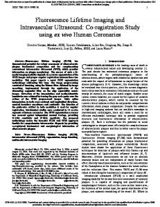
Fig. 2. (a) Conventional and (b) sectioned images of a pollen grain.
laser output 共800 mW, 860 nm兲 was directed through an isolator and focused by a 4.5 mm focal length aspheric lens into the 75 cm length of photonic crystalline fiber (PCF, Crystal Fiber AS, NL-740-2.0). The spatially coherent continuum output from the fiber (average power 220 mW, pulse width ⬍20 ps) was collimated, and the visible component of a typical spectrum is inset in Fig. 1 (note that the response of the Ocean Optics spectrometer—Model HR2000 CGUV-NIR—used is reduced at longer wavelengths, and the continuum extends into the infrared). A bandpass filter was used to remove the large infrared component of the beam before launching into the singlemode delivery fiber to the Nipkow disk microscope. The excitation wavelength could be widely tuned depending on the particular fluorophore of interest, but was in practice limited by the dichroic and filters mounted inside the Nipkow disk unit. The CSU10 is typically equipped with multibandpass dichroics that have three passbands at 488, 568, and 647 nm, matched to the lines of argon- and krypton-ion lasers. It was therefore convenient to use these wavelengths from the TCS, although the multiband dichroic was far from optimal, since the width of each passband for the fluorescence was only 40 nm, resulting in less than 50 W at the sample for an applied average power of 2 – 3 mW. The multibandpass dichroic could be replaced with a single-cutoff dichroic mirror. Figure 2 shows a comparison between unsectioned wide-field intensity and Nipkow-sectioned images of a stained pollen grain excited with the TCS in the spectral range 550– 570 nm and detected over the range 590– 650 nm. These images were recorded with a ⫻100 microscope objective and illustrate the superior image quality and out-of-focus light rejection achieved by using the Nipkow disk microscope. The acquisition time for these images was approximately 1 s, although this could be reduced to hundreds of milliseconds when the higher power levels available from the argon-ion laser or the frequencydoubled Ti:sapphire laser were used. We note that filtered supercontinua generated in microstructured fibers have previously been applied to fluorescence intensity imaging10,13 and FLIM10 in standard confocal scanning microscopes and that the TCS provides opportunities for simultaneous excitation of multiple fluorophores and rapid spectral tuning, including under electronic control, for in situ measurements of the excitation spectra of fluorophores, e.g., in cells.10 The importance of optical sectioning is emphasized when performing FLIM, for which out-of-focus light not only degrades the spatial resolution but can also
December 15, 2005 / Vol. 30, No. 24 / OPTICS LETTERS
degrade the lifetime contrast.7 The advantage of acquiring sectioned FLIM images is apparent in Fig. 3, for which the FLIM images were acquired by using a time-gated FLIM system based on a GOI (Model HRI from Kentech Instruments Ltd.), which is described in Refs. 4 and 5. Figures 3(a) and 3(c) show conventional unsectioned FLIM images, and 3(b) and 3(d) show their Nipkow-sectioned counterparts. Sectioning has increased the spatial information and enhanced the fluorescence lifetime contrast between the different pollen grains. These FLIM images were obtained in 4 s (acquiring eight time-gated images with a 1 ns gate width and a 500 ms integration time), with excitation and emission spectral passbands of 430– 470 nm and ⬎490 nm, respectively). No significant photobleaching was observed. Figure 4 shows an example of this system applied to biological tissue. The FLIM images are of a Tromastained mouse embryo section acquired with [Fig. 4(a)] ⫻10 and [Fig. 4(b)] ⫻40 magnification objectives. The acquisition time was 30 s (using eight time gates of 1 ns width) with excitation and emission spectral passbands of 550–570 and 590– 650 nm, respectively. The short-lifetime objects are unstained red blood cells in the aorta. The oval objects lower in the frames are vertebral bodies. In conclusion, we have demonstrated the use of a TCS applied to tandem scanning optically sectioned FLIM using a Nipkow disk microscope. The sectioned fluorescence intensity and lifetime images display improved image quality and contrast compared with their conventional (nonsectioned) counterparts. We believe that this approach has the potential to acquire optically sectioned FLIM images at higher rates than scanning confocal microscopy, and the TSC addresses one of the key disadvantages of the Nipkow disk microscope, namely, the limited availability of spatially coherent and ultrafast light sources in the visible spectrum. This becomes particularly promising as TCS technology is further developed, exploiting low-cost high-average-power fiber lasers14 to provide higher average powers and therefore faster imaging rates.
Fig. 3. Comparison of (a), (c) conventional and (b), (d) Nipkow-sectioned FLIM images of pollen grains at two image planes separated by 10 m
3355
Fig. 4. FLIM images of Troma-stained mouse embryo section with a (a) ⫻10 and (b) ⫻40 microscope objective
Funding for this research is gratefully acknowledged from the UK Department of Trade and Industry (DTI), the European Community (Framework VI Integrated Project ‘Integrated technologies to in vivo molecular imaging’ contract number LSHG-CT-2003503259), the Higher Education Funding Council for England [JIF (V) Award], the UK Engineering and Physical Sciences Research Council (EPSRC), GlaxoSmithKline Research and Development Ltd., and the Wellcome Trust. D. M. Grant acknowledges an EPSRC studentship. D. Schimpf acknowledges the Friedrich Ebert Foundation. D. S. Elson’s email address is
[email protected]. References 1. W. Becker, A. Bergmann, M. A. Hink, K. Konig, K. Benndorf, and C. Biskup, Microsc. Res. Tech. 63, 58 (2004). 2. M. Kollner and J. Wolfrum, Chem. Phys. Lett. 200, 199 (1992). 3. E. Wang, C. M. Babbey, and K. W. Dunn, J. Microsc. 218, 148 (2005). 4. J. Requejo-Isidro, J. McGinty, I. Munro, D. S. Elson, N. Galletly, M. J. Lever, M. A. A. Neil, G. W. H. Stamp, P. M. W. French, P. A. Kellet, J. D. Hares, and A. K. L. Dymoke-Bradshaw, Opt. Lett. 29, 2249 (2004). 5. D. S. Elson, I. Munro, J. Requejo-Isidro, J. McGinty, C. Dunsby, N. Galletly, G. W. Stamp, M. A. A. Neil, M. J. Lever, P. A. Kellett, A. Dymoke-Bradshaw, J. Hares, and P. M. W. French, New J. Phys. 6, 180 (2004). 6. A. V. Agronskaia, L. Tertoolen, and H. C. Gerritsen, J. Phys. D 36, 1655 (2003). 7. J. Siegel, D. S. Elson, S. E. D. Webb, D. ParsonsKaravassilis, S. Leveque-Fort, M. J. Cole, M. J. Lever, P. M. W. French, M. A. A. Neil, R. Juskaitis, L. O. Sucharov, and T. Wilson, Opt. Lett. 26, 1338 (2001). 8. M. A. A. Neil, R. Juškaitis, and T. Wilson, Opt. Lett. 22, 1905 (1997). 9. M. Petran, M. Hadravsky, M. D. Egger, and R. Galambos, J. Opt. Soc. Am. 58, 661 (1968). 10. C. Dunsby, P. M. P. Lanigan, J. McGinty, D. S. Elson, J. Requejo-Isidro, I. Munro, N. Galletly, F. McCann, B. Treanor, B. Onfelt, D. M. Davis, M. A. A. Neil, and P. M. W. French, J. Phys. D 37, 3296–3303 (2004). 11. J. Bewersdorf, R. Pick, and S. W. Hell, Opt. Lett. 23, 655 (1998). 12. M. Straub and S. W. Hell, Appl. Phys. Lett. 73, 1769 (1998). 13. G. McConnell, Opt. Express 12, 2844 (2004). 14. D. Grant, E. Auksorius, D. N. Schimpf, P. M. P. Lanigan, P. A. A. De Beule, J. McGinty, D. S. Elson, C. Dunsby, J. Requejo-Isidro, I. Munro, N. Galletly, G. W. H. Stamp, M. A. A. Neil, P. M. W. French, and P. Courtney, presented at Focus on Microscopy, Jena, Germany, March 20–23, 2005.