Dec 2, 2011 - cinnarizine by a factorial design. J Microencapsul. 2007;24:253â62. 10. Prajapati ST, Patel LD, Patel DM. Gastric floating matrix tablets:.
AAPS PharmSciTech, Vol. 13, No. 1, March 2012 ( # 2011) DOI: 10.1208/s12249-011-9730-y
Research Article Optimization Studies on Gastroretentive Floating System Using Response Surface Methodology Rajendra Awasthi,1 Giriraj T. Kulkarni,1,4 Vivek K. Pawar,2 and Garima Garg3
Received 4 April 2011; accepted 17 November 2011; published online 2 December 2011 Abstract. The aim of the present investigation was to develop and optimize gastroretentive floating system of amoxicillin for the efficient treatment of peptic ulcer induced by Helicobacter pylori infection. Floating microballoons were developed using central composite design (CCD), and optimization was done by employing response surface methodology. The selected independent variables were cellulose acetate phthalate, drug–Eudragit S100 ratio, and the ratio of dichloromethane/ethanol/isopropyl alcohol. The selected dependent variables were yield, mean particle size, buoyancy, encapsulation efficiency, and drug release within 8 h. A quadratic polynomial model was generated which had linear, interaction, and quadratic terms to predict and evaluate the independent variables with respect to the dependent variables. Results showed that selected independent variables significantly affect the yield (30.53–82.71%), particle size (31.62–47.03 μm), buoyancy (42.68–95.75%), encapsulation efficiency (56.96–93.13%), and cumulative drug release from the microballoons (34.01–74.65%). The interaction and quadratic terms were also found to affect the process variables. An excellent agreement was found between the actual value and predicted value. In conclusion, it can be said that CCD is a valuable second-degree design to develop and optimize GFS of amoxicillin which in turn provides a basis to localize the drug release in the gastric region for effective treatment of H. pylori-mediated infection. KEY WORDS: amoxicillin; central composite design; floating microballoons; gastroretentive floating system; H. pylori; response surface methodology.
body. To fulfill all these objectives, an optimized gastroretentive floating system (GFS) should possess physical properties like being smaller in size, high buoyancy, and encapsulation efficacy along with the controlled release of drug in the gastric environment. Gastroretentive systems produce a lasting local action on the gastroduodenal wall (4,5). Floating systems do not bind with mucosal surfaces in the body and reduce the safety problems associated with mucoadhesive systems (6). Multiparticulate systems avoid the “all or none” gastric emptying process of the single unit system (7,8). Keeping in view all these factors, amoxicillin-loaded GFS was designed and optimized to increase the residence time of the drug in the stomach without contact with the mucosa. Response surface methodology (RSM) has applications in the particular situations where several input variables potentially influence some performance measure or quality characteristic of the process. Thus, performance measure or quality characteristic is called the response. Under RSM, different designs such as factorial design, central composite design (CCD), Box–Behnken design, and simplex lattice design are used for optimization (9,10). In the present study, CCD was selected for the development of GFS. The selected independent variables in CCD were cellulose acetate phthalate (CAP), Eudragit S100, and dichloromethane/ ethanol/isopropyl alcohol ratio, and the dependent variables were yield, mean particle size, buoyancy, encapsulation efficiency, and cumulative drug release within 8 h. Levels of independent variables were decided according to preliminary trials done.
INTRODUCTION Helicobacter pylori infection is one of the major causes of peptic ulcer and gastric adenocarcinoma worldwide. More than 50% of the population is recognized to have H. pylori infection (1). Even though currently available antibiotics have a significant effect against H. pylori, its complete eradication has remained a challenge. This is mainly due to drawbacks of available conventional dosage forms (2,3). H. pylori lives deep in the gastric mucosal region, where the conventional dosage forms are unable to produce a localized release of antibiotic. Hence, a minimum inhibitory concentration of antibiotic does not get maintained in the gastric mucosa which ultimately leads to failure of therapy. To overcome this, there is a need to develop drug delivery systems with ability to release the drug specifically in the stomach. An optimum gastroretentive floating system can be defined as a system which is retained in the stomach for sufficient time against all the physiological barriers and finally metabolized in the 1
Laureate Institute of Pharmacy, Kathog 177 101 Teh, Dehra( Dist Kangra, Himachal Pradesh, India. 2 Kusum Heathcare, 2E/22, Jhandewalan Ext, 110055, New Delhi( India. 3 Department of Pharmaceutical Technology, Meerut Institute of Engineering and Technology, NH-58 Bypass, Baghpat Crossing, Meerut 250 005, Uttar Pradesh, India. 4 To whom correspondence should be addressed. (e-mail: gtkulkarni@ gmail.com)
85
1530-9932/12/0100-085/0 # 2011 American Association of Pharmaceutical Scientists
Awasthi et al.
86 MATERIALS AND METHODS Materials Amoxicillin was a generous gift from Win-Medicare (Meerut, India). Eudragit S100 was purchased from Evonik Degussa (Mumbai, India). CAP and polyvinyl alcohol were purchased from Central Drug House (New Delhi, India). All the other reagents and solvents used were of analytical grade. CCD The CCD is comprised of a central core of two level factorial designs (2n), one central point, and 2n axial or outer points. Three factors with five levels were used to explore the complete design. The experimental design is given in Table I. Mathematical Model A nonlinear quadratic polynomial model was generated for precise evaluation of the effects of independent variables on dependent variables using Design Expert v7.1.5 software (Stat-Ease, Inc., Minneapolis, Minnesota). Since the microballoons were formed at only a specific level of CAP, Eudragit S 100 and ethanol, polynomial models, were generated for the dependent variables by considering only the level of CAP (X1), Eudragit S 100 (X2), and ethanol in the solvent system (X3), as independent variables. Yi ¼ b0 þ b1 X1 þ b2 X2 þ b 3 X3 þ b4 X1 X2 þ b5 X1 X3 þ b 6 X2 X3 þ b 7 X1 2 þ b 8 X2 2 þ b 9 X3 2
ð1Þ
where Yi is the level of response variable; βi is the regression coefficient; X1, X2, and X3 stand for the main effect; X1X2, X2X3, and X1X3 are the interaction terms of the main effects; and X12, X22, and X32 are quadratic terms of the independent variables that are used to simulate the curvature in the designed space.
Preparation of the Floating Microballoons Floating microballoons were prepared using modified emulsion-solvent diffusion method (11–13). Accurately weighed quantity of drug, Eudragit S100, and CAP were dissolved in a solvent mixture containing dichloromethane, ethanol, and isopropyl alcohol. This mixture was introduced into the 100 ml aqueous solution of 0.5% w/v polyvinyl alcohol. The resultant solution was stirred at 300 rpm using a precision digital stirrer (LT400A, Yamato, Japan) for 1 h, and the temperature was maintained at 40°C. Due to fast evaporation of ethanol and isopropyl alcohol, a polymeric shell was developed enclosing dichloromethane. After 1 h, the formed microballoons were filtered and dried at 40°C to form inner gaseous phase by slow evaporation of enclosed dichloromethane.
Characterization of Developed Floating Microballoons Prepared floating microballoons were evaluated for the selected dependant variables such as percentage yield, average particle size, encapsulation efficiency, buoyancy, and in vitro drug release. The surface of microballoons was characterized by scanning electron microscopy, and physical state of the entrapped drug was determined by X-ray diffraction study.
Table I. CCD with the Levels of Independent Variables
Formulation code AMX1 AMX2 AMX3 AMX4 AMX5 AMX6 AMX7 AMX8 AMX9 AMX10 AMX11 AMX12 AMX13 AMX14 AMX15 Levels for independent variables Codes for different levels of independent variables −1 +1 0 −a +a CAP cellulose acetate phthalate
Concentration of CAP −1 +1 −1 +1 −1 +1 −1 +1 −a +a 0 0 0 0 0 CAP (g) 0.6 0.8 0.7 0.53 0.87
Drug/Eudragit S100
Dichloromethane/ethanol/ isopropyl alcohol
−1 −1 +1 +1 −1 −1 +1 +1 0 0 −a +a 0 0 0
−1 −1 −1 −1 +1 +1 +1 +1 0 0 0 0 −a +a 0
Drug/Eudragit S100 (g)
Dichloromethane/ethanol/ isopropyl alcohol (ml) 6:5:2 8:7:2 7:6:2 5:4:2 9:8:2
1:4 1:6 1:5 1:3 1:7
Optimization Studies on Gastroretentive Floating System
87
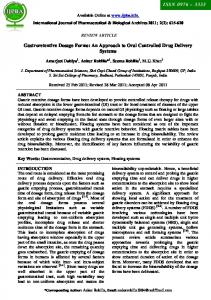
Fig. 1. Scanning electron micrographs of floating microballoons showing general appearance (a) and (b), surface morphology (c), and internal hollow structure (d). Scales are given on individual micrograph
X-ray diffraction analysis of pure amoxicillin and the optimized formulation was done by X-ray powder diffractometer (PW 3040/ 60 Xpert PRO, Panlytical, Netherlands). The X-ray diffraction patterns were recorded using Cu Kα radiations (λ=1.5405980Ả), a current of 30 ma, and a voltage of 40 kv. The samples were analyzed over 10–40 2θ range with a scan step size of 0.02 and 0.50 s per step.
samples for SEM were prepared by adhering the microballoons on a double adhesive tape stuck to an aluminum stub. The stubs were then coated with silver under an argon atmosphere using a high-vacuum evaporator (Polaron SEM coating system). The internal cavity of the microballoons was examined by cutting into two sections diametrically with a sharp surgical steel blade. The coated sample was then randomly scanned, and photomicrographs were taken with a scanning electron microscope (EVO-50, ZEISS; UK).
Scanning Electron Microscopy
Encapsulation Efficiency
The external and internal morphology of the microballoons was studied by scanning electron microscopy (SEM). The
Accurately weighed (10 mg) microballoons were crushed and dispersed into 25 ml phosphate buffer (pH 7.4) for
X-ray Diffraction Studies
Fig. 2. X-ray diffraction patterns of pure amoxicillin (A) and drug-loaded floating microballoons (B)
Awasthi et al.
88 Table II. Results of In Vitro Characterization of Microballoons Formulation code
Shape
Mean particle size (μm)
Yield (%)
Buoyancy (%)
Encapsulation efficiency (%)
AMX1 AMX2a AMX3a AMX4a AMX5 AMX6 AMX7 AMX8 AMX9a AMX10 AMX11 AMX12 AMX13 AMX14 AMX15
Spherical – – – Spherical Spherical Spherical Spherical – Spherical Spherical Spherical Spherical Spherical Spherical
31.62 – – – 38.11 32.04 37.74 39.83 – 33.57 35.90 39.69 47.03 36.10 38.90
77.63 – – – 30.53 40.36 82.71 79.74 – 72.71 79.27 80.54 72.69 79.22 80.25
91.28 – – – 86.75 72.85 84.52 77.94 – 52.63 43.10 62.82 42.68 59.05 95.75
93.02±3.74 – – – 56.96±7.45 65.79±0.79 92.14±10.4 92.22±5.26 – 73.50±4.99 87.32±5.28 83.19±13.05 57.43±4.56 91.47±7.69 93.13±10.02
a
Microballoons did not form due to polymer precipitation
determination of encapsulation efficiency. The prepared mixture was shaken for 24 h. After 24 h, the solution was filtered, and the filtrate was analyzed for the drug content by a UV spectrophotometer at 227 nm after suitable dilution (14). The percentage encapsulation was calculated as follows:
where Wf and Ws are the weights of floating and settled microballoons, respectively.
Encapsulation efficiencyð%Þ ¼ ½Da =Dt � � 100
The drug release rate from different formulations (AMX1– AMX15) was determined using USP type II apparatus (TDT08L, Electrolab, Mumbai, India). Dissolution medium (SGF, pH 1.2, 500 ml) containing 0.02% Tween 20 filled in the dissolution vessel, and the temperature was maintained at 37± 0.5°C. Microballoons equivalent to 50 mg of amoxicillin were placed in the dissolution vessel, and the paddle was rotated at 50 rpm. Aliquots were withdrawn every 15 min in the first hour and then every hour till the 4th hour followed by the 6th and 8th hours. Samples were then analyzed by a UV spectrophotometer at 228 nm. The study was conducted in triplicate.
where, Da is the actual amount of drug present in the prepared microballoons, and Dt is the theoretical amount of drug added in the preparation of microballoons. Buoyancy Study Fifty milligrams of prepared microballoons was placed in 100 ml simulated gastric fluid (SGF, pH 1.2) containing 0.02% Tween 20. The mixture was stirred at 100 rpm on a magnetic stirrer. After 8 h, the supernatant SGF was filtered through a microporous filter paper (0.2 μm) to separate floating microballoons. The settled microballoons were collected separately. Both floating and settled microballoons were dried at 40°C. The fractions of microballoons were weighed, and the buoyancy was determined by the following formula (15): Percentage buoyancy ¼ ½Wf =Wf þ Ws � � 100
In Vitro Drug Release Study
Statistical Treatment of the In Vitro Release Data The drug release data were statistically analyzed by two-way analysis of variance (ANOVA). The p value of