Research Paper
Research Paper
Human Vaccines & Immunotherapeutics 9:10, 2253–2262; October 2013; © 2013 Landes Bioscience
Optimized and enhanced DNA plasmid vector based in vivo construction of a neutralizing anti-HIV-1 envelope glycoprotein Fab Kar Muthumani1*, Seleeke Flingai1, Megan Wise1, Colleen Tingey1, Kenneth E Ugen2,3, and David B Weiner1 Department of Pathology and Laboratory Medicine; University of Pennsylvania School of Medicine; Philadelphia, PA USA; 2Department of Molecular Medicine; University of South Florida Morsani College of Medicine; Tampa, FL USA; 3Center for Molecular Delivery; University of South Florida; Tampa, FL USA
1
Keywords: DNA vaccine, VRC01 mAb, HIV-1 neutralization, monoclonal antibodies, electroporation
Monoclonal antibody preparations have demonstrated considerable clinical utility in the treatment of specific malignancies, as well as inflammatory and infectious diseases. Antibodies are conventionally delivered by passive administration, typically requiring costly large-scale laboratory development and production. Additional limitations include the necessity for repeat administrations, and the length of in vivo potency. Therefore, the development of methods to generate therapeutic antibodies and antibody like molecules in vivo, distinct from an active antigen-based immunization strategy, would have considerable clinical utility. In fact, adeno-associated viral (AAV) vector mediated delivery of immunoglobulin genes with subsequent generation of functional antibodies has recently been developed. As well, anon-viral vector mediated nucleic acid based delivery technology could permit the generation of therapeutic/ prophylactic antibodies in vivo, obviating potential safety issues associated with viral vector based gene delivery. This delivery strategy has limitations as well, mainly due to very low in vivo production and expression of protein from the delivered gene. In the study reported here we have constructed an “enhanced and optimized” DNA plasmid technology to generate immunoglobulin heavy and light chains (i.e., Fab fragments) from an established neutralizing anti-HIV envelope glycoprotein monoclonal antibody (VRC01). This “enhanced” DNA (E-DNA) plasmid technology includes codon/ RNA optimization, leader sequence utilization, as well as targeted potentiation of delivery and expression of the Fab immunoglobulin genes through use of “adaptive” in vivo electroporation. The results demonstrate that delivery by this method of a single administration of the optimized Fab expressing constructs resulted in generation of Fab molecules in mouse sera possessing high antigen specific binding and HIV neutralization activity for at least 7 d after injection, against diverse HIV isolates. Importantly, this delivery strategy resulted in a rapid increase (i.e., in as little as 48 h) in Fab levels when compared with protein-based immunization. The active generation of functional Fab molecules in vivo has important conceptual and practical advantages over conventional ex vivo generation, purification and passive delivery of biologically active antibodies. Further study of this technique for the rapid generation and delivery of immunoglobulin and immunoglobulin like molecules is highly relevant and timely.
Introduction Targeted monoclonal antibodies (mAbs) represent one of the most important therapeutic advances of the past 25 y. This type of immune based therapy is now used routinely against a host of autoimmune diseases, cancers, as well as infectious diseases.1-4 For malignancies, many of the immunoglobulin (Ig) based therapies currently used are in combination with targeted cytotoxic chemotherapy regimens. This combination approach has significantly improved overall survival of patients.5,6 Multiple mAb preparations have been licensed for use against specific cancers, including Rituxan (Rituximab), a chimeric mAb targeting CD20 for the treatment of Non-Hodgkins lymphoma7,8 and Ipilimumab (Yervoy),9 a human mAb that blocks CTLA-4 and which has been used for the treatment of melanoma and other
malignancies.10-15 Additionally, Bevacizumab (Avastin) is another prominent humanized mAb that targets VEGF and tumor neovascularization and has been used for the treatment of colorectal cancer.16 Perhaps the most high profile mAb for treatment of a malignancy is Trastuzumab (Herceptin), a humanized preparation targeting Her2/neu, and which has been demonstrated to have considerable efficacy against breast cancer in a subset of patients.16 Furthermore, a host of mAbs are in use for the treatment of autoimmune and specific blood disorders.17 In addition to cancer treatments, passive transfer of polyclonal Igs mediate protective efficacy against a number of infectious diseases including diphtheria, hepatitis A and B, rabies, tetanus, chicken-pox and respiratory syncytial virus (RSV).3,18-21 In fact, several polyclonal Ig preparations provide temporary protection against specific infectious agents in individuals traveling to
*Correspondence to: Kar Muthumani; Email:
[email protected] Submitted: 08/08/13; Revised: 09/06/13; Accepted: 09/15/13 http://dx.doi.org/10.4161/hv.26498 www.landesbioscience.com Human Vaccines & Immunotherapeutics 2253
disease endemic areas, in circumstances when there is insufficient time for protective Igs to be generated through active vaccination. Furthermore, in children with immune deficiency, Palivizumab (Synagis), a mAb which targets RSV infection, has been demonstrated to clinically protect against RSV.17 The clinical impact of mAb therapy is impressive. However, issues remain that limit the use of this therapeutic approach. Some of these include the high cost of production of these complex biologics that can limit their use in the broader population, particularly in the developing world, where they could have a great impact. Furthermore, the typical requirement for repeat administrations of the mAbs to attain and maintain efficacy can be an impediment in terms of logistics and patient compliance. Additionally, the long-term stability of these antibody formulations is frequently short and less than optimal. Along these lines, a few recent studies have suggested that gene therapy vectors could be utilized for in vivo Ig delivery and production. Seminal studies performed in the HIV model by the Johnson laboratory22 initially established that the adeno-associated viral vector (AAV) is a virally based vector system that can deliver antibodies in animal models. Novel recent studies have extended this work.23-25 These viral vector based studies have advanced the field greatly and suggest, in at least animal models, that in vivo mAb production could be long lived through the use of this delivery method. In this regard, we reasoned that a nucleic acid DNA approach might have some additional important prophylactic and therapeutic contributions in certain therapeutic situations. As non-viral vector associated DNA delivery is independent of prior serological Ig status, it may be useful in specific situations where pre-existing serology against a viral vector might be a disadvantage in attaining product efficacy. DNA expression, elicited by this delivery strategy, is not permanent which implies that it may have logistical and safety advantages in situations where only temporary immunity is important and desired. This DNA delivery technology is also highly cost effective since the treatment (i.e., expression plasmid) is produced for human delivery using a bacterial platform that typically has significantly lower production costs. However, major technological hurdles have limited the DNA delivery platform as an in vivo gene delivery system. In the past, protein expression from DNA plasmid delivery suffered from low in vivo transfection efficiencies.26 In the past several years, there has been a great deal of work performed to improve the in vivo production of protein from the DNA delivery platform. Strategies to enhance the efficacy of E-DNA have included codon optimization for expression in human cells,27,28 RNA optimization to improve mRNA stability, as well as more efficient translation at the ribosomal level. Also, the addition of specific leader sequences to enhance translation efficiency and the creation of synthetic inserts further enhanced production in vivo.2932 Importantly, our work, among others, has further improved in vivo delivery by utilizing enhanced adaptive in vivo electroporation (EP) delivery protocols and formulations.33,34 Our group has reported a dramatic improvement in in vivo expression of such EP delivered designer plasmids, resulting in the ability to drive immune responses in humans to a level equivalent to that of viral vector delivery.35-37 Using these advancements in the DNA
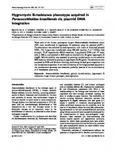
plasmid delivery platform and the in vivo murine system, we report on the generation of a synthetic Fab, and the evaluation of the potential biological activity of this preparation. The mAb selected for this proof-of-concept study was the anti-HIV broadly neutralizing human Ig designated VRC01.38,39 Specifically, this mAb binds to the CD4 binding site of the envelope (Env) glycoprotein gp120 and has been demonstrated to neutralize 90% of all HIV isolates against which it has been tested.40,41 However, current vaccine design has not been able to develop an immunogen that is able to routinely elicit such an HIV-1 neutralizing Ig. Therefore, strategies to generate, by active vaccination, neutralizing human mAbs against this or other epitopes are highly desirable. Likewise, alternative methods other than active vaccination to generate expression of biologically active Igs are important.39 Following analysis of the published nucleotide sequence for this mAb, we generated synthetic genes encoding the light and heavy chains for the Fab fragment of the full length VRC01 and cloned the final sequence into a human CMV driven promoter (cytomegalovirus) expression system. Expression of the plasmid (i.e., generation of the Fab) was then measured in vitro. Subsequently, we demonstrated significant expression of the specific Fab fragments (in as little as 2 d) in vivo in the sera of mice that were administered the Fab-encoding construct (designated pHIV-1 Env Fab). Specifically the mice were administered a single dose the pHIV-1 Env Fab and subsequently attained sera Fab concentrations of 1–3 μg/mL for at least 10 d, with associated HIV neutralizing activity against 4 viruses from different clades manifesting differences in neutralization sensitivities. In sum, the enhanced DNA strategy was able to express levels of biologically active Fabs of potential importance for an extended period of time in a small animal model. Results Generation of anti-HIV-1 Env-Fab expressing constructs. The cDNAs for both the VH and VL-Ig chains coding sequences for the anti- HIV-1 Env broadly neutralizing human mAb VRC01 were obtained from the VRC (Vaccine Research Center, NIH) through the NIH AIDS Research and Reference Reagent Program and subsequently cloned into a pVax1 vector. Several modifications, as indicated in the Materials and Methods section, were incorporated into the expression vectors in order to maximize and optimize the production of biologically active Ig molecules. Specifically, these modifications included codon and RNA optimization and stabilization, enhanced leader sequence utilization, increased plasmid production and facilitated in vivo plasmid delivery through EP. The constructs generated were placed under the control of an immediate early promoter from CMV, which is important for proper and efficient expression in mammalian cells and tissues. The schematic maps of the construct used in this study are indicated in Figure 1A and B. Ig production by transfected cells. To evaluate the expression of pHIV-1Env-Fab, the constructs were transfected into 293T cells. An ELISA immunoassay, using a consensus HIV-1 clade B gp120 protein as the binding antigen, confirmed the presence of the anti-HIV-1 Env-Fab in the supernatant from the transfected
2254 Human Vaccines & Immunotherapeutics
Volume 9 Issue 10
293 T cells as early as 24 h post transfection (Fig. 1C). High OD450nm values (i.e., ranging from approximately 0.5 to 0.8) were detected in cell extracts from 24 to 72 h post transfection and subsequently reached a peak and plateau at 48 h. These results confirmed the specificity of the anti-HIV-1 Env Fab for the HIV Env glycoprotein. Statistical analysis of the data presented in Figure 1C indicates that OD450nm values for sera from pHIV-1 Env-Fab injected mice are significantly elevated (P < 0.05, student t test) compared with pVax1 control from the 22 through 72 h time points measurements. In vivo characterization of HIV-1 Env Fab. To demonstrate in vivo Fab production from the DNA plasmids, mice were administered the pHIV-1 Env Fab by the intramuscular route followed by enhanced delivery through EP. A single injection of the DNA plasmids was performed and sera was collected at 12 h as well as at days 1, 2, 3, 4, 7, and 10 following administration. Sera (at a dilution of 1:100 dilution) were then subsequently evaluated for Ig/Fab levels by ELISA analysis, as indicated in Figure 2A. Data in this figure are Figure 1. Generation and confirmation of expression of pHIV-1Env-Fab. (A and B) Circular plasmid presented from individual mice in both maps of pHIV-1 Env Fab heavy(H) and light (L) chain expressing constructs. Constructs were designed the pVax1 and HIV-1 Env-Fab groups using VRC01 heavy (H) and light (L) variable chain Ig genes. Several modifications were included when constructing the plasmids in order to increase the level of expression, as shown in the Figure and as OD450nm, which is proportional to described in the Materials and Methods and Results section. The Fab VL and VH fragment genes, as the level of Ig/Fab. These data demonshown, were cloned separately between the BamH1 and Xho1 restriction sites of the pVax1 vector. (C) strate that the relative levels of Fab after In vitro expression of pHIV-1 Env Fab. The graph indicates the temporal kinetics of expression of the a single administration of pHIV-1EnvpHIV-1 Env Fab after transfection of 293T cells. The values indicated, indicative of expression, are mean Fab become detectable on day 1 and the OD450nm ± SD values of triplicate wells. As a control, 293T cells were also transfected with the pVax1 backbone. A statement on statistical analysis is included in the Results section. subsequently increases over time. For comparative purposes, a single administration/immunization of rgp120, as Additional analyses were performed to ensure the quality as described in the Materials and Methods was made into Balb/C mice with subsequent sera collection and analysis (at 1:100 sera well as quantity of the recombinant Fab produced by the DNA dilution) over time by ELISA in order to determine the extent delivery technology. Specifically, immunoblot analysis was perand longevity of specific anti-gp120 antibody levels. Figure 2B formed using electrophoresed and blotted HIV-1 rgp120 prosummarizes these results. In this protein delivery study, antigen tein and probed with sera from pHIV-1Env-Fab mice 48 h post specific Ig levels over background were only detectable 10 d after administration (Fig. 2C). The blot indicated a band appropriate immunization. This is in contrast to the Fab levels elicited by for the molecular weight of gp120 as well as the ability of gp120, pHIV-1 Env Fab administration (Fig. 2A) where OD450nm specific Fab to bind to the protein. Likewise, human Fab quantivalues attained at least 0.1 OD450 nm units by day 1 post tation, by ELISA, was performed and presented as a function of administration and plateaued at day 10 at levels between 0.28 time (i.e., days) after plasmid administration (Fig. 2D). In sum and 0.35 OD450 nm units. Therefore, the delivery of pHIV-1 then, these results indicate that the levels of Fab generated peaked Env Fab resulted in a more rapid generation of specific Fab than at 2–3 µg/ml. These findings demonstrated the correct polypepconventional protein immunization. This finding underscores tide assembly of the VH and VL chains of the generated VRC01 the potential clinical utility of this novel DNA plasmid delivery based Fab, as well as the ability to recognize and bind specifically to the HIV-1 Env protein. method for generation of biologically active Ig.
www.landesbioscience.com Human Vaccines & Immunotherapeutics 2255
mice were significantly elevated (P < 0.05, student t test) compared to pVax1 injected mice, from the day 2 through 10 time point measurements. Binding of Fab/Igs to cells expressing different HIV-1 Env proteins: FACS based analysis. Sera from the mice administered pHIV-1Env-Fab were also used to test binding of the generated Fab to different HIV-1 Env proteins transiently expressed by 293 T cells. The native form of the VRC01-mAb was used as a positive control, to ensure proper expression and detection of the Env proteins on the surface of the cells. As indicated earlier, the “irrelevant/unrelated” Ig (Ig-E1M2) was used as a negative control. As demonstrated in Figure 3A and B, there was essentially only background staining by different Igs/Fabs to pVax1 (i.e., lacking the Env insert) transfected cells. However, for both the purified VRC01 mAb and sera from pHIV-1Env-Fab administered mice there was significant positive staining of transfected cells expressing the consensus clade A Env plasmid (pConEnv-A) as well as an optimized clade C plasmid (pOpt-Env-A) expressing and Env from the primary HIV-1 isolate pQ23Env17. Moreover, sera from pIgFigure 2. Measurement of temporal generation of anti HIV-1 Env specific Fab following administration of pHIV-1 Env Fab. (A) Time course of generation of anti-HIV1 Env Fab. After administration E1M2 administered mice failed to demof pHIV-1 Env Fab, production of the specific Fab was measured in the sera over 10 d ßat a final onstrate staining of any of the HIV1 Env dilution of 1:100 by ELISA and presented as OD450nm. Sera from pVax1 administered mice were transfected cells above background levels. used as a negative control. (B). Comparative measurement of anti-gp120 antibody responses after FACS analysis indicating these results immunization with recombinant gp120 (rgp120). As described in Materials and Methods mice were are provided in Figure 3A. A representaimmunized with a single injection of rgp120 followed by measurement of production of anti-gp120 antibodies up to 10 d and presented as OD450nm values. PBS was used as a negative control injective graph demonstrating the data from tion for this study. (C) Confirmation of HIV1Env-Fab binding by immunoblot analysis. As indicated the FACS analysis (i.e., Fig. 3A) for this in Materials and Methods, either 5 or 10 µg of gp120 were subjected to SDS-PAGE and nitrocelexperiment is provided in Figure 3B. lulose blotting followed by incubation of the blots with sera from pHIV-1 Env Fab administered Statistical analyses of data presented in mice. The immunoblot indicates that the experimental sera recognized bound rgp120, confirming Figure 3B indicated no significant differthe specificity of the generated Fab. (D) Temporal quantitation of human Fab, measured as IgG1 in mouse sera following pHIV-1Env-Fab administration. IgG1 was measured by a standard ELISA kit, at ence (P < 0.05, student t test) in specific the time points indicated, and expressed as Fab (µg/mL) ± SD. Sera from pVax1-administered mice binding between native VRC01 antibody were used as a negative control. Sera samples were analyzed at the time points indicated on the xand sera from pHIV-1 Env-Fab injected axis. The arrow shown in the graphs displayed in (A), (B) and (D) indicate the point of DNA plasmid mice to the envelope glycoprotein generadministration. Statements on statistical analysis are included in the Results section. ated by pCon-Env-A. However, binding of VRC01 antibody to the envelope glyStatistical analyses of the data presented in Figure 2 are as fol- coprotein generated by pOpt-Env-A was significantly higher (P < lows. For data summarized in Figure 2A, OD450nm values for 0.05, student t test) than binding by sera from pHIV-1 Env-Fab the sera from the pHIV-1 Env-Fab injected mice were statistically injected mice. Statistical analyses of data presented in Figure 3B elevated (P < 0.05, student t test) compared with the sera from indicated no significant difference (P < 0.05, student t test) in pVax1 injected mice from the days 1 through 10 measurement specific binding between native VRC01 antibody and sera from time points. For data summarized in Figure 2B, OD450nm pHIV-1 Env-Fab injected mice to the envelope glycoprotein genvalues from the rpg120 group were significantly elevated (P < erated by pCon-Env-A. However, binding of VRC01 antibody to 0.05, student t test) compared with PBS control from the day the envelope glycoprotein generated by pOpt-Env-A was signifi10 through 14 time point measurements. For data summarized cantly higher (P < 0.05, student t test) than binding by sera from in Figure 2D, OD450nm values from pHIV-1 Env-Fab injected pHIV-1 Env-Fab injected mice.
2256 Human Vaccines & Immunotherapeutics
Volume 9 Issue 10
Figure 3. FACS binding analysis of anti- HIV-1 Env Fab to clade A HIV Env glycoprotein. (A) FACS scans indicating binding of anti-HIV1Env-Fab to HIV-1 clade A Env glycoprotein. DNA expressing either a consensus (pCon-Env-A) or “optimized” (pOpt-Env-A) HIV-1 clade A envelope was transfected into 293T cells. Two days post transfection, cells were stained with either purified native VRC01 Ig, sera generated from pHIV-1 Env Fab (collected 48 h after a single plasmid administration) or control Ig generated from pIgG-E1M2 administration. Sera and VRC01 antibody were diluted 1:4 or 1:100, respectively in 50 µl of PBS and incubated at room temperature for 30 min. Cells were then stained with the appropriate secondary phycoerythrin (PE) conjugated Igs and subsequently gated for FACS analysis as singlet and live cells. The percent binding of positive cells is indicated in each of the scans. (B) Graphical representation of the FACS binding data. The number of stained cells (i.e., indicative of expression levels) in each of the Ig/sera tested groups was divided by the background staining values and presented as percent of specific binding on the y-axis, as a function of the different HIV clade A Env preparations tested. A statement on statistical analysis is included in the Results section.
HIV neutralizing activity of Ig produced by pHIV-1 Env Fab. In order to assess the potential HIV-1 neutralizing activity of the HIV-Env Fab produced in this study, a luminescence based neutralization assay using TZM-Bl target cells was performed. The TZM-Bl target cells were infected with the 4 different pseudotyped HIV viral isolates in the absence or presence of the experimental and control sera, as described in the Materials and Methods section. Figure 4 depicts the neutralization curves for sera from pHIV-1 Env Fab injected mice against the HIV pseudotyped viruses. Specifically tested were the HIV-1 tier 1 viruses Bal26 and SF162S (both clade B), as well as Q23Env (clade A). In addition, the sera were also tested against the HIV-1 clade C tier 2 virus ZM53M. The data are presented as percent neutralization/inhibition of HIV infection. The hatched horizontal lines in the graphs indicate the 50% neutralization/inhibition level in the assay. A positive neutralization control mAb (data not shown) was utilized in this study to confirm the utility and validity of this assay method. Briefly, the positive control neutralizing mAb was able to inhibit infection of the all four of the viral pseudotypes by at least 50%.
Sera from the pHIV-1 Env Fab administered mice demonstrated an increase in HIV neutralizing activity over time following plasmid administration, with percent neutralization reaching 50% by day 2 for Bal25, Q23Env17 and SF162S. As well, plateau percent neutralization values for these 3 viruses were approximately 62, 60 and 70% respectively. For the ZM53M the 50% neutralization threshold was not reached until 3 d and plateau neutralization did not exceed 50%. This less robust neutralization profile, compared with the other 3 tested, is likely reflective of it being a less neutralizable Tier 2 virus. In sum, the Fab generated in this study was able to effectively neutralize a range of HIV-1 isolates. Statistical analyses of data presented in Figure 4 are as follows. Based on Kruskal-Wallis non-parametric analysis, only HIV neutralization levels for the ZM53M Clade C virus (Fig. 4D), induced by sera from pHIV-1 Env-Fab injected mice, was significantly different from the other viruses tested (Fig. 4A–C). This difference was demonstrated in the time (days) required to achieve 50% neutralization, as well as in the maximally attained level of neutralization.
www.landesbioscience.com Human Vaccines & Immunotherapeutics 2257
employed a number of strategies to enhance Fab expression that had not previously been applied to antibody-encoding plasmids, such as codon optimization and improved EP conditions that increased in vivo expression many fold over that attained in prior studies. Notably, the sera concentration of VRC01 Fab in pHIV-1 Env Fab administered mice peaked at 2–3 μg/mL at day 12 post-injection. This range is comparable to a number of mAbs currently licensed by the FDA, suggesting that our antibody approach can produce significant and biologically relevant levels of antibodies in this small animal model. In particular, Ustekinumab (trade name: Stelara) and Golimumab (Simponi), two antibodies indicated for use against autoimmune diseases such as plaque psoriasis and arthritis, attain mean ± SD sera concentrations of 0.31 ± 0.33 μg/mL and 1.8 ± 1. 1 μg/mL, respectively, after passive administration. Furthermore, the very successful TNF inhibitor Adalimumab (Humira) attains, after administration, a mean serum concentration of around 6 μg/mL In this regard, our results, which represent an improvement over prior work, are important and further optimization of such antibody vectors likely can lead to enhanced in vivo results. Figure 4. Time course of neutralization of HIV-1 isolates by sera from pHIV-1Env-Fab adOur findings also illustrate the ability to ministered mice. Sera used for analysis of neutralization activity sera were collected at the more rapidly produce Fabs in vivo, after a time points indicated in the graphs. The neutralization analysis was conducted in TZM-BL single EP enhanced administration of pHIVcells using a panel of HIV-1 pseudotyped viruses: Bal26 (A; clade B, Tier 1), Q23Env17 (B; 1Env Fab, compared with Igs produced by clade A, Tier 1), SF162S (C; clade B, Tier 1), and ZM53M (D; clade C, Tier 2). Cells were infected at an MOI of 0.01 as delineated in the Materials and Methods section and incubated in the conventional protein (i.e. vaccine) administrapresence of sera (final dilution of 1:50) containing Fab generated from pHIV-1 Env Fab adtion (Fig. 2A and B). In addition, the ability to ministration. Percent neutralization values are shown, the calculation of which is described generate functional protective Ig-like molecules in Materials and Methods. As well, horizontal lines are provided in each of the graphs, against difficult vaccine targets is addressed by indicating the approximate time points at which the experimental sera mediated 50% viral this strategy. To date, inducing HIV-1 neutralneutralization. A statement on statistical analysis is included in the Results section. izing antibodies following active vaccination has been incredibly difficult, and during primary Discussion infection, neutralizing antibodies do not often develop until years after transmission.47-50 With this DNA plasmid approach, Monoclonal antibody therapy has had considerable success in we observed neutralization titers within 1–2 d post delivery with ameliorating a number of human malignancies and other ill- peak neutralizing Fab sera concentrations (3.31 ± 0.13 μg/mL) nesses.42-45 It is important to consider strategies to make this occurring one-week post-administration (Fig. 2D). This level of technology more economically attractive and to increase the in Ig is relatively similar to the 8.3 μg/mL concentration that has vivo half-life of the mAbs, coupled with improvements in deliv- been demonstrated to provide complete protection from infecery effectiveness.43,44,46 In this regard new technologies for mAb tion in a recent study.23 Our proof-of-concept study highlights or Ig like molecule delivery are important. In this proof-of-con- the rapid induction of biologically active Ig fragments and procept study, we used a DNA plasmid-based strategy to generate, vides clear avenues for improving levels of production in future in mice, Fab fragments from the broadly neutralizing HIV-1 investigations. human mAb VRC01 as a model system to investigate imporThe generation of balanced neutralization has been a diffitant strategies for Ig delivery and generation of in vivo biological cult challenge for the development of an effective viral vaccine activity. Our investigation expands significantly upon an earlier formulation. Accordingly, we characterized the neutralizing antistudy published by Tjelle et al. in which several reporter mAbs body titer and the responses against HIV-1 primary isolates that were generated in vivo, at low levels, following EP-mediated were elicited by HIV-1Env-Fab DNA administration. Sera were delivery of DNA plasmids encoding Ig genes.26 In our study, we tested against a panel of different viral tier 1, and 2 isolates that
2258 Human Vaccines & Immunotherapeutics
Volume 9 Issue 10
represent examples from clades A, B, and C. The results indicate generation of potent neutralizing activity against these viruses (Fig. 4). An important conclusion of this study is that this DNA plasmid-based method described warrants additional investigation on the generation of specific and biologically active Fab or Ig molecules in vivo. This approach bypasses the need to use conventional antigen-based vaccination for Ig generation. In this regard, further engineering to include Fc receptor fragments would allow for potentially important and efficacious Fc-mediated biological effects such as antibody dependent cell cytotoxicity (ADCC), complement dependent cytotoxicity (CDC), and FcRn receptormediated half-life enhancement to be investigated. In principle, this EP-based delivery of Ig genes can be used for the production of any established mAb (i.e., antibody for which the DNA sequence can be obtained), and for the prophylaxis and/or treatment of any number of diseases that would benefit from the biologic activity of particular antibodies. As such, this approach obviates the need to generate and purify Igs made in vitro. This is relevant since in vitro produced Igs typically lack critical posttranslational modifications, required for proper function, that would normally occur when produced in vivo in the hosts.51 The “active” generation of antibodies via the DNA plasmid-based strategy allows for natural antibody production in vivo. Most importantly, this approach could be extremely useful in disease settings for which the desired antibodies may not be produced effectively by standard antigen-based immunization methods, such as is the case for some HIV-1 neutralizing mAbs. Given the results of this proof-of-concept study, the generation of mAbs through DNA plasmid/EP enhanced delivery warrants further evaluation to fully determine its potential utility. Materials and Methods Cells and reagents. 293T and TZM-Bl cells were maintained in Dulbecco’s Modified Eagle’s medium (DMEM; GibcoInvitrogen) supplemented with 10% fetal bovine serum (FBS) and antibiotics and passaged upon confluence.52 Recombinant HIV-1 p24 and gp120 Env (rgp120) proteins were obtained from Protein Science Inc. and peroxidase-conjugated streptavidin from Jackson Laboratory. Cell lines and other reagents listed were obtained from the AIDS Research and Reference Reagent Program, Division of AIDS, NIAID, NIH. Animals, protein and plasmid administration and delivery. Female BALB/c mice (8 weeks of age) were purchased from Taconic Farms. All experiments with experimental animals were conducted in accordance with the University of Pennsylvania Animal Care and Use Committee guidelines. For these administrations, 25 μg of plasmid DNA in a 50 μl volume (pVax1 or pHIV-1Env-Fab) was injected intramuscularly (IM) followed by EP mediated enhanced delivery by the MID-EP system (CELLECTRA®; Inovio Pharmaceuticals). Pulsing parameters for delivery were: 3 pulses of 0.5 Amp constant current, 1 s apart and 52 ms in length. Each animal received a single administration of either experimental or control plasmid formulations by methods described previously.29 For the protein immunization analysis
HIV-1 recombinant gp120 (rgp120), from the JRFL strain was used (purchased from Immune Technology Corp). In the protein immunization study, a single 25 μg dose of the rgp120 was mixed with TiterMax adjuvant and injected subcutaneously. Sera from the pHIV-1 Env Fab or rgp120-administered mice were collected at different time points depending on the particular analysis (see figure legends). Construction of HIV-1Env-Fab plasmid DNA. The HIV-1 Env-Fab sequences (VH and VL) from the anti-Env VRC01 human mAb were generated by use of synthetic oligonucleotides with several modifications.29 An efficient IgE leader sequence was incorporated into the Env antigen gene sequences in order to improve expression. The resulting modified and enhanced HIV-1Env-Fab DNA immunogens were codon-and RNAoptimized, followed by cloning into the pVax1 expression vector by GenScript, with subsequent large-scale production of these constructs. The VH and VL genes were inserted between the BamH1 and Xho1 restriction sites. Purified plasmid DNA was then formulated in water for subsequent administration into mice. As a negative control plasmid, pIgG-E1M2, which generates an “irrelevant”/control Ig, was used. HIV-1Env-Fab expression and immunoblot analysis. The 293T cell line was utilized for expression analysis using the non-liposomal FuGENE®6 transfection reagent, by methods as recommended by the manufacturer. Briefly, cells were seeded at 50–70% confluence (1–3 ´ 105 cells/2 mL per well in 35 mm culture dish) 24 h before subsequent transfection with 5 μg of the pVax1 control or pHIV-1Env-Fab. Supernatants were collected at various time points up to 70 h and assessed for levels of specific Fab molecules by standard ELISA methods. Supernatants from pVax1transfected cells were used as a negative control. In addition, 293 T cells were transfected with a gene for the HIV gp160 Env protein. Further confirmation of recognition of native HIV-1 Env protein by the generated Fab was performed by immunoblot analysis. For this study, rgp120, described above, underwent electrophoresis using 12% SDS-PAGE. The gel was blotted onto a nitrocellulose membrane (Millipore) and blocked with 5% w/v nonfat dry milk in PBS-T (0.05%). The nitrocellulose was then subsequently cut into individual strips for analysis. Sera from pHIV-1 Env Fab administered mice, were collected 48 h after administration, diluted to 1:100 in PBS and reacted with individual nitrocellulose strips for 1 h. Subsequently, strips were washed 4 times with Tris-buffered saline containing 0.2% Tween, reacted with a peroxidase-coupled antiserum against mouse IgG (Jackson Laboratories), and incubated with diaminobenzidine substrate (Sigma), allowing for the visualization of proper binding of the generated HIV-1 Env Fab to gp120.53 Ig binding analysis: ELISA. Confirmation of binding of DNA plasmid generated Fab or anti-rgp120 antibody to rgp120 by ELISA was evaluated as described previously.46 Ig binding assays were performed with sera from individual animals administered either pHIV-1 Env Fab, pVax1 or rgp120 protein. Again, for this basic Ig immunoassay analysis sera samples were collected 48 h after the single DNA plasmid administration. Briefly, 96-well high-binding polystyrene plates (Corning) plates were
www.landesbioscience.com Human Vaccines & Immunotherapeutics 2259
coated overnight at 4 °C with clade B HIV MN rgp120 (2 μg/ mL), diluted in PBS. The following day, plates were washed with PBS-T (PBS, 0.05% Tween 20), blocked for 1 h with 3% BSA in PBS-T, and incubated with 1:100 dilutions of serum from immunized and naïve mice for 1 h at 37 °C. Bound IgG was detected using goat anti-mouse IgG-HRP (Research Diagnostics) at a dilution of 1:5000. Bound enzyme was detected by the addition of the chromogen substrate solution TMB (R&D Systems), and read at 450 nm on a Biotek EL312e Bio-Kinetics reader. All sera samples were tested in duplicate.54 An additional immunoassay analysis was performed which quantified the Fab concentrations in sera from pHIV-1 Env Fab administered mice using a commercial IgG1 quantitation ELISA kit. This analysis was performed by manufacturer’s specifications. Flow cytometric analysis (FACS). For flow cytometry analyses (FACS) 293T cells were transfected with either a concensus clade A Env plasmid (pCon-Env-A) or an optimized clade A plasmid (pOpt-Env-A) expressing an Env from a primary viral isolate (Q23Env17). Transfection was performed by standard methods.55 After confirmation of transfection, cells were washed with ice-cold buffer A (PBS/0.1% BSA/0.01% NaN3) and incubated for 20 min at 4 °C with a 1:100 dilution of primary Ig (either purified VRC01 or sera from mice injected with either pHIV-1 Env Fab or control pIgG-E1M2 plasmid, collected 48 h after plasmid administration). This was followed by washing and incubation for another 20 min with 50 μl of a 1:100 diluted fluorescent-labeled secondary Igs conjugated to phycoerythrin (PE). Cells were then washed and immediately analyzed on a flow cytometer (Becton Dickinson FACS). All incubations and washes were performed at 4 °C with ice-cold buffer A. Cells were gated on singlets and live cells. To assess GFP expression GFPpositive cells was performed with a FACS-LSR instrument using CellQuest software (BD Bioscience). Data were analyzed with Flow Jo software. In the figures presented in the Results section both the FACS scans as well as graphical numerical values are provided. Single-cycle HIV-1 neutralization assay. Fab mediated HIV-1 neutralization was measured with a TZM-BI (HeLa cell derived) based assay in which a reduction in luciferase gene expression is used as an endpoint for the level of neutralization. This was measured following a single round of infection with Env-pseudotyped virus in the presence or absence of experimental or control sera. This method was performed as previously described.44 The TZM-Bl cells are engineered to express CD4 and CCR5 and contain reporter genes for firefly luciferase. In
this assay, sera from mice administered pVax1 only or pHIV1Env Fab were diluted 1:50 in wells followed by addition of pseudotyped HIV-1 Bal26, Q23Env17, SF162S, or ZM53M cell free virus, at a multiplicity of infection (MOI) of 0.01. Both Bal26 and SF162S are clade B tier 1 viruses, with the tier status indicating that the viruses have high or above average sensitivity to neutralization. Q23Env17 and ZM53M are clade A, Tier 1 and clade C, Tier 2 viruses, respectively. Tier 2 status indicates that the virus has average or moderate sensitivity to neutralization. Subsequently in this assay, 104 TZM-BL cells were added to each well, incubated for 48 h, lysed and followed by subsequent addition of 100 μl of Bright-Glo substrate (Luciferase Assay System, Promega), followed by luciferase quantitation using a luminometer. The readout of this assay is RLU (relative light units). The percentages of RLU reduction were calculated as (1−[mean RLU of experimental samples-controls/mean RLU from controls-no addition control wells]) ´ 100. HIV-1 neutralization was then expressed as percent decrease in RLU, which is indicative of the percent inhibition of infection. Statistical analysis. Statistical analysis, using either a student t test or the Kruskal-Wallis non-parametric test, was performed utilizing the Graph Pad Prism v.5.0 program (GraphPad Software, Inc.). P values of P < 0.05 are considered to be statistically significant. Disclosure of Potential Conflicts of Interest
The laboratory of Weiner DB has grant funding and collaborations as well as service in scientific review, consulting and advising capacities for commercial entities and therefore there exists possible conflicts associated with this work with Pfizer, Inovio, BMS, Virxsys, Ichor, Merck, Althea, VGXI, J&J, Aldevron, and possibly others. The authors have no other relevant affiliations or financial involvement with any organization or entity with a financial interest in or financial conflict with the subject matter or materials discussed in the manuscript apart from those disclosed. No writing assistance was utilized in the production of this manuscript. Acknowledgments
This work was supported by grants funded to Weiner DB through the National Institutes of Health including NIHCFAR, NIAIDS-HVDDT and DARPA-PROTECT award. Muthumani K was supported by a National Institute of HealthCenter for AIDS Research Training Supplemental Grant -5-P30-AI-045008-13 and DARPA-PROTECT award.
2260 Human Vaccines & Immunotherapeutics
Volume 9 Issue 10
References 1. Burton DR, Weiss RA. AIDS/HIV. A boost for HIV vaccine design. Science 2010; 329:770-3; PMID:20705840; http://dx.doi.org/10.1126/science.1194693 2. Donnelly JJ, Barnett SW, Dorenbaum A, Stamatatos L. Envelope-based HIV vaccines. Science 2002; 297:1277-8, author reply 1277-8; PMID:12194177; http://dx.doi.org/10.1126/science.297.5585.1277 3. Mascola JR. Vaccines: engineering immune evasion. Nature 2006; 441:161; PMID:16688158; http:// dx.doi.org/10.1038/441161a 4. Fauci AS, Johnston MI, Dieffenbach CW, Burton DR, Hammer SM, Hoxie JA, Martin M, Overbaugh J, Watkins DI, Mahmoud A, et al. HIV vaccine research: the way forward. Science 2008; 321:5302; PMID:18653883; http://dx.doi.org/10.1126/science.1161000 5. Firer MA, Gellerman G. Targeted drug delivery for cancer therapy: the other side of antibodies. J Hematol Oncol 2012; 5:70; PMID:23140144; http://dx.doi. org/10.1186/1756-8722-5-70 6. Alley SC, Okeley NM, Senter PD. Antibody-drug conjugates: targeted drug delivery for cancer. Curr Opin Chem Biol 2010; 14:529-37; PMID:20643572; http:// dx.doi.org/10.1016/j.cbpa.2010.06.170 7. Silverman DH, Delpassand ES, Torabi F, Goy A, McLaughlin P, Murray JL. Radiolabeled antibody therapy in non-Hodgkins lymphoma: radiation protection, isotope comparisons and quality of life issues. Cancer Treat Rev 2004; 30:165-72; PMID:15023434; http:// dx.doi.org/10.1016/j.ctrv.2003.07.006 8. Silverman GJ. Anti-CD20 therapy and autoimmune disease: therapeutic opportunities and evolving insights. Front Biosci 2007; 12:2194-206; PMID:17127456; http://dx.doi.org/10.2741/2222 9. Patel SP, Woodman SE. Profile of ipilimumab and its role in the treatment of metastatic melanoma. Drug Des Devel Ther 2011; 5:489-95; PMID:22267918 10. Agarwala SS, Ribas A. Current experience with CTLA4-blocking monoclonal antibodies for the treatment of solid tumors. J Immunother 2010; 33:55769; PMID:20551840; http://dx.doi.org/10.1097/ CJI.0b013e3181dcd260 11. Murillo O, Arina A, Hervas-Stubbs S, Gupta A, McCluskey B, Dubrot J, Palazón A, Azpilikueta A, Ochoa MC, Alfaro C, et al. Therapeutic antitumor efficacy of anti-CD137 agonistic monoclonal antibody in mouse models of myeloma. Clin Cancer Res 2008; 14:6895-906; PMID:18980984; http://dx.doi. org/10.1158/1078-0432.CCR-08-0285 12. Grosso JF, Jure-Kunkel MN. CTLA-4 blockade in tumor models: an overview of preclinical and translational research. Cancer Immun 2013; 13:5; PMID:23390376 13. McCormack PL, Keam SJ. Bevacizumab: a review of its use in metastatic colorectal cancer. Drugs 2008; 68:487-506; PMID:18318567; http://dx.doi. org/10.2165/00003495-200868040-00009 14. Hudis CA. Trastuzumab--mechanism of action and use in clinical practice. N Engl J Med 2007; 357:3951; PMID:17611206; http://dx.doi.org/10.1056/ NEJMra043186 15. Shadman KA, Wald ER. A review of palivizumab and emerging therapies for respiratory syncytial virus. Expert Opin Biol Ther 2011; 11:1455-67; PMID:21831008; http://dx.doi.org/10.1517/1471259 8.2011.608062 16. Martin DF, Maguire MG, Ying GS, Grunwald JE, Fine SL, Jaffe GJ; CATT Research Group. Ranibizumab and bevacizumab for neovascular age-related macular degeneration. N Engl J Med 2011; 364:1897908; PMID:21526923; http://dx.doi.org/10.1056/ NEJMoa1102673 17. Chan AC, Carter PJ. Therapeutic antibodies for autoimmunity and inflammation. Nat Rev Immunol 2010; 10:301-16; PMID:20414204; http://dx.doi. org/10.1038/nri2761
18. Isbister GK, Woods D, Alley S, O’Leary MA, Seldon M, Lincz LF. Endogenous thrombin potential as a novel method for the characterization of procoagulant snake venoms and the efficacy of antivenom. Toxicon 2010; 56:75-85; PMID:20338189; http://dx.doi. org/10.1016/j.toxicon.2010.03.013 19. Okeley NM, Miyamoto JB, Zhang X, Sanderson RJ, Benjamin DR, Sievers EL, Senter PD, Alley SC. Intracellular activation of SGN-35, a potent antiCD30 antibody-drug conjugate. Clin Cancer Res 2010; 16:888-97; PMID:20086002; http://dx.doi. org/10.1158/1078-0432.CCR-09-2069 20. Throsby M, van den Brink E, Jongeneelen M, Poon LL, Alard P, Cornelissen L, Bakker A, Cox F, van Deventer E, Guan Y, et al. Heterosubtypic neutralizing monoclonal antibodies cross-protective against H5N1 and H1N1 recovered from human IgM+ memory B cells. PLoS One 2008; 3:e3942; PMID:19079604; http://dx.doi.org/10.1371/journal.pone.0003942 21. van den Brand JM, Kreijtz JH, Bodewes R, Stittelaar KJ, van Amerongen G, Kuiken T, Simon J, Fouchier RA, Del Giudice G, Rappuoli R, et al. Efficacy of vaccination with different combinations of MF59adjuvanted and nonadjuvanted seasonal and pandemic influenza vaccines against pandemic H1N1 (2009) influenza virus infection in ferrets. J Virol 2011; 85:2851-8; PMID:21209108; http://dx.doi. org/10.1128/JVI.01939-10 22. Johnson PR, Schnepp BC, Zhang J, Connell MJ, Greene SM, Yuste E, Desrosiers RC, Clark KR. Vectormediated gene transfer engenders long-lived neutralizing activity and protection against SIV infection in monkeys. Nat Med 2009; 15:901-6; PMID:19448633; http://dx.doi.org/10.1038/nm.1967 23. Balazs AB, Chen J, Hong CM, Rao DS, Yang L, Baltimore D. Antibody-based protection against HIV infection by vectored immunoprophylaxis. Nature 2012; 481:81-4; PMID:22139420; http://dx.doi. org/10.1038/nature10660 24. Limberis MP, Adam VS, Wong G, Gren J, Kobasa D, Ross TM, Kobinger GP, Tretiakova A, Wilson JM. Intranasal antibody gene transfer in mice and ferrets elicits broad protection against pandemic influenza. Sci Transl Med 2013; 5:87ra72; PMID:23720583; http:// dx.doi.org/10.1126/scitranslmed.3006299 25. Shimada M, Abe S, Takahashi T, Shiozaki K, Okuda M, Mizukami H, Klinman DM, Ozawa K, Okuda K. Prophylaxis and treatment of Alzheimer’s disease by delivery of an adeno-associated virus encoding a monoclonal antibody targeting the amyloid Beta protein. PLoS One 2013; 8:e57606; PMID:23555563; http:// dx.doi.org/10.1371/journal.pone.0057606 26. Tjelle TE, Corthay A, Lunde E, Sandlie I, Michaelsen TE, Mathiesen I, Bogen B. Monoclonal antibodies produced by muscle after plasmid injection and electroporation. Mol Ther 2004; 9:328-36; PMID:15006599; http://dx.doi.org/10.1016/j.ymthe.2003.12.007 27. Shedlock DJ, Silvestri G, Weiner DB. Monkeying around with HIV vaccines: using rhesus macaques to define ‘gatekeepers’ for clinical trials. Nat Rev Immunol 2009; 9:717-28; PMID:19859066; http:// dx.doi.org/10.1038/nri2636 28. Kutzler MA, Weiner DB. DNA vaccines: ready for prime time? Nat Rev Genet 2008; 9:776-88; PMID:18781156; http://dx.doi.org/10.1038/nrg2432 29. Mallilankaraman K, Shedlock DJ, Bao H, Kawalekar OU, Fagone P, Ramanathan AA, Ferraro B, Stabenow J, Vijayachari P, Sundaram SG, et al. A DNA vaccine against chikungunya virus is protective in mice and induces neutralizing antibodies in mice and nonhuman primates. PLoS Negl Trop Dis 2011; 5:e928; PMID:21264351; http://dx.doi.org/10.1371/journal. pntd.0000928 30. Yan J, Yoon H, Kumar S, Ramanathan MP, Corbitt N, Kutzler M, Dai A, Boyer JD, Weiner DB. Enhanced cellular immune responses elicited by an engineered HIV-1 subtype B consensus-based envelope DNA vaccine. Mol Ther 2007; 15:411-21; PMID:17235321; http://dx.doi.org/10.1038/sj.mt.6300036
31. Morrow MP, Yan J, Pankhong P, Shedlock DJ, Lewis MG, Talbott K, Toporovski R, Khan AS, Sardesai NY, Weiner DB. IL-28B/IFN-lambda 3 drives granzyme B loading and significantly increases CTL killing activity in macaques. Mol Ther 2010; 18:171423; PMID:20571540; http://dx.doi.org/10.1038/ mt.2010.118 32. Lang Kuhs KA, Ginsberg AA, Yan J, Wiseman RW, Khan AS, Sardesai NY, O’Connor DH, Weiner DB. Hepatitis C virus NS3/NS4A DNA vaccine induces multiepitope T cell responses in rhesus macaques mimicking human immune responses [corrected]. [corrected]. Mol Ther 2012; 20:669-78; PMID:21952169; http://dx.doi.org/10.1038/mt.2011.188 33. Bagarazzi ML, Yan J, Morrow MP, Shen X, Parker RL, Lee JC, Giffear M, Pankhong P, Khan AS, Broderick KE, et al. Immunotherapy against HPV16/18 generates potent TH1 and cytotoxic cellular immune responses. Sci Transl Med 2012; 4:ra138; PMID:23052295; http://dx.doi.org/10.1126/scitranslmed.3004414 34. Hirao LA, Draghia-Akli R, Prigge JT, Yang M, Satishchandran A, Wu L, Hammarlund E, Khan AS, Babas T, Rhodes L, et al. Multivalent smallpox DNA vaccine delivered by intradermal electroporation drives protective immunity in nonhuman primates against lethal monkeypox challenge. J Infect Dis 2011; 203:95102; PMID:21148501; http://dx.doi.org/10.1093/ infdis/jiq017 35. Laddy DJ, Yan J, Khan AS, Andersen H, Cohn A, Greenhouse J, Lewis M, Manischewitz J, King LR, Golding H, et al. Electroporation of synthetic DNA antigens offers protection in nonhuman primates challenged with highly pathogenic avian influenza virus. J Virol 2009; 83:4624-30; PMID:19211745; http:// dx.doi.org/10.1128/JVI.02335-08 36. Hirao LA, Wu L, Satishchandran A, Khan AS, Draghia-Akli R, Finnefrock AC, Bett AJ, Betts MR, Casimiro DR, Sardesai NY, et al. Comparative analysis of immune responses induced by vaccination with SIV antigens by recombinant Ad5 vector or plasmid DNA in rhesus macaques. Mol Ther 2010; 18:156876; PMID:20551910; http://dx.doi.org/10.1038/ mt.2010.112 37. Fagone P, Shedlock DJ, Bao H, Kawalekar OU, Yan J, Gupta D, Morrow MP, Patel A, Kobinger GP, Muthumani K, et al. Molecular adjuvant HMGB1 enhances anti-influenza immunity during DNA vaccination. Gene Ther 2011; 18:1070-7; PMID:21544096; http://dx.doi.org/10.1038/gt.2011.59 38. Zhou T, Georgiev I, Wu X, Yang ZY, Dai K, Finzi A, Kwon YD, Scheid JF, Shi W, Xu L, et al. Structural basis for broad and potent neutralization of HIV-1 by antibody VRC01. Science 2010; 329:811-7; PMID:20616231; http://dx.doi.org/10.1126/science.1192819 39. Wu X, Yang ZY, Li Y, Hogerkorp CM, Schief WR, Seaman MS, Zhou T, Schmidt SD, Wu L, Xu L, et al. Rational design of envelope identifies broadly neutralizing human monoclonal antibodies to HIV-1. Science 2010; 329:856-61; PMID:20616233; http://dx.doi. org/10.1126/science.1187659 40. Li Y, O’Dell S, Walker LM, Wu X, Guenaga J, Feng Y, Schmidt SD, McKee K, Louder MK, Ledgerwood JE, et al. Mechanism of neutralization by the broadly neutralizing HIV-1 monoclonal antibody VRC01. J Virol 2011; 85:8954-67; PMID:21715490; http://dx.doi. org/10.1128/JVI.00754-11 41. Li Y, O’Dell S, Wilson R, Wu X, Schmidt SD, Hogerkorp CM, Louder MK, Longo NS, Poulsen C, Guenaga J, et al. HIV-1 neutralizing antibodies display dual recognition of the primary and coreceptor binding sites and preferential binding to fully cleaved envelope glycoproteins. J Virol 2012; 86:1123141; PMID:22875963; http://dx.doi.org/10.1128/ JVI.01543-12
www.landesbioscience.com Human Vaccines & Immunotherapeutics 2261
42. Jiang XR, Song A, Bergelson S, Arroll T, Parekh B, May K, Chung S, Strouse R, Mire-Sluis A, Schenerman M. Advances in the assessment and control of the effector functions of therapeutic antibodies. Nat Rev Drug Discov 2011; 10:101-11; PMID:21283105; http:// dx.doi.org/10.1038/nrd3365 43. Komoto M, Nakata B, Amano R, Yamada N, Yashiro M, Ohira M, Wakasa K, Hirakawa K. HER2 overexpression correlates with survival after curative resection of pancreatic cancer. Cancer Sci 2009; 100:1243-7; PMID:19432892; http://dx.doi.org/10.1111/j.13497006.2009.01176.x 44. Aldrich JF, Lowe DB, Shearer MH, Winn RE, Jumper CA, Kennedy RC. Vaccines and immunotherapeutics for the treatment of malignant disease. Clin Dev Immunol 2010; 2010:697158; PMID:20936120; http://dx.doi.org/10.1155/2010/697158 45. Xu M, Li J, Gulfo JV, Von Hofe E, Humphreys RE. MHC class II allosteric site drugs: new immunotherapeutics for malignant, infectious and autoimmune diseases. Scand J Immunol 2001; 54:39-44; PMID:11439146; http://dx.doi.org/10.1046/j.13653083.2001.00964.x 46. Kutzler MA, Robinson TM, Chattergoon MA, Choo DK, Choo AY, Choe PY, Ramanathan MP, Parkinson R, Kudchodkar S, Tamura Y, et al. Coimmunization with an optimized IL-15 plasmid results in enhanced function and longevity of CD8 T cells that are partially independent of CD4 T cell help. J Immunol 2005; 175:112-23; PMID:15972637 47. Aasa-Chapman MM, Hayman A, Newton P, Cornforth D, Williams I, Borrow P, Balfe P, McKnight A. Development of the antibody response in acute HIV-1 infection. AIDS 2004; 18:371-81; PMID:15090788; http://dx.doi.org/10.1097/00002030-20040220000002
48. Gray ES, Moore PL, Choge IA, Decker JM, BibolletRuche F, Li H, Leseka N, Treurnicht F, Mlisana K, Shaw GM, et al.; CAPRISA 002 Study Team. Neutralizing antibody responses in acute human immunodeficiency virus type 1 subtype C infection. J Virol 2007; 81:6187-96; PMID:17409164; http:// dx.doi.org/10.1128/JVI.00239-07 49. Richman DD, Wrin T, Little SJ, Petropoulos CJ. Rapid evolution of the neutralizing antibody response to HIV type 1 infection. Proc Natl Acad Sci U S A 2003; 100:4144-9; PMID:12644702; http://dx.doi. org/10.1073/pnas.0630530100 50. Wei X, Decker JM, Wang S, Hui H, Kappes JC, Wu X, Salazar-Gonzalez JF, Salazar MG, Kilby JM, Saag MS, et al. Antibody neutralization and escape by HIV-1. Nature 2003; 422:307-12; PMID:12646921; http:// dx.doi.org/10.1038/nature01470 51. Kamionka M. Engineering of therapeutic proteins production in Escherichia coli. Curr Pharm Biotechnol 2011; 12:268-74; PMID:21050165; http://dx.doi. org/10.2174/138920111794295693 52. Muthumani K, Choo AY, Shedlock DJ, Laddy DJ, Sundaram SG, Hirao L, Wu L, Thieu KP, Chung CW, Lankaraman KM, et al. Human immunodeficiency virus type 1 Nef induces programmed death 1 expression through a p38 mitogen-activated protein kinase-dependent mechanism. J Virol 2008; 82:1153644; PMID:18799583; http://dx.doi.org/10.1128/ JVI.00485-08
2262 Human Vaccines & Immunotherapeutics
53. Muthumani K, Choo AY, Hwang DS, Premkumar A, Dayes NS, Harris C, Green DR, Wadsworth SA, Siekierka JJ, Weiner DB. HIV-1 Nef-induced FasL induction and bystander killing requires p38 MAPK activation. Blood 2005; 106:2059-68; PMID:15928037; http://dx.doi.org/10.1182/blood2005-03-0932 54. Muthumani K, Bagarazzi M, Conway D, Hwang DS, Manson K, Ciccarelli R, Israel Z, Montefiori DC, Ugen K, Miller N, et al. A Gag-Pol/Env-Rev SIV239 DNA vaccine improves CD4 counts, and reduce viral loads after pathogenic intrarectal SIV(mac)251 challenge in rhesus Macaques. Vaccine 2003; 21:629-37; PMID:12531331; http://dx.doi.org/10.1016/S0264410X(02)00571-6 55. Muthumani K, Shedlock DJ, Choo DK, Fagone P, Kawalekar OU, Goodman J, Bian CB, Ramanathan AA, Atman P, Tebas P, et al. HIV-mediated phosphatidylinositol 3-kinase/serine-threonine kinase activation in APCs leads to programmed death-1 ligand upregulation and suppression of HIV-specific CD8 T cells. J Immunol 2011; 187:2932-43; PMID:21856939; http://dx.doi.org/10.4049/jimmunol.1100594
Volume 9 Issue 10