doi:10.1006/mthe.2002.0610, available online at http://www.idealibrary.com on IDEAL
ARTICLE
Optimized Regulation of Gene Expression Using Artificial Transcription Factors Reza Yaghmai* and Garry R. Cutting McKusick-Nathans Institute of Genetic Medicine, Johns Hopkins Hospital, 600 North Wolfe St., Blalock 1008, Baltimore, Maryland 21287-4922, USA *To whom correspondence and reprint requests should be addressed. Fax: (410) 955-0484. E-mail:
[email protected].
A major focus in the basic science of gene therapy is the study of factors involved in targetspecific regulation of gene expression. Optimization of artificial or “designer” transcription factors capable of specific regulation of target genes is a prerequisite to developing practical applications in human subjects. In this paper, we present a systematic and combinatorial approach to optimize engineered transcription factors using designed zinc-finger proteins fused to transcriptional effector domains derived from the naturally occurring activators (VP16 or P65) or repressor (KRAB) proteins. We also demonstrate effective targeting of artificial transcription factors to regulate gene expression from three different constitutive viral promoters (SV40, CMV, RSV). Achieving a desired level of gene expression from a targeted region depended on several variables, including target site affinities for various DNA-binding domains, the nature of the activator domain, the particular cell type used, and the position of the target site with respect to the core promoter. Hence, several aspects of the artificial transcription factors should be simultaneously evaluated to ensure the optimum level of gene expression from a given target site in a given cell type. Our observations and our optimization approach have substantial implications for designing safe and effective artificial transcription factors for cell-based and therapeutic uses. Key Words: gene activation, gene expression regulation, zinc fingers, transcription factors, transactivators, genetic transcription, artificial genes, gene therapy
INTRODUCTION Understanding the influences involved in regulating gene expression in a target-specific fashion is a critical part of developing practical applications in gene therapy. Despite the sequencing and analysis of the genomes of various organisms, including the human [1,2], the mechanisms by which a particular cell type achieves coordinated and specific regulation of genes remain largely unknown. Hence, a major focus in the field of gene therapy [3] and in the new genomics era of biology is to describe the functional regulation of gene expression. The highly complex process of eukaryotic gene expression involves integration of various signal transduction pathways that can be regulated at many levels including the assembly of general (or basic) transcription factors (GTFs) [4], the recruitment of the transcriptional machinery to the promoter [5,6], the utilization of shared subunits between cofactor complexes involved in activation or repression [7,8], the organization of nuclear architecture and chromatin targeting and remodeling [9,10], and the integration of transcription into other nuclear events [11].
MOLECULAR THERAPY Vol. 5, No. 6, June 2002 Copyright © The American Society of Gene Therapy 1525-0016/02 $35.00
Targeted transcriptional regulation in nature is commonly achieved through the action of proteins known as transcription factors that contain at least two functionally separable domains: a specific DNA-binding domain capable of recognizing particular regulatory elements in the target genes, and an effector domain capable of regulating or recruiting various components of the multiprotein machinery that directs transcription. Thus factors affecting GTF recruitment, assembly, stabilization, and/or modification of the RNA polymerase II can all serve to regulate transcriptional outcome from a given promoter [4,11,12]. The highly adaptable Cys2-His2 zinc-finger proteins form the largest family of eukaryotic transcription factors that includes nearly half of all the transcriptional activators in the human genome [13]. Zinc fingers have thus provided a very attractive framework for the design and selection of novel DNA-binding proteins to create a variety of artificial transcription factors [14–19]. Substituting zinc fingers as the DNA-binding domains in artificial transcription factors allows for direct recognition of endogenous target sites, thus avoiding manipulation of the target DNA (for example, obviating
685
ARTICLE
doi:10.1006/mthe.2002.0610, available online at http://www.idealibrary.com on IDEAL
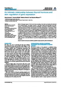
the need to engineer specific target sites like the tetracycline operator or the Gal4-binding site). Designed zinc fingers with engineered DNA sequence specificities have been successfully used to regulate endogenous chromosomal genes [15,16,19–21]. There have also been many recent advances in regulating gene expression in a temporal and quantitative manner using different inducible systems under the control of specific molecules (for example, Tet repressor-based systems, ecdysone-regulated gene switch) [22,23]. Such expression systems consist of chimeric transactivators and responsive target promoters of heterologous bacteFIG. 1. Structure of effector and reporter constructs. Effectors are constructed by fusing three or six zinc fingers rial, insect, or artificially designed (3Zn or 6Zn), SV40 nuclear localization signal (NLS), and the different activators shown. The effectors are placed components [24]. Specific mole- in a vector (pCDNA) just downstream of a T7/CMV promoter. The reporter constructs are as labeled. Arrowheads cules can regulate gene expres- represent zinc-finger target sites containing 9 bp or 18 bp (two copies of the 9 bp). sion by modulating the binding of a transactivator to its cognate promoter and bringing the transactivation domain into intended target must be prevented [15]. Thus it is very contact with a minimal promoter. Further improvement important to know the best predictor of optimized gene and refinement of expression systems are achieved by expression with respect to both target site affinity and incorporating a variety of well-characterized tissue-spespecificity in any given expression system. Second, we cific promoters or using modified constitutive viral promust achieve a desired level of gene expression in a given moter and enhancer elements including simian virus 40 cell type. Overexpression of a particular gene may not be (SV40), cytomegalovirus (CMV), and the Rous sarcoma desirable. The functional activity of a given effector virus long terminal repeat (RSV LTR). The major goals of domain fused to the DNA-binding domain of artificial any regulated expression system compatible with humantranscription factors may vary considerably in different therapeutic use include achieving a desired level of gene cell types. Furthermore, overexpression of a particular expression in a given cell type, being able to upregulate effector domain may result in “squelching” [25], which is and downregulate gene expression considerably from the seen as a consequence of titrating components of the transame target site (or transgenic construct), and ultimately scriptional machinery from a given cellular milieu. To being able to target endogenous promoters without ensure optimized gene expression in a given cell type, we manipulating the target DNA. Despite all the recent must assess the nature of the effector domain. Third, we progress, there are no expression systems that accommust regulate the activity of only the targeted gene and plish all the noted goals. Artificial transcription factors avoid altering the level of expression of the neighboring hold the greatest promise for developing practical expresgenes. If artificial transcription factors behave as true sion systems for use in gene therapy. enhancers, they may affect the expression of adjacent In gene therapy applications, it is important to address genes. For instance, the “classic” or “general” activation a number of different functional issues to ensure the opti- domains derived from naturally occurring transcriptional mum design of any artificial transcription factor. There activators like the herpes simplex VP16, were originally are at least three major issues facing the use of artificial suggested to activate transcription from proximal (or “protranscription factors in gene therapy applications. First, moter”) and from remote (or “enhancer”) positions [26]. we must maintain a high level of DNA-binding specificity. More recently, the classic activators were also suggested to The artificial transcription factors must have sufficient tar- have a relative insensitivity to the position of activatorget site affinity while simultaneously maintaining a high binding element [27,28]. Hence, for practical applications degree of target site specificity. However, exceeding a cer- of artificial transcription factors in gene therapy, several tain level of target site affinity is undesirable because of aspects of the design must first be experimentally tested increased binding to nonspecific DNA [21]. Any undesired to ensure optimum level of gene expression from a given pleiotropic effects on expression of genes other than the target in a given cell type.
686
MOLECULAR THERAPY Vol. 5, No. 6, June 2002 Copyright © The American Society of Gene Therapy
doi:10.1006/mthe.2002.0610, available online at http://www.idealibrary.com on IDEAL
In this study, we used engineered transcription factors based on a consensus zinc-finger-framework sequence [29] fused to classic regulator domains to address each of the noted major issues facing the artificial transcription factors in gene therapy applications. We also examined the utility of artificial transcription factors as regulators of gene expression from constitutive viral promoter–enhancer pairs. Previously, considerable gene activation had only been demonstrated from a viral promoter such as the SV40 promoter (pSV40) alone [30,31]. Our results reveal a substantial increase in relative activation, above the extent of gene activation achieved by the presence of both the viral promoter and enhancer pairs. We also showed that optimized activation of gene expression from a targeted region depended on several variables, all of which must be empirically evaluated in a given expression system, including target site affinities for various DNA-binding domains, the nature of the activator domain, the particular cell type used, and the position of the target site with respect to the core promoter. By engineering target sites at various positions and orientations with respect to the SV40 core promoter element, we demonstrated substantial increase in gene activation using a target position just upstream of the core promoter elements (that is, TATA box). Thus, even when using a classic activator like VP16, the zincfinger artificial transcription factors did not behave like true enhancer elements. Finally, we discuss several important implications of our observations and our combinatorial optimization approach for designing safe and effective artificial transcription factors for therapeutic and cell-based applications.
RESULTS
AND
DISCUSSION
Differential Effects of Three- and Six-Finger Proteins Previous studies have shown that a variety of variables can substantially influence the biological properties of artificial zinc-finger proteins, including different selection strategies, different framework sequence of zinc fingers, and different linker peptides used to generate larger polydactyl proteins [14–17,32,33]. In gene therapy applications, it is critical to maintain a high level of DNAbinding specificity, thus only regulating the expression of the target gene(s). To increase the specificity of artificial zinc fingers with the potential for genome-specific addressing, polydactyl proteins containing multiple fused zinc-finger domains capable of binding 15- to 20-bp target sequences have been successfully used [15–17,31,33]. To compare the effects of three- versus six-finger proteins in our expression system, we created various effector constructs as shown in Fig. 1, and directly examined the effect of the number of zinc fingers on target site binding affinity and on reporter gene activation. The approximate equilibrium dissociation constants (Kd) of purified zinc-finger proteins to the duplex MOLECULAR THERAPY Vol. 5, No. 6, June 2002 Copyright © The American Society of Gene Therapy
ARTICLE
oligonucleotides containing the 9-bp target or the 18-bp target were measured by protein titration with gel-shift assays from at least two independent experiments as described [29,34]. The starting unmodified three-finger protein (3Zn) had a dissociation constant (Kd) value of ~ 10 nM against the 9-bp (or the 18-bp) target, which is consistent with the previous characterization of this protein. When using a six-finger protein against the 18-bp target, we noted an ~ 20 times increase in affinity (~ 0.5 nM versus 10 nM). Modification of 3Zn protein with the addition of the SV40 nuclear localization signal (3ZnNLS), and subsequent fusion with three or four tandem repeats of the VP16’s minimal activation domain (3ZnNLS-VP3 or 3Zn-NLS-VP4), did not alter the apparent Kd. Similar to the three-finger proteins, various modifications of the six-finger protein (6Zn-NLS) by fusion with activator domains did not change the apparent Kd. However, the affinity of the six-finger protein for the single 9-bp half-site target was ~ 50 times lower (~ 25 nM versus 0.5 nM). Hence, the highest target-binding affinities were observed for the fusion proteins (effectors shown in Fig. 1) containing six-finger proteins against the 18-bp target. Next, we examined the functional outcome of various effectors containing six-finger proteins (Fig. 2A) and three-finger proteins (Fig. 2B), using the different reporter constructs shown. Our general findings included the following: 1) activation required the presence of the zincfinger DNA target sequence (only basal level of expression was seen in the absence of the effector target site; see pSV40-luc); 2) the presence of the target sequence alone did not alter the basal level of expression (see 18bppSV40-luc with No Zn); 3) activation required the presence of core promoter elements (see 18bp-luc; without the pSV40, the target had no effect in the presence of effectors containing VP3 or VP4); 4) the most effective activation occurred with 6Zn-NLS-VP4 from the reporter containing the 18-bp target just upstream of the pSV40, matching the extent of activation achieved by the reporter containing the combination of the SV40 promoter and SV40 enhancer sequences; and 5) the same effector (6Zn-NLS-VP4) was unable to activate gene expression from a single 9-bp target corresponding to the half-site of the six-finger protein domain. Effectors containing three-finger proteins were unable to activate transcription from the 9-bp target (Fig. 2B). However, the three-finger fusion proteins activated transcription from the 18-bp target (composed of two identical copies of the 9-bp target). Our observation of a positive correlation between affinity and function in our expression system is consistent with several other studies [20,21,33]. The main conclusion from this section of our study is that the zinc-finger artificial factors binding with a Kd in the single-digit nanomolar range (generally, ~ 1–10 nM) provide the most effective regulation. Hence this level of target site
687
ARTICLE
doi:10.1006/mthe.2002.0610, available online at http://www.idealibrary.com on IDEAL
affinity may be the best predictor of the optimum functional outcome while maintaining a high degree of target site specificity. For gene therapy applications, it is important to note that factors binding with considerably better affinities may be undesirable because of increased binding to nonspecific DNA [21]. In addition, substantially increased binding affinities have been shown to repress targeted gene expression even in the absence of any effector domain [33], particularly if the binding site is located within ~ 70 bp around the TATA box and the transcriptional start site [35]. It has also long been known that use of multiple target sites results in higher transcription levels. Various explanations include more effective target localization and recruitment [36], synergistic effect of activation surfaces on the transcriptional machinery [12], FIG. 2. Transcriptional regulation mediated by the indicated effectors. (A) Transcriptional regulation mediated and cooperative DNA binding by the various effector expression plasmids containing six zinc fingers (6Zn). HeLa cells were cotransfected with [37]. When using duplex oligonu- the indicated effector expression plasmids, reporter constructs, or empty vector (No Zn), and a -gal-expressing control vector. Normalized luciferase activity in total cell extracts was measured ~ 48 hours after transfeccleotides containing the 18-bp tion. Each bar represents the mean value (± 1 SEM) of three independent experiments. (B) Transcriptional regtarget DNA against the three-fin- ulation mediated by the indicated effector expression plasmids containing three zinc fingers (3Zn). ger proteins, gel-shift assays revealed two complexes reflecting the binding of one or two three-finger proteins (data not shown to be effective in transcriptional activation in HEK shown). Thus, with both three-finger proteins bound to 293 cells [19]. the 18-bp target, one effectively doubles the number of At least three tandem repeats of the VP16’s minimal activator surfaces present because each three-finger proactivation domain were required to achieve a substantial tein is covalently linked to the VP16 domains. The same activation of transcription (Figs. 2A and 2B). The addisynergistic effect of the additional domains of VP16 (3Zn- tion of one activation domain of VP16 doubled the NLS-VP3 versus 3Zn-NLS-VP4 in Fig. 2B) is also observed extent of reporter activation (VP3 versus VP4 in Figs. 2A when using six-finger fusion proteins with the 18-bp tar- and 2B). The VP3 and VP4 domains also synergistically get (6Zn-NLS-VP3 versus 6Zn-NLS-VP4 in Fig. 2A, and activated the transcription from a reporter construct consee the next section). taining both the pSV40 and the SV40 enhancer, suggesting independent mechanisms of activation between Differential Effects of Effector Domains the SV40 enhancer and the VP16’s acidic domains. Our To examine further the effect of the activator domain on results demonstrating the synergistic effect of the VP16’s transcription, we created several fusion proteins using minimal activation domain further support the suggesvarious tandem repeats of the minimal activation domain tion that a particular “classic” activator like VP16 is able of VP16 and P65 [26]. The P65 minimal activation to interact with multiple sites on the transcriptional domain and the two tandem repeats of the VP16’s minmachinery [27,28]. Synergistic increase in the potency imal activation domain did not activate transcription of a multimerized VP16’s activation domain has been above levels generated by the reporter vector lacking the previously reported for a different class of zinc-finger target site for the zinc-finger binding (Fig. 2A). proteins [38]. The current view is that small combinaAdditionally, the zinc-finger proteins without an effections of ubiquitous signal- and tissue-specific activators tor domain failed to activate transcription (data not can synergistically regulate gene expression through mulshown). Larger versions of the P65 activator containing tiple interactions with the general machinery and/or other specificity domains (amino acids 288–548) were between various coactivators [12,36]. Thus the presence
688
MOLECULAR THERAPY Vol. 5, No. 6, June 2002 Copyright © The American Society of Gene Therapy
doi:10.1006/mthe.2002.0610, available online at http://www.idealibrary.com on IDEAL
of individual activators or a particular combination of activators may be required for effective gene activation from a given promoter. Our observations have important implications for designing artificial transcription factors for use in gene therapy. It is very important to be able to regulate the activity of gene expression at a required level. For instance, overexpression of a particular gene using the most potent activator may not be desirable. Hence, empirical determination of the potency of gene activation associated with various “designer” effector domains allows for choosing the most appropriate effector in a given cell type. It is also highly desirable to be able to regulate the concentration of the artificial transcription factor. To this end, various systems to regulate levels of zinc finger–based artificial transcription factors have been developed [reviewed in 18]. Differential Effects of the Cellular Milieu A general mechanism of synergistic and promoter-selective activation by various activators is the cooperative recruitment of distinct subcomplexes containing key components of the transcriptional machinery like TFIIB and TFIID [39–41]. The function of various effector domains may depend on other key factors (for example, other coactivators) that are required for stimulation of transcription in a specific context of other enhancerbinding proteins [12]. Effective recruitment of key transcription factors (or subcomplexes) may thus require
creation of a stereospecific interface among different activators for proper docking and assembly of the preinitiation complex (PIC) at a given promoter site [12]. Furthermore, it is well known that the same given promoter sequence, such as the widely used viral promoters pSV40, pCMV, and pRSV, has very different potencies of activation in various cell types. Hence, selective determinants of particular functional outcomes from a given promoter depend not only on the promoter elements (providing binding sites for key factors) but also on the presence of various key factors in a particular cellular milieu. Consistent with this notion, we observed significantly different activation potency associated with the same zinc finger–activator fusion protein from the same promoter target site in two different cell types (Fig. 3). The SV40 promoter (pGL3-promoter or the pGL3-control vectors) is ~ 3 times more active in the CRL8805 cells compared with the HeLa cells (data not shown). We observed about 3–5 times less activation by VP16’s tetrameric repeat domain (VP4) in the CRL8805 cells, suggesting differential availability of key transcription factors that may serve as rate-limiting steps for effective recruitment by acidic activators. Our suggestion is also supported by previous “activator bypass” experiments in which the effect of activators can be mimicked by increasing the concentration of the transcriptional machinery [27,28]. In gene therapy applications, it is thus critical to assess the effect of a particular “designer” transcription factor directly in the target cell of interest. The same “general” or “classic” activator may have a considerably different extent of gene activation from the same promoter sequence in a different cell type. Depending on the targeted cell type, the nature and the particular design of the “effector” domain can subsequently be optimized to obtain the desired specific targeted regulation of gene expression. For instance, fusion of the zinc-finger DNAbinding domains with tissue-specific regulators rather than the “general” factors can be empirically assessed to ensure target-specific gene regulation in a given cell type.
FIG. 3. Different extent of transcriptional regulation in two cell types. The same effector containing six zinc fingers, a nuclear localization signal from SV40, and a tetrameric repeat of VP16 minimal activation domain (6Zn-NLS-VP4), or no effector (empty vector or No Zn) is used with the same reporter construct (18-bp target just upstream of the pSV40-luciferase vector) in two cell types (HeLa and CRL8805 cells). Cells were cotransfected with the indicated reporter vector, the effector expression plasmid, and a -gal-expressing control vector. Normalized luciferase activity in total cell extracts was measured ~ 48 hours after transfection. Relative transcriptional activation (fold activation) was determined by dividing the luciferase activity measured in the presence of the effector to the level of activation without the effector (No Zn). Each bar represents the mean value (± 1 SEM) of three independent experiments.
MOLECULAR THERAPY Vol. 5, No. 6, June 2002 Copyright © The American Society of Gene Therapy
ARTICLE
Differential Effects of Target Position with Respect to the Core Promoter Site To determine the effect of target position and orientation relative to the SV40 promoter (pSV40) on reporter activation by the zinc finger–containing effector protein (6Zn-NLS-VP4), a series of reporter constructs were made (Fig. 4A). In all cases, a single target site containing the 18-bp recognition sequence of the same six-finger
689
ARTICLE
doi:10.1006/mthe.2002.0610, available online at http://www.idealibrary.com on IDEAL
FIG. 4. Effect of position and orientation of the effector target site on gene activation. (A) Structures of effector and reporter constructs. The arrowhead represents the 18-bp DNA target of the effector containing six zinc fingers, a nuclear localization signal from SV40, and a tetrameric repeat of VP16 minimal activation domain (6Zn-NLS-VP4). (B) Effect of position and orientation of the effector target site relative to the SV40 promoter on transcriptional activation. HeLa cells were cotransfected with the indicated reporter vector, the effector expression plasmid, and a -gal-expressing control vector. Normalized luciferase activity in total cell extracts was measured ~ 48 hours after transfection. Relative transcriptional activation (fold activation) was determined by dividing the normalized luciferase activity measured in the presence of the effector to the level of activation without the effector (“No Zn” represents the empty vector). Each bar represents the mean value (± 1 SEM) of three independent experiments.
effector protein was used in various positions and orientations with respect to the pSV40 TATA box [42]. The activator domain used in all cases was the four tandem repeats of VP16’s minimal activation domain (VP4). We used the 18-bp target and the six zinc fingers fused to VP4, because this combination resulted in maximum level of gene activation. Our design also allowed us to assess the effect of target position in episomal reporter genes with a relative lack of fully organized nucleosomes and higherorder structures found in chromatin [43]. Our results showed a substantial difference in the extent of gene activation that correlated with the position of the target site with respect to the TATA box of the pSV40 (Fig. 4B). The “classic” or “general” activation domains like the herpes simplex VP16 were originally suggested to activate transcription from proximal (or “promoter”) and from remote (or “enhancer”) positions using fusion proteins bearing a Gal4 DNA-binding domain [26]. Our data revealed that maximum activation occurred in the reporter construct carrying the 18-bp target, ~ 150 bp upstream of the pSV40 TATA box. The reverse orientation of the target site also resulted in important gene activation (11–17 times was not statistically different from 20–34 times). Placing the target ~ 350 bp upstream or ~100 bp downstream of the TATA box (after the transcription initiation site) resulted in substantial reduction of activation (~ 3–4 times versus 20–34 times). Note that a more distant upstream position of the target site (~ 350 bp) resulted in a comparable extent of activation to the closer, but downstream
690
location of the target (~ 100 bp). In either orientation, there was no effective gene activation when the target was placed ~ 2000 bp downstream of the TATA box, where the endogenous SV40 enhancer is fully capable of effective activation. These data suggest that the mechanism of gene activation from distance- and orientation-independent enhancers (like the SV40 enhancer) are complementary but distinct from typical activators that are only capable of stimulating gene activation from proximal promoter positions. In fact, it has been shown that the core 72-bp repeats of the SV40 enhancer functions, in part, by facilitating nuclear transport [44]. Other factors operating at enhancers located at remote positions from the core promoter may directly influence gene activation from proximal promoter sites, presumably by looping out of the DNA between the remote enhancer and the proximal promoter sites [5,26]. Our results also support the structural, biochemical, and genetic studies describing the mechanisms of oriented assembly of the PIC for productive transcription [45–49]. These studies suggest that alternate conformations of the TFIIB factor, a key general transcription factor recruited by VP16, can provide the basis for the proper oriented assembly and subsequent productive function of the PIC. Factor TFIIB is composed of two domains, which are engaged in an intramolecular interaction that is disrupted upon interaction with the activation domain of the VP16 protein. Hence, interaction with VP16 can properly nucleate the assembly of the PIC
MOLECULAR THERAPY Vol. 5, No. 6, June 2002 Copyright © The American Society of Gene Therapy
doi:10.1006/mthe.2002.0610, available online at http://www.idealibrary.com on IDEAL
A
ARTICLE
B
FIG. 5. Regulation of SV40 expression system. (A) Structures of effectors and SV40 reporter constructs. Effectors are constructed by fusing six zinc fingers (6Zn), SV40 nuclear localization signal (NLS), and four tandem repeats of the VP16’s minimal activation domain (VP4), or KRAB repressor–coding domains derived from amino acids 1–99 of the KRAB domain of KOX-1. The effectors are placed in a vector (pCDNA) just downstream of a CMV promoter. The reporter constructs are as labeled. (B) Regulation of SV40 expression system. Transcriptional regulation mediated by the various effectors is depicted. HeLa cells were cotransfected with the indicated reporter constructs, the effector expression plasmids, or empty pCDNA vector (No Zn), and a -gal-expressing control vector. Normalized luciferase activity in total cell extracts was measured ~ 48 hours after transfection. Each bar represents the mean value (± 1 SEM) of three independent experiments.
in a directional (upstream to downstream) fashion. Our results are thus consistent with a model that requires proper localization of the VP16 domains for most effective activation. The observation of relative difference of target position on gene activation has important implications for designing more effective artificial transcription factors independently of the issue of target accessibility to certain chromatin regions [19]. Hence, proper target site location must be considered even for nonprotein artificial factors based on synthetic polyamides [50]. Finally, because of the striking correlation between target position and relative gene activation (Fig. 4B), our study suggests a novel approach toward functional dissection of a promoter sequence, locating the approximate position of the core promoter elements required in productive PIC assembly. Targeted Regulation of Gene Expression from Constitutive Viral Systems To test the effects of zinc finger–based artificial transcription factors on the extent of transcription activation or repression from a reporter vector containing both the pSV40 and the SV40 enhancer, we fused six zinc fingers (6Zn) to four tandem repeats of the minimal activator domains derived from VP16 or to the Krüppel-associated box (KRAB) repressor (effectors; Fig. 5A). The two effectors (activator or repressor) were expressed from a CMV promoter (pCMV). Note that the two SV40-based luciferase reporter vectors differ only in the presence of a single 18-bp target sequence of the zinc fingers just upstream of the pSV40 (Fig. 5A). Reporter expression with either the negative control (“No Zn” in Fig. 5B) or just using 6Zn-NLS without an effector domain did not significantly differ from the basal level (data not shown). When using the zinc finger–activator fusion protein (6Zn-NLS-VP4), we observed a significant increase (~ 5
MOLECULAR THERAPY Vol. 5, No. 6, June 2002 Copyright © The American Society of Gene Therapy
times) in reporter activation in the presence of the target sequence (Fig. 5B). The zinc finger–repressor fusion protein (KRAB-NLS-6Zn) brought about significant repression of gene expression (~ 3 times) in a targetdependent fashion (Fig. 5B). The maximum level of repression was ~ 70% decrease in luciferase expression compared with controls. Placing the KRAB domain at the end of the fusion protein further decreased the effectiveness of the repression (data not shown). Note that there is more than an order of magnitude difference between the “on” state (with activator) and the “off” state (with repressor) of the same reporter construct bearing the 18-bp target of the zinc finger–effector proteins (Fig. 5B). Thus the zinc finger–based artificial gene switch provides a very effective means of modulating the level of gene expression in the presence of both the pSV40 and the SV40 enhancer. The same zinc finger–effector fusion proteins were also effective in regulating transcription from a reporter vector containing both the pCMV and the CMV enhancer (Fig. 6B). To avoid competition between the same promoters, we expressed the effectors shown in Fig. 6A by the pSV40 plus the SV40 enhancer. The luciferase reporter vectors vary only in the presence of a single 18bp target sequence of the zinc fingers just upstream of the CMV enhancer and pCMV (Fig. 6A). For the negative control (“No Zn” in Fig. 6B), or in the absence of the 18bp target sequence, similar levels of luciferase expression were observed (Fig. 6B). In the presence of the zinc finger–activator fusion protein (6Zn-NLS-VP4), we observed a significant increase (~ 2–3 times) in reporter activation (Fig. 6B). The zinc finger–repressor fusion protein (KRABNLS-6Zn) also brought about significant repression of gene expression (at least 50%; Fig. 6B). There is more than a fivefold difference in gene expression between the “on” state (with activator) and the “off” state (with
691
ARTICLE
doi:10.1006/mthe.2002.0610, available online at http://www.idealibrary.com on IDEAL
A
B
FIG. 6. Regulation of CMV expression system. (A) Structures of effectors and CMV reporter constructs. Effectors are constructed by fusing six zinc fingers (6Zn), SV40 nuclear localization signal (NLS), and four tandem repeats of the VP16’s minimal activation domain (VP4), or KRAB repressor coding domains derived from amino acids 1–99 of the KRAB domain of KOX-1. The effectors are placed in a vector (pGL3) under the expression of the SV40 promoter plus the SV40 enhancer. The reporter constructs are as labeled. (B) Regulation of CMV expression system. Transcriptional regulation mediated by the various effectors is depicted. HeLa cells were cotransfected with the indicated reporter constructs, the effector expression plasmids, or empty pGL3 vector (No Zn), and a -gal-expressing control vector. Normalized luciferase activity in total cell extracts was measured ~ 48 hours after transfection. Each bar represents the mean value (± 1 SEM) of three independent experiments.
repressor) from the same reporter construct. Finally, we also obtained similar fold activation (~ 3 times) when using a targeted RSV LTR-driven luciferase reporter vector under the influence of the RSV LTR (data not shown). The potential sources for the apparent differences between the effect of the zinc finger–effector proteins on various reporters (comparing Fig. 5B with Fig. 6B) include: (1) different distances of the target from the core promoter elements; (2) different mechanisms or functional potencies of various promoters in HeLa cells; (3) different interactions between the particular effectors used (VP16 and KRAB) with various regulatory elements (different enhancers and promoters) in a given cell type (HeLa cells); and (4) different levels of zinc finger–effector fusion proteins present in each case (in that the effectors are under the expression of different promoters). Despite the noted differences between the extent of gene regulation, it is clear that even using a suboptimal expression system, we were able to substantially regulate gene expression from the constitutive viral promoter–enhancer pairs. In summary, designing more effective artificial transcription factors with a diverse range of applications in basic and applied sciences across many areas of biotechnology including medical, agricultural, functional genomics, and gene therapy remains a major focus of research in the genomics era. We have examined several aspects of gene expression using artificial transcription factors. Achieving optimized regulation of gene expression from a targeted region of a given promoter involves multiple levels of complexity, each of which must be empirically evaluated, including the following: 1) binding affinities and specificities of zinc finger–effector fusion proteins to a given target; 2) nature of the effector domains fused to the zinc fingers; 3) cellular milieu with the availability of various key factors present; 4)
692
effects of target site position with respect to the PIC assembly site; and 5) accessibility of factors to the target site in endogenous chromatin location.
MATERIALS
AND
METHODS
Construction of polydactyl zinc-finger proteins. The studies reported here use a previously designed and characterized zinc-finger-framework sequence based on a consensus sequence [29,34]. The starting plasmid pG5, containing the gene encoding a three-finger protein recognizing the 9-bp target sequence 5⬘-GAGGCAGAA-3⬘, was provided by Jeremy Berg (Johns Hopkins, Baltimore, MD). The construction, expression, purification, and crystallization of this protein bound to its target DNA have been described [34]. Each zinc finger of this protein is identical in sequence except for particular changes in its DNA recognition region, which spans seven residues. Genes encoding six tandem zinc fingers were made by ligation of two identical three-finger-encoding genes using Cfr10I and XmaI as described [29]. The resulting six-finger coding sequence preserved the conserved canonical “TGEKP” linker residues between each finger. Hence, the 18-bp DNA target containing the six-finger binding site consists of two identical 9-bp half-sites (5⬘-GAGGCAGAAGAGGCAGAA-3⬘). The DNA sequences of all constructs were confirmed by dideoxy sequencing. DNA-binding properties of purified proteins were assessed using gel mobility-shift assays essentially as described [29,51]. Radioactive gel signals from labeled double-stranded DNA oligonucleotides containing the 9-bp target, or the 18-bp target, were quantitated with a PhosphorImager (Molecular Dynamics) and recorded on X-ray films. The KALEIDAGRAPH program (Synergy Software, Reading, PA) was used to fit the data and determine the equilibrium dissociation constants. Construction of zinc finger–effector domain fusion proteins. To construct artificial transcription factors, standard cloning procedures were used to make fusions between various zinc-finger-coding regions (three or six fingers), a nuclear localization signal (NLS) from simian virus 40 (SV40) [52], and coding regions derived from VP16’s or P65’s minimal activation domains [26] (Fig. 1). Various tandem repeats consisting of two (VP16X2 or VP2), three (VP16X3 or VP3), and four (VP16X4 or VP4) of the VP16’s minimal activation domain, comprising amino acids 437–447 [26], were generated from pairs of complementary oligonucleotides. Particular fragments with various tandem repeats (two, three, or four) were then fused to the 3⬘ end of coding regions of zinc fingers (three or six) containing a NLS from SV40 as shown in Figure 1. Similarly, the minimal activation domain derived from amino acids 520–550 of P65 [26]
MOLECULAR THERAPY Vol. 5, No. 6, June 2002 Copyright © The American Society of Gene Therapy
doi:10.1006/mthe.2002.0610, available online at http://www.idealibrary.com on IDEAL
was cloned to the 3⬘ end of a six-finger-NLS coding construct. The same strategy was used to clone the KRAB repressor coding domains derived from amino acids 1–99 of the KRAB domain of KOX-1 [53], the NLS from SV40, and the same six-finger coding region noted above (6Zn; Fig. 5A). The zinc finger–effector fusion constructs were cloned in the eukaryotic expression vector pCDNA 3 (Invitrogen, Carlsbad, CA), just downstream of a T7/pCMV-containing region. The most effective zinc finger–activator fusion in pCDNA 3 (6Zn-NLS-VP4) was used to examine the extent of targeted gene activation from a pSV40 promoter and SV40 enhancerdriven luciferase reporter vector (pGL3-control; Promega, Madison, WI) by incorporating a single 18-bp target sequence ~ 20 bp upstream of the pSV40. The same zinc finger–activator fusion was also used to examine the extent of targeted gene activation from a RSV LTR-driven luciferase reporter vector (derived from pRc/RSV; Invitrogen) by incorporating a single 18-bp target sequence ~ 20 bp upstream of the LTR. In the case of CMV testing, the zinc finger–effector fusion constructs were cloned just downstream from pSV40 in a pGL3-derived vector (pGL3-control; Promega) containing both the SV40 promoter and enhancer sequences (Fig. 6A). The effector vectors were then used to examine the extent of targeted gene activation from a pCMV-driven luciferase reporter vector (derived from pCDNA 3) by incorporating the 18-bp target sequence ~ 35 bp upstream of the 5⬘ end of the hCMV enhancer–promoter pair. DNA sequences of all constructs were verified by DNA sequencing. Construction of luciferase reporter plasmids. Reporter plasmids with different target sites containing a single 9-bp DNA target (5⬘-GAGGCAGAA-3⬘), or a single 18-bp target with two identical 9-bp half-sites (5⬘GAGGCAGAAGAGGCAGAA-3⬘), were constructed by PCR cloning with inclusion of the target sequences into the appropriate PCR primers. Targets were incorporated into various pGL3 luciferase reporter vectors (Promega), containing the firefly luciferase gene. The reporter vectors used in this study include pGL3-basic vector (which lacks eukaryotic promoter and enhancer sequences), the pGL3-promoter vector (which contains a SV40 promoter just upstream of the luciferase gene), and the pGL3-control vector containing both the SV40 promoter and enhancer sequences. Targets were engineered into different positions and orientations with respect to the SV40 promoter. Single 18-bp target sites were also engineered into a pCMV-driven luciferase vector (derived from pCDNA 3) at ~ 500 bp away from the TATA box, and near the LTR of a RSV-driven luciferase vector (derived from pRc/RSV; Invitrogen) at ~ 300 bp away from the TATA box. Sequences of all the PCR-cloned fragments containing various zinc-finger target sites were confirmed by DNA sequencing. Luciferase assays. For all transfections involving HeLa and CRL8805 (American Type Culture Collection, Manassas, VA), cells were used at a confluency of ~ 40–60%. Cells were transfected with 300 ng of the RSVor pGL3-derived plasmids (reporters or effectors or empty vector), 100 ng of pCDNA 3-derived plasmids (reporters or effectors or empty vector), and 100 ng of internal standard plasmid in all cases (-gal-containing vector from Invitrogen) in a well of a six-well dish by using lipofectamine 2000 reagent (Gibco/BRL) for the CRL 8805 cells, or lipofectin and “PLUS” reagents (GIBCO/BRL) for HeLa cells. Cell extracts were prepared ~ 48–72 hours after transfection. Luciferase activity was measured with luciferase assay reagent (Promega), -galactosidase activity with Galacto-Light (Tropix, Bedford, MA), in a Victor microplate luminometer (Perkin-Elmer Wallac, Gaithersburg, MD). Luciferase activity was normalized to the galactosidase activity to correct for transfection efficiency. Student’s t-test was used to determine statistical significance for the differences noted.
ACKNOWLEDGMENTS We thank Jeremy Berg and members of his lab (Holly Berkovits, Greg Gatto, and Derek Jantz) for providing us with the starting plasmid (pG5), purified protein sample, and technical assistance with determination of the equilibrium dissociation constants. We acknowledge Harry Dietz and Ada Hamosh for their contribution and critical reading of the manuscript. This work was supported by grants from the National Institutes of Health and C. F. Foundation to G.R.C. RECEIVED FOR PUBLICATION JANUARY 23; ACCEPTED APRIL 2, 2002.
MOLECULAR THERAPY Vol. 5, No. 6, June 2002 Copyright © The American Society of Gene Therapy
ARTICLE
REFERENCES 1. Lander, E. S., et al. (2001). Initial sequencing and analysis of the human genome. Nature 409: 860–921. 2. Venter, J. C., et al. (2001). The sequence of the human genome. Science 291: 1304–1351. 3. Verma, I. M., and Somia, N. (1997). Gene therapy—promises, problems and prospects. Nature 389: 239–242. 4. Hampsey, M. (1998). Molecular genetics of the RNA polymerase II general transcriptional machinery. Microbiol. Mol. Biol. Rev. 62: 465–503. 5. Ptashne, M., and Gann, A. (1997). Transcriptional activation by recruitment. Nature 386: 569–577. 6. Sauer, F., and Tjian, R. (1997). Mechanisms of transcriptional activation: differences and similarities between yeast, Drosophila, and man. Curr. Opin. Genet. Dev. 7: 176–181. 7. Rachez, C., et al. (1999). Ligand-dependent transcription activation by nuclear receptors requires the DRIP complex. Nature 398: 824–828. 8. Naar, A. M., et al. (1999). Composite co-activator ARC mediates chromatin-directed transcriptional activation. Nature 398: 828–832. 9. Cremer, T., and Cremer, C. (2001). Chromosome territories, nuclear architecture and gene regulation in mammalian cells. Nat. Rev. Genet. 2: 292–301. 10. Wolffe, A. P., and Hansen, J. C. (2001). Nuclear visions: functional flexibility from structural instability. Cell 104: 631–634. 11. Hirose, Y., and Manley, J. L. (2000). RNA polymerase II and the integration of nuclear events. Genes Dev. 14: 1415–1429. 12. Carey, M. (1998). The enhanceosome and transcriptional synergy. Cell 92: 5–8. 13. Tupler, R., Perini, G., and Green, M. R. (2001). Expressing the human genome. Nature 409: 832–833. 14. Segal, D. J., and Barbas, C. F. (2000). Design of novel sequence-specific DNA-binding proteins. Curr. Opin. Chem. Biol. 4: 34–39. 15. Kang, J. S., and Kim, J. S. (2000). Zinc finger proteins as designer transcription factors. J. Biol. Chem. 275: 8742–8748. 16. Bartsevich, V. V., and Juliano, R. L. (2000). Regulation of the MDR1 gene by transcriptional repressors selected using peptide combinatorial libraries. Mol. Pharmacol. 58: 1–10. 17. Moore, M., Klug, A., and Choo, Y. (2001). Improved DNA binding specificity from polyzinc finger peptides by using strings of two-finger units. Proc. Natl. Acad. Sci. USA 98: 1437–1441. 18. Pabo, C. O., Peisach, E., and Grant, R. A. (2001). Design and selection of novel Cys2His2 zinc finger proteins. Annu. Rev. Biochem. 70: 313–340. 19. Liu, P. Q., et al. (2001). Regulation of an endogenous locus using a panel of designed zinc finger proteins targeted to accessible chromatin regions. Activation of vascular endothelial growth factor A. J. Biol. Chem. 276: 11323–11334. 20. Zhang, L., et al. (2000). Synthetic zinc finger transcription factor action at an endogenous chromosomal site. Activation of the human erythropoietin gene. J. Biol. Chem. 275: 33850–33860. 21. Beerli, R. R., Dreier, B., and Barbas, C. F. (2000). Positive and negative regulation of endogenous genes by designed transcription factors. Proc. Natl. Acad. Sci. USA 97: 1495–1500. 22. Saez, E., et al. (2000). Identification of ligands and coligands for the ecdysone-regulated gene switch. Proc. Natl. Acad. Sci. USA 97: 14512–14517. 23. Baron, U., and Bujard, H. (2000). Tet repressor-based system for regulated gene expression in eukaryotic cells: principles and advances. Methods Enzymol. 327: 401–421. 24. Fussenegger, M. (2001). The impact of mammalian gene regulation concepts on functional genomic research, metabolic engineering, and advanced gene therapies. Biotechnol. Prog. 17: 1–51. 25. Gill, G., and Ptashne, M. (1988). Negative effect of the transcriptional activator GAL4. Nature 334: 721–724. 26. Seipel, K., Georgiev, O., and Schaffner, W. (1992). Different activation domains stimulate transcription from remote (‘enhancer’) and proximal (‘promoter’) positions. EMBO J. 11: 4961–4968. 27. Gaudreau, L., et al. (1999). Transcriptional activation by artificial recruitment in yeast is influenced by promoter architecture and downstream sequences. Proc. Natl. Acad. Sci. USA 96: 2668–2673. 28. Nevado, J., Gaudreau, L., Adam, M., and Ptashne, M. (1999). Transcriptional activation by artificial recruitment in mammalian cells. Proc. Natl. Acad. Sci. USA 96: 2674–2677. 29. Desjarlais, J. R., and Berg, J. M. (1993). Use of a zinc-finger consensus sequence framework and specificity rules to design specific DNA binding proteins. Proc. Natl. Acad. Sci. USA 90: 2256–2260. 30. Pomerantz, J. L., Sharp, P. A., and Pabo, C. O. (1995). Structure-based design of transcription factors. Science 267: 93–96. 31. Liu, Q., Segal, D. J., Ghiara, J. B., and Barbas, C. F. (1997). Design of polydactyl zincfinger proteins for unique addressing within complex genomes. Proc. Natl. Acad. Sci. USA 94: 5525–5530. 32. Moore, M., Choo, Y., and Klug, A. (2001). Design of polyzinc finger peptides with
693
ARTICLE
doi:10.1006/mthe.2002.0610, available online at http://www.idealibrary.com on IDEAL
structured linkers. Proc. Natl. Acad. Sci. USA 98: 1432–1436. 33. Kim, J. S., and Pabo, C. O. (1998). Getting a handhold on DNA: design of poly-zinc finger proteins with femtomolar dissociation constants. Proc. Natl. Acad. Sci. USA 95: 2812–2817. 34. Kim, C. A., and Berg, J. M. (1996). A 2.2 Å resolution crystal structure of a designed zinc finger protein bound to DNA. Nat. Struct. Biol. 3: 940–945. 35. Kim, J. S., and Pabo, C. O. (1997). Transcriptional repression by zinc finger peptides. Exploring the potential for applications in gene therapy. J. Biol. Chem. 272: 29795–29800. 36. Ptashne, M., and Gann, A. (1998). Imposing specificity by localization: mechanism and evolvability. Curr. Biol. 8: R812–R822. 37. Giniger, E., and Ptashne, M. (1988). Cooperative DNA binding of the yeast transcriptional activator GAL4. Proc. Natl. Acad. Sci. USA 85: 382–386. 38. Emami, K. H., and Carey, M. (1992). A synergistic increase in potency of a multimerized VP16 transcriptional activation domain. EMBO J. 11: 5005–5012. 39. Gonzalez-Couto, E., Klages, N., and Strubin, M. (1997). Synergistic and promoter-selective activation of transcription by recruitment of transcription factors TFIID and TFIIB. Proc. Natl. Acad. Sci. USA 94: 8036–8041. 40. Grondin, B., and DeLuca, N. (2000). Herpes simplex virus type 1 ICP4 promotes transcription preinitiation complex formation by enhancing the binding of TFIID to DNA. J. Virol. 74: 11504–11510. 41. Chi, T., Lieberman, P., Ellwood, K., and Carey, M. (1995). A general mechanism for transcriptional synergy by eukaryotic activators. Nature 377: 254–257. 42. Pauly, M., Treger, M., Westhof, E., and Chambon, P. (1992). The initiation accuracy of the SV40 early transcription is determined by the functional domains of two TATA elements. Nucleic Acids Res. 20: 975–982.
694
43. Kadonaga, J. T. (1998). Eukaryotic transcription: an interlaced network of transcription factors and chromatin-modifying machines. Cell 92: 307–313. 44. Dean, D. A., Dean, B. S., Muller, S., and Smith, L. C. (1999). Sequence requirements for plasmid nuclear import. Exp. Cell Res. 253: 713–722. 45. Kays, A. R., and Schepartz, A. (2000). Virtually unidirectional binding of TBP to the AdMLP TATA box within the quaternary complex with TFIIA and TFIIB. Chem. Biol. 7: 601–610. 46. Littlefield, O., Korkhin, Y., and Sigler, P. B. (1999). The structural basis for the oriented assembly of a TBP/TFB/promoter complex. Proc. Natl. Acad. Sci. USA 96: 13668–13673. 47. Hayashi, F., et al. (1998). Human general transcription factor TFIIB: conformational variability and interaction with VP16 activation domain. Biochemistry 37: 7941–7951. 48. Hawkes, N. A., Evans, R., and Roberts, S. G. (2000). The conformation of the transcription factor TFIIB modulates the response to transcriptional activators in vivo. Curr. Biol. 10: 273–276. 49. Zhang, D. Y., et al. (2000). Intramolecular interaction of yeast TFIIB in transcription control. Nucleic Acids Res. 28: 1913–1920. 50. Mapp, A. K., Ansari, A. Z., Ptashne, M., and Dervan, P. B. (2000). Activation of gene expression by small molecule transcription factors. Proc. Natl. Acad. Sci. USA 97: 3930–3935. 51. Desjarlais, J. R., and Berg, J. M. (1992). Redesigning the DNA-binding specificity of a zinc finger protein: a data base-guided approach. Proteins 12: 101–104. 52. Kalderon, D., Roberts, B. L., Richardson, W. D., and Smith, A. E. (1984). A short amino acid sequence able to specify nuclear location. Cell 39: 499–509. 53. Margolin, J. F., et al. (1994). Kruppel-associated boxes are potent transcriptional repression domains. Proc. Natl. Acad. Sci. USA 91: 4509–4513.
MOLECULAR THERAPY Vol. 5, No. 6, June 2002 Copyright © The American Society of Gene Therapy