Oct 15, 2014 - in the porcine animal model. First, trainees practiced with animals without using a model of injured (ureteroscopy, management of guide wires ...
Am J Clin Exp Urol 2014;2(3):258-265 www.ajceu.us /ISSN:2330-1910/AJCEU0001181
Original Article Description and validation of realistic and structured endourology training model Federico Soria1, Esther Morcillo1, Juan Luis Sanz3, Alberto Budia2, Alvaro Serrano3, Francisco M Sanchez-Margallo4 Department of Endoscopy, Minimally Invasive Surgery Centre Jesús Usón, Cáceres, Spain; 2Department of Urology, La Fe University Hospital, Valencia, Spain; 3Department of Urology, Guadalajara University Hospital, Madrid, Spain; 4Minimally Invasive Surgery Centre Jesús Usón, Cáceres, Spain 1
Received June 22, 2014; Accepted July 7, 2014; Epub October 2, 2014; Published October 15, 2014 Abstract: Purpose: The aim of the present study was to validate a model of training, which combines the use of non-biological and ex vivo biological bench models, as well as the modelling of urological injuries for endourological treatment in a porcine animal model. Material and Methods: A total of 40 participants took part in this study. The duration of the activity was 16 hours. The model of training was divided into 3 levels: level I, concerning the acquisition of basic theoretical knowledge; level II, involving practice with the bench models and level III, concerning practice in the porcine animal model. First, trainees practiced with animals without using a model of injured (ureteroscopy, management of guide wires and catheters under fluoroscopic control) and later practiced in lithiasic animal model. During the activity, an evaluation of the face and content validity was conducted, as well as constructive validation provided by the trainees versus experts. Evolution of the variables during the course within each group was analysed using the Student’s t test for paired samples, while comparisons between groups, were performed using the Student’s t test for unpaired samples. Results: The assessments of face and content validity were satisfactory. The constructive validation, “within one trainee” shows that were statistical significant differences between the first time the trainees performed the tasks in the animal model and the last time, mainly in the knowledge of procedure and Holmium laser lithotripsy cathegories. At the beginning of level III, there are also statistical significant differences between trainee’s scores and the expert’s scores.Conclusions: This realistic Endourology training model allows the acquisition of knowledge and technical and non-technical skills as evidenced by the face, content and constructive validity. Structured use of bench models (biological and non biological) and animal model simulators increase the endourological basic skills. Keywords: Endourology, training, animal model, bench model, validation
Introduction Nowadays the need for training in endourology is a matter of fact, specifically at the level of medical residents. Endourology training during residency has traditionally relied on live patients in the operating room under the strict supervision of an attending urologist. This apprentice-type of training requires a large caseload and often involves substantial additional operating room time devoted to education [1]. Residents training are essential because these techniques are part of the daily urological armamentarium, although the learning process has a relatively long learning curve, and in early
stages the iatrogenic conditions risk is high [2], due to the fact that the endourological techniques are notably difficult to perform. The complications, which are not avoidable sometimes even in experienced hands, depends significantly on the number of procedures performed and the urologist skills [3]. Economic pressures to decrease operating times, as well as an increasingly litigious climate, have reduced the time dedicated to resident training [4-8]. For these reasons, it is becoming more and more difficult for the urologist in training to acquire experience in a time-efficient manner
Endourology training model
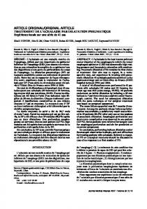
Figure 1. A. Bench model. B. Exvivo model.
[9]. In an effort to address this aspect of learning for endourology, surgical educators have developed alternative methods of training [6]. Over the last two decades, training models have been developed that are based on bench models, training with cadavers, animal models, and, recently, VR systems (virtual reality) [5]. The objective of the study was present and to validate our endourology training model. It is based on three levels, which combines the use of non-biological and ex vivo biological bench models, as well as the animal model combined with the modelling of urological injuries for endourological treatment in a porcine animal model. Material and methods
Trainees spent an equal amount time on the simulators, live animals models, and practice the same tasks in the course of the training program. “Principles of laboratory animal care” (NIH publication No. 86-23, revised 1985) were followed, as well as the current version of the European Union Laws on the Protection of Animals used for scientific purposes. Training design The training was carried out in the experimental operating theatre with all equipment needed available. To make the procedure as realistic as possible, the simulators were placed on an operating table and covered with drapes.
Subjects
Training model was distributed into three levels:
The participants included urology residents and a board-certified urologist with no previous experience in semi-rigid ureteroscopy (survey evaluated). A total of 40 participants took part in this study. The total duration of the activity was 16 hours (divided in four sessions). Trainees practiced in pairs, with each group being supervised by an experienced endourologist.
Level I. Acquisition of basic theoretical knowledge. At this level, theoretical sessions (videos and lectures) related to techniques are addressed, including indications regarding ureterorenoscopy (URS) technique and complications; instrumentation; comparative anatomy; intracorporeal lithotripsy; and endourological management of ureteral stricture.
259
Am J Clin Exp Urol 2014;2(3):258-265
Endourology training model
Figure 2. Practices in animal model.
The duration is approximately 20% of the total training activity duration. This level was evaluated using a test. Level II. Practice using bench models. First of all, bench models were used (ETXY-Uro Adam©, ProDelphus, Brazil), to allow the completion of an urethrocystoscopy and the ureteral orifices cannulation with a guide wire followed by a semi-rigid URS. Then a second simulator was used. The simulator consists in a porcine renoureteral unit from a slaughterhouse, into which ureteral lithiasis has been introduced at the mid ureteral level [10]. This simulator enables the trainee to practice laser lithotripsy, basket removal of stone fragments, and anti-migration device handling. The duration of this second level is approximately 20% of the total training activity. (Figure 1). Level III. Practice with live animal models. The animal model used was a female porcine. The practices carried out using the animal model include the following: -Urethrocystoscopy. -Ureteral orifices cannulation and subsequent ureteroscopy. To carry out these practices in lived porcine model, the trainees have 2 porcine models at their disposal.
260
Model 1. In the left nephroureteral unit, no actuation is carried out, thereby enabling the practice of basic ureteroscopy (Figure 2). In the right nephroureteral unit, an ureteropelvic junction (UPJ) obstruction model was created [11], 3 weeks before the training. To perform the UPJ obstruction model, first it was necessary the bipolar coagulation with laparoscopic forceps the adventitial layer of the UPJ and after partial occluding the ureteral lumen using laparoscopic approach an 3/0 polyglicolic acid ligature. The UPJ model enables the following practice techniques: the manipulation of the upper urinary tract using endoscopic-fluoroscopic control, manipulation and proper selection of guide wires and catheters, negotiation of ureteral curves and bends; instruction regarding the upper urinary tract, endourological treatment of a strictures; laser endopyelotomy, and subsequent placement of a JJ ureteral stent. Model 2. The model 2 consists in a bilateral ureteral lithiasic animal model. These models were created surgically introducing artificial ureteral stones in both renal pelvises, one week prior to the training activity. Both ureters were stented with JJ ureteral stents to prevent renal colic and the animals were treated with analgesics during this time. So, trainees can practice laser lithotripsy, manipulation of migration of lithiasic fragments and their removal. The duration of this third level was approximately 60% of the total training activity (9,5 hours), distributed uniformly between model 1 and 2. All the practices performed in this training activity were under the supervision of an expert endourologist (more than 200 URS performed). Evaluations Trainees and 10 experts’ endourologists had evaluated the realism of the activities using multi-item questionnaires that were specifically design for this simulation training (concerning face validity and content validity). Participants
Am J Clin Exp Urol 2014;2(3):258-265
Endourology training model Table 1. Face and content validation Non biological bench model
Ex vivo biological bench model
Learning skills
4.5 ± 0.8
4.3 ± 0.6
Reality bench models
3.8 ± 1.2
4.3 ± 0.8
Ease of use
4.2 ± 0.9
4.2 ± 0.1
Allows instrumentalization?
4.1 ± 0.7
4.2 ± 0.9
Global: 4.04 ± 0.31a,b
Global: 4.25 ± 0.13a,c
Face validation. (Mean ± SD) (0-5) LEVEL II
LEVEL III
Animal model
Learning skills
4.9 ± 0.3
Similarity to human anatomy (Reality)
3.6 ± 0.7
Does it help to lower the iatrogenic?
4.8 ± 0.4
Ease of use
4.4 ± 0.9
Allows instrumentalization?
4.7 ± 0.4 Global: 4.48 ± 0.49b,c
Content validation. (Mean ± SD) (0-5) Utility for training in endourology
4.8 ± 0.4
Range of exercises
4.8 ± 0.4
Effectiveness for skill acquisition
4.9 ± 0.3
Level II assessment
4.2 ± 0.6
Level III assessment
4.9 ± 0.3
Suitable for skills assessment during training
5.0 ± 0.0
Do you think this training model contributes to the reduction of medical iatrogenic?
5.0 ± 0.0 Global: 4.82 ± 0.27
Same superscripts in the values indicate significant differences (p