original papers Adv Clin Exp Med 2013, 22, 1, 27–31 ISSN 1899–5276
© Copyright by Wroclaw Medical University
Andrzej Wojnar1, A–D, F, Bartosz Puła2, A–D, F, Marzena Podhorska-Okołów2, A, C–F, Piotr Dzięgiel2, 3, A, C–F
Discrepancies Between HER2 Assessment from Core Needle Biopsies and Surgical Specimens of Invasive Ductal Breast Carcinoma* Niezgodność oceny receptora HER2 w materiale z biopsji gruboigłowej oraz materiału operacyjnego raka przewodowego gruczołu piersiowego Lower Silesian Oncology Center, Wrocław, Poland Department of Histology and Embryology, Wroclaw Medical University, Wrocław, Poland 3 Department of Physiotherapy, Wroclaw University School of Physical Education, Wrocław, Poland 1 2
A – research concept and design; B – collection and/or assembly of data; C – data analysis and interpretation; D – writing the article; E – critical revision of the article; F – final approval of article; G – other
Abstract Background. The assessment of HER2 status is particularly important for qualifying patients for trastuzumab treatment of invasive ductal breast carcinoma (IDC). HER2 assessment in core needle biopsies (CNBs) of IDC could contribute to a better therapy schedule. Objectives. The study aimed at examining the relationship between HER2 immunohistochemistry assessment scores in paired CNBs and whole tissue sections of IDC. Material and Methods. The study was performed on paired samples of CNBs and whole tissue sections from 49 IDC patients operated on at the Lower Silesian Oncology Center in Wrocław, Poland. Results. Discrepancies in HER2 scores were noted in eleven (22.45%) of the paired samples analyzed. Three cases (6.12%) were underscored in the CNB specimens as compared to the surgical HER2 specimens, whereas eight cases (16.33%) were overscored in the CNB specimens. Conclusions. Based on the high level of discrepancy between the tested pairs of IDC tissues, the authors recommend caution in assessing HER2 in CNB tissue specimens as a standard procedure. Wherever possible whole tissue sections should be utilized for HER2 assessment (Adv Clin Exp Med 2013, 22, 1, 27–31). Key words: breast cancer, HER2, needle core biopsy.
Streszczenie Wprowadzenie. Ocena ekspresji receptora HER2 jest ważnym elementem kwalifikacji pacjentek chorych na raka przewodowu gruczołu piersiowego (IDC) do terapii trastuzumabem. Ocena receptora HER2 w materiale z biopsji gruboigłowej (CNB) guzów IDC mogłaby przyczynić się do lepszego planowania terapii przeciwnowotworowej. Cel pracy. Zbadanie zależności między oceną ekspresji receptora HER2 w materiale IDC z biopsji gruboigłowej oraz tkankach pobranych operacyjnie. Materiał i metody. Badanie przeprowadzono na parach tkanek IDC pobranych metodą biopsji gruboigłowej oraz operacyjnie od 49 pacjentek operowanych w Dolnośląskim Centrum Onkologii. Wyniki. Zanotowano rozbieżność w ocenie ekspresji receptora HER2 w jedenastu (22,25%) spośród analizowanych par przypadków. Trzy (6,12%) przypadki miały niedoszacowaną ocenę, a osiem (16,33%) było przeszacowanych w materiale CNB w porównaniu z klasycznymi próbkami HER2. Wnioski. Na podstawie uzyskanych wyników oraz dużej rozbieżności między badanymi parami tkanek autorzy zalecają ostrożność w ocenie receptora HER2 w materiale CNB w standardowym postępowaniu w przypadku możliwości użycia do oceny preparatów HER2 przygotowanych z materiału operacyjnego (Adv Clin Exp Med 2013, 22, 1, 27–31). Słowa kluczowe: rak gruczołu piersiowego, HER2, biopsja gruboigłowa. * The study was supported financially by the Wrocław Research Centre EIT+.
28 Breast cancer poses a serious health problem worldwide. In 2008, approximately 450,000 new cases of this malignancy were diagnosed in Europe and more than 140,000 patients died of the disease [1]. Therefore, an early diagnosis and effective treatment of the disease are immensely important. In breast cancer diagnosis, core needle biopsy (CNB) is regarded as a reliable method for tissue sampling of palpable as well as non-palpable breast lesions [2, 3]. CNB has been found to be a fast and accurate diagnostic tool allowing for fast preoperative diagnosis and preliminary selection of breast lesion treatment [3, 4]. In comparison to fine-needle aspiration biopsies (FNABs) of breast lesions, CNBs are characterized by a greater sensitivity and allow additional immunohistochemical markers to be determined, due to the amount of tumor material in the biopsied core [5, 6]. Moreover, CNBs have also been shown to yield predictive information, since assessment of estrogen receptor (ER), progesterone receptor (PR) and human epidermal growth factor receptor 2 (HER2) are possible in tissue samples obtained this way [4, 7–14]. The assessment of HER2 status is particularly important for selecting patients for trastuzumab treatment in patients showing HER2 gene amplification [15, 16]. HER2 has been found to be amplified in up to 30% of breast cancers, and its overexpression is associated with a more aggressive disease course [17, 18]. HER2 testing is performed on formalin-fixed, paraffin embedded tumor tissue. Two complementary methods used for HER2 testing are immumohistochemistry (IHC) and fluorescence in situ hybridization (FISH), which allow for examination of protein overexpression or gene amplification, respectively In the diagnostic algorithm, the IHC is performed first, in accordance with a well-established worldwide four-grade scale based on estimating the continuity and intensity of membrane reaction. If the result of the IHC are equivocal and do not allow the HER2 expression status to be established, additional FISH examinations are undertaken to determine HER2 amplification [19–21]. Assessing the HER2 status in CNBs may result in early treatment planning. Earlier studies concerning the assessment of HER2 status in CNBs and surgical tissue specimens showed some discrepancies, ranging up to 40% [14]. Therefore, the goal of this study was to assess and compare the discrepancies in HER2 testing in pairs of breast cancer specimens obtained by CNB and by standard surgical resection of the tumor.
A. Wojnar et al.
Material and Methods The Specimens The breast cancer tissues utilized in the study originated from 49 female patients diagnosed with IDC and treated at the Lower Silesian Oncology Center in Wrocław, Poland. The CNBs were performed under ultrasound guidance using a true cut needle coupled to an automated biopsy device. The number of cores taken per tumor ranged from three to five. After the CNB, 24 patients underwent quadrantectomy followed by lymphadenectomy; 25 had radical mastectomies. During both procedures surgical tissue specimens were collected before the initiation of systemic treatment. Pairs of CNBs and surgical tissue specimens were fixed in 10% buffered formalin, embedded in paraffin, cut into 4-µm thick sections and mounted on SuperfrostPlus slides (Mänzel Glässer, Braunschwig, Germany). The slides were stained with hematoxylin and eosin (H&E) and HER2 using the Pathway HER-2/neu (4B5) Kit (Ventana, Tuscon, USA) in an automated immunostainer (Benchmark System, Ventana) using the protocol recommended by the manufacturer.
HER2 Assessment The CNB and surgical specimen slides were evaluated by two independent pathologists (AW and PD) under a BX-41 microscope (Olympus, Tokyo, Japan). A four-grade scoring system developed by the American Society of Clinical Oncology (ASCO) and College of American Pathologists (CAP) was used to evaluate HER2 expression, which was encoded as follows: 0 (no staining), 1+ (incomplete, weak membrane staining regardless of the proportion of tumor cells stained), 2+ (nonuniform complete membrane staining or staining with obvious circumferential distribution in at least 10% of the tumor cells, or intense, complete membrane staining ≤ 30% of the invasive tumor cells), 3+ (intense membrane staining in > 30% of the invasive tumor cells) [20]. In cases where the two pathologists differed with regard to the HER2 score, the slides were carefully reviewed under a double-headed microscope until a consensus was achieved.
Results Among the CNB specimens 26 cases (53.1%) were scored 0 and 23 (46.9%) were scored 1+. None of the analyzed CNB specimens had a score of 2+ or 3+. Among the surgical specimens 32 cases
29
HER2 Assessment in Invasive Ductal Breast Carcinoma
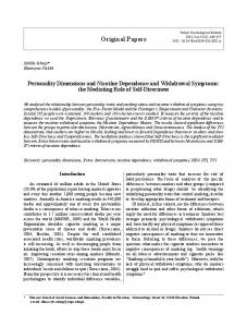
Fig. 1. HER2 IHC staining in CNB (A, C) and surgical specimens (B, D) as examples of underscoring (A, B) and overscoring (C, D) of HER2 assessment Ryc. 1. Reakcje IHC HER2 w materiale z biopsji gruboigłowej (A, C) i operacyjnym (B, D) oraz przykłady niedoszacowania (A, B) oraz przeszacowania (C, D) oceny HER2
(65.3%) were scored 0, 15 (30.6%) were rated 1+ and two (4.1%) were scored as 2+. None of the cases received a score of 3+. The two cases that were scored 2+ in the surgical specimens of IDC underwent subsequent FISH testing, but the final results were negative. In the CNB specimens as compared to the surgical HER2 specimens, three cases (6.12%) were underscored, whereas eight cases (16.33%) were overscored (Fig. 1). Overall, discrepancies between the HER2 scores were observed in 11 cases (22.45%), which are listed in Table 1.
Discussion Because HER2 expression status is of great importance for selecting therapy for breast cancer patients, early information concerning its overexpression could result in a better therapy schedule for traztuzumab treatment [15, 16]. Nonetheless, concerns may arise, as some earlier studies dealing with HER2 expression in CNB specimens and whole tissue sections reported poor concordance (60% and 80%) between the two types of breast cancer specimens [13, 14]. Such vast discrepancies, similar to those observed in the current study, are not acceptable from the clinical point of view. Interestingly, studies performed on larger cohorts of patients reported higher concordance rates, where the discrepancies in HER2 testing reached only 1.2% and 2% [7, 9]. Recent studies have shown that many factors may contribute to discrepancies in HER2 IHC assessment. The pathologist’s experience seems to
Table 1. List of cases where discrepancies were noted between the HER2 scores from CNBs and surgical specimens Tabela 1. Lista przypadków, w których zanotowano rozbieżność w ocenie materiału CNB oraz chirurgicznego Case (Przypadek)
1 2 3 4 5 6 7 8 9 10 11
HER2 score (Punktacja HER2)
FISH
CNB specimen
surgical specimen
0 0 1+ 1+ 1+ 1+ 1+ 1+ 1+ 1+ 1+
2+ 1+ 2+ 0 0 0 0 0 0 0 0
– –
account for up to one third of the discrepancies in the final HER2 scores, as shown by the large multicenter study by Umemura et al. [22]. In addition to this, in one fourth of the instances of discrepancies, the difference was attributed to the staining procedures only [22]. A combination of the two factors was found in 41.7% of the cases of discrepancies [22]. Other studies also identify these factors to be key in overall HER2 staining assessment [23–25]. In the current study, the slides were evaluated by two experienced pathologists (>10 years of experience in HER2 assessment) at a large pathology center (more than 800 HER2 assessments
30
A. Wojnar et al.
annually), and all the staining was performed using the same automated staining devices and the Ventanas Pathway HER-2/neu protocol, which in the authors’ opinion should have enhanced the study’s reproducibility. Another key factor that may be responsible for the discrepancies noted in breast cancer specimens is the morphological heterogeneity of the tumor itself, as the authors reported earlier regarding ER and PR expression [26]. To some extent the use of tissue micro-arrays (TMAs) may reproduce the conditions (limited amounts and random parts of the tumor) obtained in the CNB specimens [27, 28]. In numerous studies, the use of TMAs for HER2 assessment showed great concordance with HER2 IHC scores noted in whole tissue specimens, but some studies reported a discordance in HER2 scores and lower specifity and sensitivity in TMAs when compared to the results obtained in whole tissue sections [29]. Similar findings were noted in the study by Lin et al., who found that HER2 and PR expression are underestimated in TMAs [30]. A study by Tamaki et al. compared the outcomes of ER, PR and HER2 status in CNBs de-
pending on the number of cores obtained from the tumor [31]. The rate of HER2 assessment concordance between CNBs and whole tissue sections strongly depended on the number of cores utilized. For one core the concordance rate reached 85.6%; for two cores it was 91.4%; and for three and four cores it reached 100% concordance. Using three or four cores was initially recommended for breast cancer biopsies in a pioneer study comparing different needle calibers and excursions [31, 32]. In the current study the number of cores taken during the biopsies ranged from three to five, which according to the earlier studies should be optimal. In summary, this study showed discrepancy rates reaching 22.45% in HER2 IHC scoring between CNBs and whole tissue specimens, although the slides were evaluated by two experienced pathologists and a reasonable number of cores (3–5) were taken during the initial biopsy of each tested tumor. Based on these findings, the authors recommend caution when HER2 assessment is conducted using CNB tissue specimens in cases where whole tissue sections could be utilized for HER2 assessment.
Acknowledgements The authors thank Mrs. Teresa Klepuszewska for her technical assistance.
References [1] Ferlay J, Autier P, Boniol M, Heanue M, Colombet M, Boyle P: Estimates of the cancer incidence and mortality in Europe in 2006. Ann Oncol 2007, 18, 581–592. [2] Pettine S, Place R, Babu S, Williard W, Kim D, Carter P: Stereotactic breast biopsy is accurate, minimally invasive, and cost effective. Am J Surg 1996, 171, 474–476. [3] Bilous M: Breast core needle biopsy: issues and controversies. Mod Pathol 2010, Suppl 2, 36–45. [4] Wong AY, Salisbury E, Bilous M: Recent developments in stereotactic breast biopsy methodologies: an update for the surgical pathologist. Adv Anat Pathol 2000, 7, 26–35. [5] Berner A, Davidson B, Sigstad E, Risberg B: Fine-needle aspiration cytology vs. core biopsy in the diagnosis of breast lesions. Diagn Cytopathol 2003, 29, 344–348. [6] Kooistra B, Wauters C, Strobbe L: Indeterminate breast fine-needle aspiration: repeat aspiration or core needle biopsy? Ann Surg Oncol 2009, 16, 281–284. [7] Arnedos M, Nerurkar A, Osin P, A’Hern R, Smith IE, Dowsett M: Discordance between core needle biopsy (CNB) and excisional biopsy (EB) for estrogen receptor (ER), progesterone receptor (PgR) and HER2 status in early breast cancer (EBC). Ann Oncol 2009, 20, 1948–1952. [8] Cavaliere A, Sidoni A, Scheibel M, Bellezza G, Brachelente G, Vitali R, Bucciarelli E: Biopathologic profile of breast cancer core biopsy: is it always a valid method? Cancer Lett 2005, 218, 117–121. [9] Lee AH, Key HP, Bell JA, Hodi Z, Ellis IO: Concordance of HER2 status assessed on needle core biopsy and surgical specimens of invasive carcinoma of the breast. Histopathology 2012, 60, 880–884. [10] Park SY, Kim KS, Lee TG, Park SS, Kim SM, Han W, Noh DY, Kim SW: The accuracy of preoperative core biopsy in determining histologic grade, hormone receptors, and human epidermal growth factor receptor 2 status in invasive breast cancer. Am J Surg 2009, 197, 266–269. [11] Sutela A, Vanninen R, Sudah M, Berg M, Kiviniemi V, Rummukainen J, Kataja V, Karja V: Surgical specimen can be replaced by core samples in assessment of ER, PR and HER-2 for invasive breast cancer. Acta Oncol 2008, 47, 38–46. [12] Usami S, Moriya T, Amari M, Suzuki A, Ishida T, Sasano H, Ohuchi N: Reliability of prognostic factors in breast carcinoma determined by core needle biopsy. Jpn J Clin Oncol 2007, 37, 250–255. [13] Cahill RA, Walsh D, Landers RJ, Watson RG: Preoperative profiling of symptomatic breast cancer by diagnostic core biopsy. Ann Surg Oncol 2006, 13, 45–51. [14] Mann GB, Fahey VD, Feleppa F, Buchanan MR: Reliance on hormone receptor assays of surgical specimens may compromise outcome in patients with breast cancer. J Clin Oncol 2005, 23, 5148–5154. [15] Slamon D, Eiermann W, Robert N, Pienkowski T, Martin M, Press M, Mackey J, Glaspy J, Chan A, Pawlicki M, Pinter T, Valero V, Liu MC, Sauter G, von Minckwitz G, Visco F, Bee V, Buyse M, Bendahmane B, Tabah-
31
HER2 Assessment in Invasive Ductal Breast Carcinoma
-Fisch I, Lindsay MA, Riva A, Crown J: Breast Cancer International Research Group: Adjuvant trastuzumab in HER2-positive breast cancer. NEJM 2011, 365, 1273–1283. [16] Valero V, Forbes J, Pegram MD, Pienkowski T, Eiermann W, von Minckwitz G, Roche H, Martin M, Crown J, Mackey JR, Fumoleau P, Rolski J, Mrsic-Krmpotic Z, Jagiello-Gruszfeld A, Riva A, Buyse M, Taupin H, Sauter G, Press MF, Slamon DJ: Multicenter phase III randomized trial comparing docetaxel and trastuzumab with docetaxel, carboplatin, and trastuzumab as first-line chemotherapy for patients with HER2-gene-amplified metastatic breast cancer (BCIRG 007 study): two highly active therapeutic regimens. J Clin Oncol 2011, 29, 149–156. [17] Slamon DJ, Clark GM, Wong SG, Levin WJ, Ullrich A, McGuire WL: Human breast cancer: correlation of relapse and survival with amplification of the HER-2/neu oncogene. Science 1987, 235, 177–182. [18] Kallioniemi OP, Kallioniemi A, Kurisu W, Thor A, Chen LC, Smith HS, Waldman FM, Pinkel D, Gray JW: ERBB2 amplification in breast cancer analyzed by fluorescence in situ hybridization. Proc Natl Acad Sci U S A 1992, 89, 5321–5325. [19] Bartlett JM, Starczynski J, Atkey N, Kay E, O’Grady A, Gandy M, Ibrahim M, Jasani B, Ellis IO, Pinder SE, Walker RA: HER2 testing in the UK: recommendations for breast and gastric in-situ hybridisation methods. J Clin Pathol 2011, 64(8), 649–653. [20] Wolff AC, Hammond ME, Schwartz JN, Hagerty KL, Allred DC, Cote RJ, Dowsett M, Fitzgibbons PL, Hanna WM, Langer A, McShane LM, Paik S, Pegram MD, Perez EA, Press MF, Rhodes A, Sturgeon C, Taube SE, Tubbs R, Vance GH, van de Vijver M, Wheeler TM, Hayes DF: American Society of Clinical Oncology/College of American Pathologists guideline recommendations for human epidermal growth factor receptor 2 testing in breast cancer. Arch Pathol Lab Med. 2007, 131, 18–43. [21] Carlson RW, Moench SJ, Hammond ME, Perez EA, Burstein HJ, Allred DC, Vogel CL, Goldstein LJ, Somlo G, Gradishar WJ, Hudis CA, Jahanzeb M, Stark A, Wolff AC, Press MF, Winer EP, Paik S, Ljung BM: HER2 testing in breast cancer: NCCN Task Force report and recommendations. J Natl Compr Canc Netw 2006, Suppl 3, 1–22. [22] Umemura S, Osamura RY, Akiyama F, Honma K, Kurosumi M, Sasano H, Toyoshima S, Tsuda H, Ruschoff J, Sakamoto G: What causes discrepancies in HER2 testing for breast cancer? A Japanese ring study in conjunction with the global standard. Am J Clin Pathol 2008, 130, 883–891. [23] Tsuda H, Kurosumi M, Umemura S, Yamamoto S, Kobayashi T, Osamura RY: HER2 testing on core needle biopsy specimens from primary breast cancers: interobserver reproducibility and concordance with surgically resected specimens. BMC cancer 2010, 10, 534. [24] Wludarski SC, Lopes LF, Berto ESTR, Carvalho FM, Weiss LM, Bacchi CE: HER2 testing in breast carcinoma: very low concordance rate between reference and local laboratories in Brazil. Appl Immunohistochem Mol Morphol 2011, 19, 112–118. [25] Gavrielides MA, Gallas BD, Lenz P, Badano A, Hewitt SM: Observer variability in the interpretation of HER2/ neu immunohistochemical expression with unaided and computer-aided digital microscopy. Arch Pathol Lab Med 2011, 135, 233–242. [26] Wojnar A, Drozdz K, Podhorska-Okolow M, Pudelko M, Szuba A, Zabel M, Dziegiel P: Differentiated expression of estrogen receptors (ER) and progesterone receptors (PgR) in ductal breast cancers. Folia Histochem Cytobiol 2009, 47, 55–60. [27] Kobierzycki C, Pula B, Wojnar A, Podhorska-Okolow M, Dziegiel P: Tissue microarray technique in evaluation of proliferative activity in invasive ductal breast cancer. Anticancer Res 2012, 32, 773–777. [28] Nassar A, Radhakrishnan A, Cabrero IA, Cotsonis GA, Cohen C: Intratumoral heterogeneity of immunohistochemical marker expression in breast carcinoma: a tissue microarray-based study. Appl Immunohistochem Mol Morphol 2010, 18, 433–441. [29] Drev P, Grazio SF, Bracko M: Tissue microarrays for routine diagnostic assessment of HER2 status in breast carcinoma. Applied immunohistochemistry & molecular morphology. Appl Immunohistochem Mol Morphol 2008, 16, 179–184. [30] Lin Y, Hatem J, Wang J, Quinn A, Hicks D, Tang P: Tissue microarray-based immunohistochemical study can significantly underestimate the expression of HER2 and progesterone receptor in ductal carcinoma in situ of the breast. Biotech Histochem 2011, 86, 345–350. [31] Tamaki K, Sasano H, Ishida T, Miyashita M, Takeda M, Amari M, Tamaki N, Ohuchi N: Comparison of core needle biopsy (CNB) and surgical specimens for accurate preoperative evaluation of ER, PgR and HER2 status of breast cancer patients. Cancer Sci 2010, 101, 2074–2079. [32] Dronkers DJ: Stereotaxic core biopsy of breast lesions. Radiology 1992, 183, 631–634.
Address for correspondence: Piotr Dzięgiel Department of Histology and Embryology Wroclaw Medical University Chałubińskiego 6a 50-368 Wrocław Poland Tel.: +48 71 784 13 54 E-mail:
[email protected]
Conflict of interest: None declared Received: 13.07.2012 Revised: 17.09.2012 Accepted: 11.02.2013