Article pubs.acs.org/Macromolecules
Orthogonal Chemoselective Assembly of Hyaluronic Acid Networks and Nanogels for Drug Delivery Dmitri Ossipov,* Sujit Kootala, Zheyi Yi, Xia Yang, and Jöns Hilborn Division of Polymer Chemistry, Department of Materials Chemistry, Uppsala University, Uppsala, SE 75121, Sweden S Supporting Information *
ABSTRACT: Functionalization of hyaluronic acid (HA) with orthogonally reactive aldehyde and thiol groups permitted simultaneous bioconjugation and networking/nanostructuring of the HA chains for potential use as local and systemic delivery vehicles for medical therapies. In one experiment, the thiol− disulfide exchange reaction and carbazone chemistry were employed to construct a disulfide hydrogel matrix of HA macromolecules with the carbazone-linked poly(vinyl alcohol) prodrug of doxorubicin (PVA−DOX). In another experiment, orthogonal chemoselective reactions were utilized to prepare nanogel particles through conjugation of the polymeric PVA− DOX prodrug to HA and simultaneous attachment of hydrophobic fluorescent groups to the HA chains. The effect of the prodrug nanostructuring and functionalization with HA on the in vitro drug release and uptake by cancer cells was preliminary verified.
■
INTRODUCTION Combination of chemoselective reactions (also named in literature as “click” reactions) has been recognized as a powerful tool in construction of sophisticated biomolecular assemblies,1 ranging from glycopeptides,2,3 labeled peptides,4 and multimeric protein assemblies5,6 to advanced soft materials based on polymers,7 nanoparticles,8 and smart hydrogel networks.9,10 Fast kinetics, high-yields, biocompatibility, and “bioorthogonality” of chemoselective reactions allow simultaneous assembly of multicomponent systems with a defined connectivity between the components. Such reactions also provide exact spatial presentation of the components of the assembly and preserve their function/biological activity. Dual and even triple combinations of “click” and other chemoselective groups installed on polymers,7,11 oligonucleotides,12 polysaccharides,13,14 and proteins15,16 have been reported. Usually, only nucleophilic (like aminooxy and thiol) or only electrophilic (maleimide, aldehyde) chemoselective groups have been combined in one molecule to prevent crossreactivity. “Click” functionalities of the copper-catalyzed alkyne−azide cycloaddition (CuAAC) reaction were also combined with the nucleophilic or electrophilic chemoselective groups. Even though the above combinations hold great promise, we are not aware of any reports on multifuctionalization of biomacromolecules with groups of opposite electronegativity. Such dually functional biopolymers, however, may represent useful building blocks that can be conjugated to biomolecules of different types, one bearing nucleophilic groups and another one bearing electrophilic groups. This would permit higher synthetic flexibility with which broader range of © 2013 American Chemical Society
sophisticated biomolecular assemblies can be accessed with high fidelity. Smart local and systemic drug delivery systems based on hydrogels have been extensively explored because hydrogels can (i) protect drugs from hostile environment, (ii) control drug release in response to environmental stimuli (physical and chemical), and (iii) target the drug to certain organs, tissues and cells thus improving pharmacokinetic characteristic of the drug. Novel chemistry and application of orthogonal reactions to networks allows introduction of functional biomacromolecules and pH- or enzyme-sensitive linkers which can be effectively combined for fine regulation of drug delivery and release in the targeted region. Previously, we have performed dual functionalization of hyaluronic acid (HA) with orthogonally reactive hydrazide and thiol groups.14 HA is a major glucosaminoglycan component of extracellular matrices (ECM) which regulates many biological processes, including carcinogenesis that is regulated through the complex interactions of HA with its CD44 receptor.17 Up-regulation of CD44 in certain cancers18 has been utilized for active targeting to tumors through grafting of HA or anti-CD44 antibodies to various nanosized vehicles.19,20 Particularly, in our laboratory, we have demonstrated the utility of sequential hydrazone/amide and disulfide ligation reactions for the modular synthesis of local HA hydrogel-based14 and systemic HA nanoparticles-based21 drug delivery vehicles. In the hydrogel-linked prodrug Received: March 14, 2013 Revised: May 2, 2013 Published: May 13, 2013 4105
dx.doi.org/10.1021/ma400543u | Macromolecules 2013, 46, 4105−4113
Macromolecules
Article
approach, hydrophilic22 or hydrophobic drugs14 were covalently linked to HA networks and were inactive in this state. The hydrogel depot should gradually be degraded in vivo by the endogenous HA cleaving enzyme, hyaluronidase (Hase), liberating HA-drug conjugates. In the nanoparticle-linked prodrug approach, we directly synthesized HA−drug conjugates that self-associate in aqueous media. Our previous studies clearly showed that both enzymatically derived and synthetic HA carrier−drug conjugates could be selectively taken up by CD44-expressing cells. The subsequent intracellular release of the active drug provides the rational for developing HA-based delivery systems for anticancer therapeutics. To establish a library of independent chemoselective reactions in application to biomacromolecules, in this work we have introduced a combination of orthogonally reactive aldehyde and thiol groups installed on HA macromolecules. Electrophilic aldehyde can be efficiently coupled to biological amines including cysteine aminoacid, while nucleophilic thiol is particularly suitable for Michael addition or thiol−disulfide exchange reactions. The main goal of the study was to demonstrate flexibility with which the aldehyde and thiol dually functional HA derivative can be utilized in preparation of both local and systemic drug delivering vehicles. We have shown that smart drug delivering networks and amphiphilic nanoparticles can be modular synthesized in one pot basing on orthogonality of the chosen chemoselective functionalities. Particularly, anticancer drug doxorubicin was in situ linked via an acid-labile carbazone bond to the in situ forming disulfide network upon simultaneous mixing of three polymeric components in aqueous medium. By changing one of the polymeric components to a hydrophobic pyrene reagent with a sulfurcontaining leaving group, nanosized vehicles were formed instead. Controlled release of the drug from the formed nanoparticles was subsequently studied. Moreover, we have preliminarily examined the uptake of the formed nanoparticles by cancer cells.
■
the solution of HA. Molar ratio of HOBt to HA disaccharide was 1. The pH of the resultant solution was adjusted to 4.7 after which the coupling reaction was initiated by addition of solid EDC (0.5 mol equiv per HA disaccharide units) to the reaction mixture. The mixture was stirred overnight and then dialyzed (Mw cutoff = 3500) first against dilute HCl (pH 3.5) containing 0.1 M NaCl, followed by dialysis against dilute HCl, pH 3.5 two times. The dialyzed solution was lyophilized to give the intermediate HA derivative modified with disulfide and 1,2-diol groups (78% yield). The incorporation of disulfide groups was verified by 1H NMR deuterated water (Figure S1 in Supporting Information). Specifically, the peaks corresponding to the native HA protons, such as acetamide protons at 1.9 ppm, 2′-, 3′-, 4′-, 5′-, and 6′-protons of HA disaccharide unit at 3.2−4.0 ppm, as well as anomeric 1′-protons at 4.4 ppm, were compared with newly appeared peaks corresponding to the methylene protons adjacent to the disulfide bond at 3.1−2.9 ppm and 2.8−2.7 ppm, respectively. This indicated that 25% of the HA disaccharide units were cross-linked with the disulfide linker. The obtained disulfide and 1,2-diol-modified HA was dissolved in deionized water at concentration 10 mg/mL. 0.15 M aqueous sodium periodate solution was added to the above HA solution in 1:1 molar ratio of periodate to HA disaccharide units. The mixture was stirred for 10 min at room temperature, after which ethylene glycol (10 mol equiv per amount of periodate used) was added to the mixture. The mixture was stirred in the dark overnight. The reaction solution was dialyzed against pure water two times (Mw cutoff = 3500) and finally lyophilized (74% yield). The amount of aldehyde groups was obtained by reaction with tert-butyl carbazate (TBC) followed by reduction with NaBH3CN. Briefly, HA−aldehyde (∼20 mg) was dissolved in 2 mL of water and to this solution was added the 0.5 M aqueous solution of TBC (10-fold excess per molar amount of sodium periodate that was used in the preparation of HA−aldehyde derivative). The mixture was stirred for 1 h at room temperature after which 0.5 M aqueous solution of NaBH3CN (equimolar amount to that of TBC) was added to the mixture. The mixture was allowed to react for 24 h at room temperature. The TBC-modified HA was recovered by dialysis in 3500 MW cut off tubing against water twice. 1H NMR of the obtained product was examined and the peak corresponding to the tert-butyl substituent ((CH3)3COCONHNH−, δ = 1.38 ppm) was compared with the peak of HA acetamide protons at 1.9 ppm. Degree of aldehyde functionalization in the obtained HA−disulfide and aldehyde derivative (∼2%) was calculated from the amount of reacted TBC reagent (Figure S2 in the Supporting Information). For the last step, the disulfide and aldehyde-functionalized HA was dissolved in deionized water at concentration 20 mg/mL. The reaction solution was basified to 8.5 with 1 M NaOH and DTT was added to the solution. A 10-fold molar excess of DTT relative to the estimated amount of disulfide linkages in the HA derivative was used to ensure the cleavage of disulfide bond by the reagent. The mixture was stirred overnight, after which the solution was transferred to a dialysis tube (Mw cutoff = 3500). After exhaustive dialysis against dilute HCl (pH 3.5), followed by dialysis against pure water, the solution was lyophilized to give the dually clickable HA 3 (44% yield for the last step). Doxorubicin-Grafted PVA−Carbazate 4. Poly(vinyl alcohol) functionalized with carbazate groups (PVA−carb) was used for the carbazone attachment to doxorubicin molecules (see the Supporting Information) for the synthesis of PVA−carb). PVA−carb (17.6 mg, 1 μmol, 0.35 mmol of carbazate groups) was dissolved in dry DMSO (2 mL). DOX hydrochloride (1 mg, 1.7 μmol) was added to the solution followed by 4 μL of TFA. The reaction mixture was stirred at room temperature for 22 h in the dark. Excess of diethyl ether (∼100 mL) was added to the mixture to precipitate the product. After decantation, the precipitate was washed with diethyl ether and dichloromethane. It was then redissolved in water and the aqueous solution was dialyzed against water for 1 h in 1000 g/mol cut off membrane. After freeze-drying it was obtained 15.3 mg (87%) of the product 4. DS with DOX has been determined by UV−vis spectrometry measurements in PBS buffer using free DOX hydrochloride as a reference. Accordingly, DS with DOX was found to be 0.4%, i.e.
EXPERIMENTAL SECTION
Materials. Di(N-succinimidyl) carbonate (DSC), N-hydroxybenzotriazole (HOBt), and 1-ethyl-3-[3-(dimethylamino)propyl] carbodiamide (EDC), were purchased from Fluka. Trifluoroacetic acid (TFA), tert-butylcarbazate (BocNHNH2), DL-dithiothreitol, and doxorubicin hydrochloride (DOX·HCl) were purchased from Aldrich Chemical Co. Hyaluronic acid (HA) sodium salt of molecular weight 130 kDa was purchased from LifecoreBiomedical. The reagents were used as received. Tartaric acid dihydrazide 1,23 3,3′-dithiobis(propionic hydrazide) 2,24 and 2-(2-pyridinyldithio)ethyl hydrazinecarboxylate 525 were synthesized according to the literature procedures. The preparation of 2-(pyridin-2-yldisulfanyl)ethyl (pyren-1-ylmethyl)carbamate 7 was documented in our previous publication.14 All solvents were of analytical grade (p.a.) and were dried over 4 Å molecular sieves. Dialysis membranes Spectra/Por 6 (3500, g/mol cut off) were purchased from VWR International. The NMR experiments (δ scale; J values are in Hz) were carried out on Jeol JNM-ECP Series FT NMR System at a magnetic field strength of 9.4 T, operating at 400 MHz for 1H. UV−vis absorption spectra were recorded using UV−vis spectrometer (Lambda 35, PerkinElmer Instruments). Fluorescence spectra were recorded on LS 45 Luminescence Spectrometer (PerkinElmer Instruments). Aldehyde−Thiol Dually Modified HA 3. HA (MW 7500 Da) was dissolved in deionized water at concentration 10 mg/mL. Dihydrazide linkers 1 and 2 were added to the HA solution at the reagent/HA disaccharide molar ratios 0.05/1 and 0.15/1 respectively. N-Hydroxybenzotriazole (HOBt) was separately dissolved in a 1:1 (v/ v) mixture of acetonitrile−water at concentration 0.2 M and added to 4106
dx.doi.org/10.1021/ma400543u | Macromolecules 2013, 46, 4105−4113
Macromolecules
Article
Pyrene−HA−PVA−DOX Nanogel Particles 8. Dually modified HA 3 (13.6 mg, 0.039 mmol of disaccharide units, 9.8 μmol of thiol groups, 0.8 μmol of aldehyde groups) was dissolved in minimal amount of water (270 μL) and the obtained concentrated solution was diluted with 5 mL of NMP. Pyridyl disulfide modified pyrene 7 (20.6 mg, 46.3 μmol) in 1 mL of NMP was added to the solution of 3 and the combined solution was stirred for 10 min at room temperature. Colorless reaction solution was turned yellow after addition of 7 indicating the release of 2-thiopyridine as a result of the formation of new S−S bond between the HA backbone and the pyrene ligand. After that, the solution of PVA−DOX 4 (10.0 mg, 0.54 μmol, 18.63 μmol of carbazate groups, 0.8 μmol of DOX) in 2 mL of NMP was added to the reaction mixture. The mixture was further stirred in the dark at room temperature overnight. The mixture was transferred into a dialysis bag (MWCO 3500, Spectra/Por 6, VWR International) and dialyzed twice against NMP (2 × 200 mL) for 3 h to remove unreacted pyrene. HA nanogel particles (NPs) were then selfassembled in a dialysis tube during the NMP-to-water solvent exchange (5 × 2 L) for 90 min as a result of hydrophobic association of the pyrene ligands grafted to the HA backbone via disulfide bonds. The obtained dispersion of HA NPs 8 was freeze-dried and stored at −20 °C. 29.5 mg of HA NPs 8 was obtained which was slightly higher than 100% of the theoretical amount (28.8 mg). This means that some small amount (∼0.7 mg) of pyrene was still physically entrapped in the purified HA NPs. Determination of DOX by UV−vis spectrometry confirmed that PVA−DOX was quantitatively linked to the dually modified HA 3. Amount of grafted pyrene has been determined by UV−vis spectrophotometry measurements in PBS−methanol−DMSO mixture (3: 3: 4, v/v/v) using free pyrene as a reference. Accordingly, 0.1 mg of pyrene was found in 1 mg of HA NPs 8 which means that 22.5% of all HA disaccharide units were grafted with pyrene ligands. Because pyrene has been linked through disulfide bonds and the degree of thiolation in the initial HA derivative 3 was 25%, this corresponded to 90% of grafting efficiency. Release of Doxorubicin from PVA−DOX Prodrug 4 and HA NPs 8. A 1.5 mg sample of PVA−DOX prodrug 4 (0.12 μmol of DOX) was dissolved in 1 mL of either acidic acetate buffer (pH 5) or in the neutral BPS buffer (pH 7.2). The py−HA−PVA−DOX NPs 8 were similarly dissolved in either acidic acetate buffer (pH 5) or in the neutral PBS buffer (pH 7.2) at the same 0.12 μmol/mL concentration of the attached DOX as for the prodrug 4. This DOX concentration corresponded to the NPs 8 concentration of 4.4 mg/mL. The obtained solutions were transferred into dialysis membranes (3500, g/mol cut off) and dialyzed against 10 mL of the same solvent, i.e. the conjugates dissolved in the acetate buffer were dialyzed against the acetate buffer (pH 5), while the conjugates dissolved in the PBS buffer were dialyzed against the BPS buffer (pH 7.2). The membrane was permeable for the released low molecular weight DOX, but not for the particles and polymers. At certain intervals of time, 3 mL of the released media were withdrawn for UV−vis measurements. The aliquots were put back to the release media after each measurement in order to keep the volume invariable. The released amount of DOX was determined spectrophotometrically at 480 nm. After completion of the release experiments, the content of membranes with the prodrug 4 were combined with the release media and the amount of DOX in the combined mixture was set as 100%. Contrary, the dispersions of NPs 8 were taken from the membranes and diluted with PBS−methanol− DMSO (3: 3: 4, v/v/v) in order to obtain transparent solutions. The total amounts of the unreleased and the released DOX from the NPs 8 were then determined using the standard calibration curve for the DOX·HCl (Figure S3 of the Supporting Information). The percentage of the released DOX from the NPs 8 was defined as the ratio of the released absolute amount in the media to the sum of the total amounts of the unreleased and the released DOX. All the release experiments were repeated 3 times, and the results were averaged. Uptake Study by Inverted Fluorescent Microscopy. PVA− DOX prodrug 4 and the NPs 8 were dispersed in PBS at concentrations of 1.5 and 4.4 mg/mL respectively as stock solution. The dispersions were sterilized by UV light for 30 min. MDA−MB231 and MCF7 cells were cultured in 24 well plates in DMEM with
approximately one and a half of doxorubicin molecules were grafted per one PVA polymer chain. This corresponds to 80% grafting efficiency. 1H NMR spectroscopy in D2O confirmed covalent attachment of DOX to PVA. Signal at 7.39 ppm in the spectrum of 4 was assigned to aromatic protons at the C-1, 2, 3. Integration of this peak and comparing it with the integral for the methylene protons of the PVA backbone gave DS = 0.4% which is exactly the same value obtained by UV−vis spectrophotometry. DS determination by fluorescence measurements was also consistent with the UV−vis and NMR analyses. When the molar ratio between DOX hydrochloride and PVA−carb was changed from 1.7: 1 to 2.5: 1, the obtained product had DS = 0.64%, which corresponded to 2.3 molecules of doxorubicin grafted per one PVA polymer chain (92% of grafting efficiency). Pyridyl Disulfide Modified HA 6. HA (MW 150000 Da) (400 mg, 1 mmol of disaccharide repeating units) was dissolved in deionized water at the concentration of 8 mg/mL. 2-(2Pyridinyldithio)ethyl hydrazinecarboxylate 525 (49 mg, 0.2 mmol) was dissolved in 4 mL of methanol and the solution was added to the solution of HA. N-Hydroxybenzotriazole (153 mg, 1 mmol) in 5 mL of 1:1 (v/v) mixture of acetonitrile−water was also added to the HA solution under stirring. The pH of the resultant solution was adjusted to 4.8, after which EDC (48 mg, 0.25 mmol) was finally added to the mixture. The reaction mixture was stirred overnight at room temperature. After the amide coupling, the mixture was acidified to 3.5 with 1 M HCl and transferred to a dialysis tube (Mw cutoff = 3500). After exhaustive dialysis against dilute HCl (pH 3.5) containing 0.1 M NaCl, followed by dialysis against dilute HCl, pH 3.5, the solution was lyophilized to give 348 mg of HA 6. The incorporation of pyridyl group was verified by 1H NMR. Specifically, the newly appeared peaks at 7.45, 7.92−8.06, and 8.45 ppm corresponding to four aromatic protons of the pyridyl group were integrated. This indicated that 10% of HA disaccharide units were modified. Hydrogel Formation and Characterization. HA hydrogels were prepared in cylindrical glass vials (10 mm in diameter) the following precursors (3 or HA−SH, PVA−DOX 4 and 6). In a typical gelation experiment, dually modified HA 3 or HA−thiol (HA−SH) were dissolved separately in PBS (100 μL) at concentration of 50 mg/mL. PVA−DOX 4 (3.6 mg) was then added to each of the above HA− nucleophile solutions and the resulting mixture was vortexed until complete dissolution of the PVA−DOX. Finally, a solution of the pyridyl disulfide modified HA 6 (5 mg in 250 μL of PBS) was mixed with either the (3 + 4) solution or the (HA−SH + 4) solution. After brief vortexing for a few seconds, the three components mixtures were set for 24 h. Gelation time was determined by a flow test as the point at which the mixture would no longer flow under the force of gravity. The (3 + 4 + 6)-hydrogels were formed in about 25 min after mixing, while the (HA−SH + 4 + 6)-hydrogels were formed within a minute after mixing of the components. After complete cross-linking, the hydrogels were swollen in 3 mL of PBS buffer for a specified period of time. After this period, the PBS buffer was separated from the swollen hydrogels. Fresh PBS (3 mL) was again added to each hydrogel sample and incubated for another specified period of time. This washing procedure was repeated for overall 24 h. Washings from each time point were collected for UV−vis spectrophotometric determination of the amount of DOX species. To determine the amount of the DOX species retained in the hydrogels, the washed hydrogels were disassembled by breaking the disulfide cross-linkages. For that, each hydrogel sample was placed in 2.5 mL of PBS containing 12 mg of DTT and then agitated on a shaker. A total of 30−35 min was required to dissolve the hydrogels completely under these conditions. After UV−vis spectrophotometric measurement of the dissolved (3 + 4 + 6)-hydrogel, the liquefied mixture (3.3 mL) was first dialyzed twice against 50 mL of dimethyl sulfoxide (DMSO) for overall time of 2 days followed by dialysis against pure water (2 × 2 L) for 1 h. The dialysis was performed in membranes with 25 kDa cut off. The dialyzed mixture was freeze-dried to yield 11.6 mg of the mixture containing HA−PVA−DOX conjugate and the HA−hydrazide derived from 6. 4107
dx.doi.org/10.1021/ma400543u | Macromolecules 2013, 46, 4105−4113
Macromolecules
Article
10% FBS at 37 °C and 5% CO2. The cells were seeded at 5000 cells/ mL of medium (0.5 mL total volume of media per well) and grown to near 75% confluence. An aliquot (10 μL) from the stock solutions of the DOX-linked carriers was added to 0.5 mL of cell culture media containing cells. The cells were then incubated at 37 °C and 5% CO2 for 3 and 6 h. After incubation, the medium was removed and the cells were washed with 1 × PBS (pH 7.4) three times. The particle uptake was observed with a Leica epi-fluorescence microscope at 20× magnification with monochromatic filters using Texas red for DOX and DAPI for pyrene channels.
respectively in the middle of the linkers. These easily cleavable fragments are particularly useful in organic synthesis for generation of aldehyde and sulfhydryl groups. First, carbodiimide mediated simultaneous coupling of 1 and 2 to HA carboxylate groups was performed under the conditions allowing the reagents to act as intra/intermolecular crosslinkers (more than 2 mol equiv of the coupling agent per total molar amount of 1 and 2). The subsequent periodate oxidation of the appended 1,2-diols cleaved the portion of the cross-links formed by 1 with the generation of 2 mols of aldehyde groups per mole of the diol. Finally, treatment of the oxidized product with dithiothreitol (DTT) cleaved the remaining disulfide cross-links yielding the HA derivative dually functionalized with the aldehyde and thiol groups (HA−al−SH, 3 in Scheme 1). The product was characterized by 1H NMR (Figure 1). We did not observe a peak corresponding to the aldehyde proton in the spectrum of 3, because aldehydes in water exist in equilibrium with their hydrated form as well as they can reversibly form hemiacetals with numerous hydroxyl groups of HA. Instead, we observed new resonances at 5.43 and 3.63 ppm that were attributed to the protons of the hydrate and hemiacetal forms of aldehyde. Moreover, 3 was reactive toward tert-butyl carbazate (TBC) which further confirmed the presence of aldehyde groups in the derivative. HA functionalization with thiol groups (25%) was more effective than functionalization with aldehyde groups (2%) might be due to the linker 2 is longer and more flexible than the linker 1, which permitted coupling of 2 from the both terminals, while 1 could only be coupled from one hydrazide group. One Pot Aqueous Synthesis of Drug-Linked HA Hydrogel. We first evaluated the ability of the dually functionalized HA 3 to simultaneously participate in multiple orthogonal reactions affording multimodal HA hydrogel materials. In our previous work, a hydrazone network with residual free thiol groups was prepared first followed by linking of the model hydrophobic drug to the hydrogel in an organic solvent.14 In the present work we sought to achieve drug conjugation and networking reactions simultaneously in the
■
RESULTS AND DISCUSSION Synthesis of Thiol and Aldehyde Dually Modified HA. The incorporation of aldehyde and thiol groups to HA have been accomplished by using symmetrical dihydrazide linkers 1 and 2 (Scheme 1) containing 1,2-diol and disulfide fragments Scheme 1. Dual Functionalization of HA with Chemically Orthogonal Aldehyde and Thiol Groups
Figure 1. 1H NMR spectrum of aldehyde−thiol dually modified HA 3 in D2O. 4108
dx.doi.org/10.1021/ma400543u | Macromolecules 2013, 46, 4105−4113
Macromolecules
Article
Scheme 2. Synthesis of PVA−Carbazate−DOX 4 and Pyridyl Disulfide-Derivatized HA 6
Figure 2. 1H NMR spectrum of pyridyl disulfide modified HA 6 in D2O.
aqueous medium. We have considered anticancer drug doxorubicin (DOX) since its C-13 carbonyl group can be directly linked through an acid-labile hydrazone bond to an appropriate hydrazide-presenting carrier.26 To this end, polymeric poly(vinyl alcohol)−DOX (PVA−DOX) prodrug in which DOX was conjugated to the PVA backbone (MW 16000 Da) via an acid-sensitive carbazone bond, was designed (4 in Scheme 2). The reason for attachment of DOX to the neutral PVA carrier was 2-fold: (1) to increase water solubility of the parent drug and (2) to achieve easy isolation of the acidlabile polymer conjugate from the unbound free drug. For this purpose, commercial PVA was functionalized with carbazate groups to give PVA−carb derivative (see the structure in
Scheme 2 and the Supporting Information for the synthesis details). PVA−carb (Scheme 2) was treated with DOX•HCl in DMSO with trifluoroacetic acid as a catalyst, and the polymerlinked prodrug was isolated by precipitation with diethyl ether. The resulting PVA−DOX prodrug contained one or two drug molecules attached to the polymer chain (basing on measuring of UV absorbance of the prodrug at 480 nm, Figure S3 of the Supporting Information). 1H NMR of the isolated product revealed aromatic protons of the attached DOX at 7.4 ppm as well as protons of 5′-methyl group of the DOX sugar ring at 0.9 ppm (Figure S4 of the Supporting Information). The obtained PVA−DOX prodrug was readily dispersible in an aqueous medium. 4109
dx.doi.org/10.1021/ma400543u | Macromolecules 2013, 46, 4105−4113
Macromolecules
Article
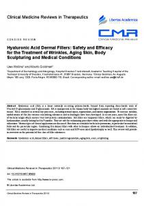
Figure 3. (a) Dually functionalized HA derivative 3 undergoes simultaneous carbazone coupling to PVA−DOX prodrug 4 and disulfide cross-linking with pyridyl disulfide-derivatized HA 6. Washing of the formed network removes the nonbound prodrug 4 which is followed by the hydrogel disassembly with dithiothreitol (DTT). DOX molecule is designated as a sphere with a label D, HA chains are shown in black and PVA chains are shown in brawn. (b) Image of the disulfide network with the carbazone-linked PVA−DOX prodrug. (c) Absorbance at 480 nm of the solutions obtained after repeated hydrogel washings in certain intervals of time. Black curve corresponds to the (HA−al−SH + 4 + 6)-hydrogel and red curve corresponds to the (HA−SH + 4 + 6)-hydrogel. (d) Image of the disassembled network.
Only a small portion of carbazate groups was used for linking of DOX to the PVA backbone in order to permit the rest of the carbazate groups to be engaged in the following chemical attachment to the aldehyde groups of the dually modified HA 3. Thiol groups of 3 were subsequently designed for the formation of a disulfide network as a result of a thiol−disulfide exchange reaction. For this purpose, HA derivative 6 carrying pyridyl disulfide groups was prepared from native high molecular weight HA (MW 150 kDa) by partial (10%) amidation of the HA carboxylate groups with 2-(2pyridinyldithio)ethyl hydrazinecarboxylate 5 (Scheme 2). Functionalization with the pyridyl disulfide groups was confirmed by 1H NMR (Figure 2). Particularly, proton signals at 8.5, 8.0, and 7.5 ppm are characteristic for the protons a, b, and c of pyridyl disulfide pendant groups. Double chemoselective reactivity of HA derivative 3 was then demonstrated by consecutive addition of the polymeric components 4 and 6 to 3 in PBS-buffered solution (Figure 3a). A hydrogel (2.9 wt % of HA) was formed in 15 min after mixing of all three components (Figure 3b). Elastic modulus (G′) of the hydrogel after 24 h of setting was 107 ± 11 Pa (Figure S5A of Supporting Information). After swelling the hydrogel in PBS, its elastic modulus decreased to 21.5 ± 6.8 Pa (Figure S5B of Supporting Information). This result is expected for a soft hydrogel such as the one formed from the low molecular weight HA derivative 3. The disulfide nature of the network cross-links was confirmed by substituting the dually modified HA 3 to its monofunctional analogue containing only 2% of aldehyde groups (it has been synthesized from native HA of MW 7.5 kDa using only dihydrazide linker 1, see Supporting
Information). When HA−al was mixed with 4 and 6 in PBS, no gel was formed even after 24 h of incubation. We have also verified the covalent attachment of the PVA− DOX prodrug 4 to the disulfide network by conducting another control experiment in which dually modified HA 3 was substituted with its monofunctional analogue containing only 25% of sulfhydryl groups (HA−SH, see the structure and synthesis details in Supporting Information). After preparation of the two disulfide networks, (HA−al−SH + 4 + 6) and (HA− SH + 4 + 6), they were incubated in PBS buffer that has been later exchanged into a fresh one. This washing cycle was repeated 7 times over 24 h. The unreacted prodrug 4 was expected to be washed out and subsequently detected in the washing solutions. Figure 3c shows quantification of DOX as a function of washing time examined by UV−vis spectroscopy. While the absorbance for the washed out DOX containing molecules reached 80% of the whole DOX amount loaded upon entrapment of the prodrug 4 in the (HA−SH + 6) hydrogel, the release of DOX species from the (HA−al−SH + 6) hydrogel was only 30% complete after 24 h. A portion of the washed out DOX species should originate from the hydrolytic release of the free DOX from both the network-linked and unlinked prodrug 4 during the time of the hydrogels incubation. According to our release experiments performed on soluble DOX conjugates in PBS (see below), it accounts for 10−15% of DOX release. Subtracting this value from the above 30 and 80%, the difference in binding capacity for HA−al−SH and HA−SH toward the prodrug 4 becomes even more evident. This clearly demonstrates the formation of carbazone linkages between the prodrug 4 and the dually functionalized HA 3. It is worth noting that both the carbazone coupling and 4110
dx.doi.org/10.1021/ma400543u | Macromolecules 2013, 46, 4105−4113
Macromolecules
Article
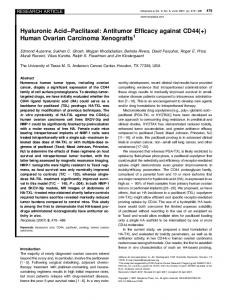
Figure 4. (a) 1H NMR spectrum of pyrene−HA−PVA−DOX 8 in D2O. The conjugate 8 was obtained upon simultaneous linking of PVA−DOX prodrug 4 and hydrophobic pyrene reagent 7 to the dually modified HA 3. Parallel carbazone coupling and thiol−disulfide exchange reactions are presented by red and blue arrows, respectively. (b) UV−vis spectra of 8 (red curve) and 25 μM pyrene (blue curve). Inset: magnified region from 270 to 700 nm showing also the absorbance of 10 μM DOX hydrochloride (green curve) for comparison. (c) TEM image of the nanoassemblies that are formed by the conjugate 8 in an aqueous medium.
dissolved hydrogel was first dialyzed against DMSO followed by repeated dialysis against water (2 × 30 min) and the dialyzed mixture was examined by 1H NMR, which confirmed the presence of the HA−PVA−DOX product (Figure S6 of Supporting Information). One Pot Synthesis of HA Nanoparticles with the Attached PVA−DOX Prodrug. Having demonstrated the success of the methodology for the production of a hydrogellinked prodrug for local therapies, we anticipated that dual clickable HA 3 can be used as a starting material to prepare multifunctional nanoparticles. Pyridyl disulfide derivative of pyrene 7 and PVA−DOX prodrug 4 were employed for linking to 3 via simultaneous thiol−disulfide exchange and carbazone coupling reactions in NMP (Figure 4a). The pyrene molecules were appended to the HA backbone to provide hydrophobic self-association of the resulting amphiphilic conjugate in an aqueous medium and at the same time to label the associates with an individual fluorescent tag (excitation and emission wavelengths of pyrene are 320 and 385 nm, while for DOX they are 488 and 590 nm respectively). The presence of all structural components (HA, PVA, pyrene, and DOX) in the obtained conjugate was confirmed by 1H NMR analysis (Figure 4a). By comparing integral of the pyrene peaks appeared between 7.2 and 8.6 ppm with the integral for the HA acetamide protons (2.0 ppm) we found the degree of modification of HA repeating units with pyrene groups (22.8%). UV−vis spectrometry was also used for quantification of pyrene grafting (Figure 4b). For this purpose, the NPs were disassembled by dispersing them in a mixture of PBS, methanol and DMSO which afforded a transparent solution. In this case,
thiol−disulfide exchange reactions were performed in situ under the same reaction conditions that are compatible for cell encapsulation. Release of HA-Functionalized Prodrug by ThiolTriggered Hydrogel Dissolution. The advantage of using thiol−disulfide cross-linking chemistry is that the formed network can be disassembled specifically under mild conditions by using reagents such as dithiothreitol (DTT). Such reduction-sensitive cleavage of the disulfide bond is useful in the design of bioresponsive biomaterials for drug delivery,27 since glutathione is an important tripeptide antioxidant that is commonly found within cells at millimolar concentrations.28 For example, copolymers with disulfide linked blocks were synthesized to self-associate into micelles29 and corresponding hydrogels in an aqueous solution. These biomaterials composed of bioreducible hydrophobic30,31 and hydrophilic domains32 can serve as water swollen vehicles for hydrophobic drugs and provide stimulus-responsive release of the drugs. On the other hand, the presence of independent easily cleavable linkage can be used in separation techniques for rapid isolation of complex bioconjugates from the parent macromolecules. In the present context, the disulfide network can be viewed as a phase that is separable from a swelling solution of the unbound molecules. For the proof-of-concept, the (3 + 4 + 6)-hydrogel was swollen in PBS containing 30 mM DTT after washing cycles. As expected, the hydrogel liquefied in 30 min (Figure 3d), additionally proving the disulfide nature of the cross-links. Such controlled network disassembly should liberate 3−4 conjugate, in which PVA backbone is linked to both DOX and HA through carbazone bonds (HA−PVA−DOX, Figure 3a). The 4111
dx.doi.org/10.1021/ma400543u | Macromolecules 2013, 46, 4105−4113
Macromolecules
Article
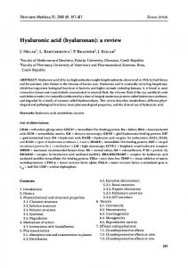
pyrene ligands did not form a hydrophobic environment which can in turn affect the pyrene spectroscopic properties. The same solvent composition has been used to dissolve not conjugated pyrene at different concentrations and obtain a linear calibration curve (Figure S7 in Supporting Information). The spectrophotometric determination revealed that 22.5% of all HA disaccharide units were modified with the pyrene ligands which was close to the value obtained from 1H NMR analysis. The formed pyrene−HA−PVA−DOX conjugate 8 selfassociates in an aqueous medium with the formation of nanoparticles according to dynamic light scattering (DLS) measurements (Figure S8A of Supporting Information). TEM studies shed light on the morphology of the forming particles. It appeared that association of py−HA−PVA−DOX conjugate 8 affords nanovesicles with average diameter of around 200 nm (Figure 4c). Formation of vesicular nanostructures can be envisaged by stacking of pyrene ligands around some imaginary sphere, thus delimiting inner and outer areas of the vesicles. HA chains may thread through the pyrene-made hydrophobic sphere (in average four pyrene ligands are attached per one HA chain) and provide stabilization of the lipid-like spherical layer. Additionally, these nanovesicles can be stabilized by means of PVA chains which covalently cross-link several HA chains. DOX molecules are in turn linked to the PVA chains and, therefore, are likely localized in the hydrophobic layer of the vesicles. Together with the nanovesicles, smaller nanoparticles were also observed (Figure S8B of Supporting Information) which could be micelles of hydrophobic core−hydrophilic shell structure. Polydispersity of functionalization of the dually modified HA 3 with aldehyde and thiol groups can be the reason for that not all PVA macromolecules were coupled to HA. The unbound PVA−DOX may therefore self-assemble into smaller micelles. In Vitro Release Study and Interaction of DOX-Linked Carriers with Cells. Attachment of the PVA−DOX prodrug 4 to HA and simultaneous grafting of HA with hydrophobic pyrene groups led to a drastic change in the in vitro release of the carbazone-linked DOX. Two trends were noteworthy: (1) Release of DOX from polymeric PVA was faster as compared with NPs 8. (2) For both types of carriers (4 and 8), the release of DOX proceeded more efficiently at acidic pH than at neutral conditions. The last observation is in accordance with the acidic lability of the carbazone bond. It therefore confirms that the drug was indeed linked chemically to the both types of the carriers. In our previous study we showed that hydrophobic and/or electrostatic loading of DOX into pyrene-grafted HA NPs resulted in the consequent rapid drug release within 5 h.21 Covalent carbazone coupling allowed increase of the release time by at least 10 times (Figure 5). We have observed the release of DOX from both types of carriers over 100 h of incubation of the conjugates. After this time, 85 ± 3% and 39 ± 10% of DOX was released from the PVA−DOX prodrug 4 at pH 5 (red solid curve in Figure 3) and pH 7.2 (red dashed curve in Figure 5) respectively. The corresponding values for the NPs 8 were 50 ± 5% (pH 5, blue solid curve in Figure 5) and 19 ± 3% (pH 7.2, blue dashed in Figure 5). Two reasons may be responsible for the increased retention of DOX in NPs 8. Apart from the covalent bonding to PVA, positively charged DOX molecules become additionally bound in 8 (i) through electrostatic interactions to HA polyanion as well as (ii) through hydrophobic and aromatic stacking interactions with the pyrene ligands appended to HA. Several studies indicated stacking interactions between pi electron
Figure 5. Profiles of DOX release from PVA−DOX prodrug 4 (red curves) and from the core−shell pyrene−HA−PVA−DOX NPs 8 (blue curves) at pH 5 (solid lines) and pH 7.2 (dashed lines).
systems of doxorubicin and other polycyclic aromatic molecules.33 Thus, methylxanthines were shown to protect cells against cytotoxic effect of DOX due to the formation of mixed aggregates with anticancer aromatic drugs, such as daunomycin, doxorubicin and mitoxantrone.33 Aromatic association of DOX with both inner and outer surfaces of carbon nanotubes have been widely studied for the delivery of the drug by nanoparticles.34 In addition, the amino sugar of DOX has a pKa value of 8.6, which confers alkaline properties onto this molecule and positive charge at neutral pH.35 Because of the positive charge of DOX, it associates at a high level with various anionic polymers such as poly(acrylic acid), γpolyglutamic acid, polyaspartate, and polyglutamate.36,37 We have also observed that conjugating of the PVA−DOX prodrug 4 to the pyrene-appended HA macromolecules endowed the prodrug with the enhanced uptake by CD44 expressing cells. MDA-MB-231 cells that overexpress CD44 receptor and MCF-7 cells that express CD44 to a low extent were used in this study. The cells were incubated with the druglinked carriers at the same DOX concentration (1.2 μg/mL) for 3, 6, and 24 h. Fluorescence microscopy studies revealed that the PVA−DOX prodrug 4 was not taken up by both type of cells after 3 and 6 h of incubation as was judged from the observation of DOX fluorescence emission at 590 nm (DOX imaging channel) (Figure 6a,b). On the contrary, NPs 8 were shown to be able to differentiate between the two types of cells. MDA-MB-231 cells were showing DOX fluorescence (Figure 6c), whereas the fluorescence in MCF-7 cells was sufficiently diminished (Figure 6d). The interaction of NPs 8 with the cells
Figure 6. Fluorescence microscopy images of CD44-overexpressing MDA-MB-231 cells (a, c) and CD44-underexpressing MCF-7 cells (b, d) incubated for 3 h with PVA−DOX prodrug 4 (a, b) and the NPs 8 (c, d). 4112
dx.doi.org/10.1021/ma400543u | Macromolecules 2013, 46, 4105−4113
Macromolecules
Article
was also observed by monitoring the fluorescence of pyrene at 380 nm (pyrene/DAPI imaging channel). It is noteworthy that pyrene fluorescence of the incubated cells was colocalized with the fluorescence corresponding to DOX molecules. Therefore, it can be concluded that HA plays an important role in differentiating the cells expressing CD44 to a different extent. Cellular studies aimed at the elucidation and quantification of the NPs uptake is under the progress and will be communicated elsewhere.
(3) Thomas, B.; Fiore, M.; Bossu, I.; Dumy, P.; Renaudet, O. Beilstein J. Org. Chem. 2012, 8, 421−427. (4) Bark, S. J.; Schmid, S.; Hahn, K. M. J. Am. Chem. Soc. 2000, 122, 3567−3573. (5) Mutter, M.; Dumy, P.; Garrouste, P.; Lehmann, C.; Mathieu, M.; Peggion, C.; Peluso, S.; Razaname, A.; Tuchscherer, G. Angew. Chem., Int. Ed. 1996, 35, 1482−1485. (6) Kochendoerfer, G. G.; et al. Science 2003, 299, 884−887. (7) Ghosh, S.; Basu, S.; Thayumanavan, S. Macromolecules 2006, 39, 5595−5597. (8) An, Z.; Tang, W.; Wu, M.; Jiao, Z.; Stucky, G. D. Chem. Commun. 2009, 6501−6503. (9) DeForest, C. M.; Polizzoti, B. D.; Anseth, K. S. Nat. Mater. 2009, 8, 659−664. (10) Wylie, R. G.; Ahsan, S.; Aizawa, Y.; Maxwell, K. L.; Morshead, C. M.; Shoichet, M. S. Nat. Mater. 2011, 10, 799−806. (11) Kempe, K.; Hoogenboom, R.; Jaeger, M.; Schubert, U. S. Macromolecules 2011, 44, 6424−6432. (12) Schoch, J.; Staudt, M.; Samanta, A.; Wiessler, M.; Jäschke, A. Bioconjugate Chem. 2012, 23, 1382−1386. (13) Wang, D.-A.; Varghese, S.; Sharma, B.; Strehin, I.; Fermanian, S.; Gorham, J.; Fairbrother, D. H.; Cascio, B.; Elisseeff, J. H. Nat. Mater. 2007, 6, 385−392. (14) Ossipov, D. A.; Yang, X.; Varghese, O.; Kootala, S.; Hilborn, J. Chem. Commun. 2010, 8368−8370. (15) Chang, P. V.; Prescher, J. A.; Hangauer, M. J.; Bertozzi, C. J. Am. Chem. Soc. 2007, 129, 8400−8401. (16) Yi, L.; Sun, H.; Itzen, A.; Triola, G.; Waldmann, H.; Goody, R. S.; Wu, Y.-W. Angew. Chem., Int. Ed. 2011, 50, 8287−8290. (17) Zöller, M. Nature Rev. Cancer 2011, 11, 254−267. (18) Girish, K. S.; Kemparaju, K. Life Sci. 2007, 80, 1921−1943. (19) Platt, V.; Szoka, F. C., Jr. Mol. Pharmaceutics 2008, 5, 474−486. (20) Ossipov, D. A. Expert Opin. Drug Delivery 2010, 7, 681−703. (21) Yang, X.; Kootala, S.; Hilborn, J.; Ossipov, D. A. Soft Matter 2011, 7, 7517−7525. (22) Varghese, O. P.; Sun, W.; Hilborn, J.; Ossipov, D. A. J. Am. Chem. Soc. 2009, 131, 8781−8783. (23) Varcruysse, K. P.; Marecak, D. M.; Marecak, J. F.; Prestwich, G. D. Bioconjugate Chem. 1997, 8, 686−694. (24) Shu, X. Z.; Liu, Y.; Luo, Y.; Roberts, M. C.; Prestwich, G. D. Biomacromolecules 2002, 3, 1304−1311. (25) Kaneko, T.; Willner, D.; Mankovic, I.; Knipe, J. O.; Braslawsky, G. R.; Greenfield, R. S.; Vyas, D. M. Bioconjugate Chem. 1991, 2, 133− 141. (26) Kratz, F. Top. Curr. Chem. 2008, 283, 73−97. (27) Meng, F.; Hennink, W. E.; Zhong, Z. Biomaterials 2009, 30, 2180−2198. (28) Carelli, S.; Ceriotti, A.; Cabibbo, A.; Fassina, G.; Ruvo, M.; Sitia, R. Science 1997, 277, 1681−1684. (29) Sun, H.; Guo, B.; Li, X.; Cheng, R.; Meng, F.; Liu, H.; Zhong, Z. Biomacromolecules 2010, 11, 848−854. (30) Sun, L.; Liu, W.; Dong, C.-M. Chem. Commun. 2011, 47, 11282−11284. (31) Liu, D.-L.; Chang, X.; Dong, C.-M. Chem. Commun. 2013, 49, 1229−1231. (32) Li, C.; Madsen, J.; Armes, S. P.; Lewis, A. L. Angew. Chem., Int. Ed. 2006, 45, 3510−3513. (33) Piosik, J.; Gwizdek-Wiśniewska, A.; Ulanowska, K.; Ochociński, J.; Czyz, A.; Wegrzyn, G. Acta Biochim. Pol. 2005, 52, 923−926. (34) Datir, S. R.; Das, M.; Singh, R. P.; Jain, S. Bioconjugate Chem. 2012, 23, 2201−2213. (35) Angerer, J., Schaller, K. H. Analyses of Hazardous Substances in Biological Materials; Wiley-VCH: Weinheim, Germany, 2001; Vol. 7. (36) Tian, Y.; Bromberg, L.; Lin, S. N.; Alan Hatton, T.; Tam, K. C. J. Controlled Release 2007, 121, 137−145. (37) Pratesi, G.; Savi, G.; Pezzoni, G. Br. J. Cancer 1985, 52, 841− 848.
■
CONCLUSIONS In conclusion, we have demonstrated the versatility of dual functionalization of macromolecules with chemically orthogonal aldehyde and thiol groups. A carbazone coupling, involving aldehyde groups, was used to attach cell-specific HA macromolecules to the neutral polymeric PVA−DOX prodrug. Simultaneously, the carbazone coupling was accompanied by a thiol−disulfide exchange reaction which transformed the forming macromolecular conjugate into either the 3-D network or to the self-associating nanostructures. The macroscopic network can be separated from the nonconjugated species and subsequently disassembled serving as a temporary separation template. On the other hand, we have shown that the drug release profile of the neutral polymeric prodrug can be changed upon anionic bioconjugation and nanostructuring. Resulting HA-presenting nanocarriers can selectively recognize CD44expressing cells.
■
ASSOCIATED CONTENT
S Supporting Information *
Synthesis procedures for PVA−carb, HA−al, and HA−SH; NMR spectra of intermediates of aldehyde−thiol dually modified HA 3 (Figures S1 and S2); standard UV−vis and fluorescence emission calibration curves for PVA−DOX 4 (Figure S3); NMR spectra of PVA−DOX 4 and HA−PVA− DOX obtained after dissolution of (3 + 4 + 6)-hydrogel (Figures S4 and S6); standard UV−vis calibration curves for pyrene (Figure S7); DLS measurement and TEM image of NPs 8 (Figure S8). This material is available free of charge via the Internet at http://pubs.acs.org.
■
AUTHOR INFORMATION
Corresponding Author
*(D.O.) E-mail:
[email protected]. Telephone: +46 18 4717335. Author Contributions
The manuscript was written through contributions of all authors. All authors have given approval to the final version of the manuscript. Notes
The authors declare no competing financial interest.
■
ACKNOWLEDGMENTS This work was supported by the European Community′s Seventh Framework Programme (BIODESIGN project).
■
REFERENCES
(1) Iha, R. K.; Wooley, K. L.; Nyström, A. M.; Burke, D. J.; Kade, M. J.; Hawker, C. J. Chem. Rev. 2009, 109, 5620−5686. (2) Galibert, M.; Renaudet, O.; Dumy, P.; Boturyn, D. Angew. Chem., Int. Ed. 2011, 50, 1901−1904. 4113
dx.doi.org/10.1021/ma400543u | Macromolecules 2013, 46, 4105−4113