© 2001 OPA (Overseas Publishers Association) N.V. Published by license under the Harwood Academic Publishers imprint, part of Gordon and Breach Publishing, a member of the Taylor & Francis Group.
Free Rad. Res., Vol. 35, pp. 119-128
Free Radic Res Downloaded from informahealthcare.com by Radboud Universiteit Nijmegen on 10/27/14 For personal use only.
Reprints available directly from the publisher Photocopying permitted by license only
Oxidative Stress Induces the Expression of the Major Histocompatibility Complex in Murine Tumor Cells MARIA R. OLWA a, ANTONIO IRADIb, FEDERICO GARRIDOc, MERCEDES RAMOSa, ANA MARIA OLTRAa, PILAR MUlqIZ a and GUILLERMO T. S~kEZb'* aDepartamento de Bioquimica y Biologia Molecular; bDepartamento de Fisiologia, Facultad de Medicina, Universitat de Valencia; CServicio de Andlisis Clfnicos, Hospital Virgen de las Nieves, Granada
Accepted by Prof. J.V. Bannister (Received 15 June 2000; In revisedform 26 October 2000) Keywords: MHC class I antigens, fibrosarcoma, 8-OHdG, oxidative stress, NF~B
The effect of t-butyl hydroperoxide (t-BOOH) on the induction of the Major Histocompatibility Complex (MHC) class I genes has been studied in two cell clones (B9 and G2) of the methylcholanthreneinduced murine fibrosarcoma GR9. These two clones were selected based on their different biological and biochemical behavior specially related to their tumor induction capability when injected into a BALB/c mouse, t-BOOH (0.125mM) induced the expression of H-2 molecules in both cell clones. In B9 cell clone, in which MHC basal expression is very low or absent, t-BOOH significantly induced H-2Kd, H-2Da and H-2La molecules. In G2 cell clone the expression of MHC class I genes was also enhanced by the xenobiotic, the effect being especially significant on the H-2La molecule which is not expressed under basal conditions. H-2 molecules expression was accompanied by the activation of the transactivator factor NF~B. These results suggest that oxidative stress may modulate the antigen expression of tumor cells and thus the immune response of the host organism. Basal levels of oxidative parameters, such as antioxidant enzymes, malondialdehyde (MDA) and the DNA damaged base 8-hydroxy-2'-deoxyguanosine (8-OHdG), showed differences between the two fibrosarcoma cell clones.
INTRODUCTION During the past years, a great a m o u n t of information has been reported supporting the involvement of reactive oxygen species (ROS) in the regulation of metabolism and gene expression. [1'21 It has also been s h o w n that m o d e r a t e concentrations of intracellular ROS m a y influence the molecular m a c h i n e r y of gene expression and the translational modification of proteins b y different mechanisms. [31 Important contributions in relation to the regulatory mechanisms involved in the adaptation of cells to oxidative stress, [4"51 together with the expression of different proto-oncogenes, h e m e oxygenase, apoptotic, g r o w t h arrest and D N A d a m a g e genes b y intracellular redox m o d u l a t i o n have
*Corresponding author. Tel.: 34-6-3864160.E-mail:
[email protected]. 119
Free Radic Res Downloaded from informahealthcare.com by Radboud Universiteit Nijmegen on 10/27/14 For personal use only.
120
M.R. OLIVAet aL
been reported I6-14] and have been of great value for the understanding of the signal transduction pathways underlying the biological response to oxidative stress. In addition, two well-defined transcription factors, nuclear factor-nB (NFnB) and activator protein-1 (AP-1), have been implicated in the inducible expression of different genes triggered by changes of the redox state of the cells. I2I NFnB is a multisubunit transactivator factor that can rapidly activate the expression of genes involved in inflammatory, immune and acute phase responses I15I such as the MHC class I genes, whose expression in tumor cells under oxidative stimulus, has not been reported before. MHC class I genes represent a set of genes that synthesize products specializing in the processing and presentation of endogenous or exogenous antigens to the immune system. Due to the central role of MHC molecules in antigen presentation including tumor antigens, [16"17] any alteration in MHC class I gene expression may have profound implications in tumor development. Indeed, in a high percentage of human and murine tumors screened, complete locus [18'191 a n d / o r allelic losses of HLA and H-2 expression have been described and recently reviewed. I2°1 These facts prompted us to investigate the possible susceptibility of the histocompatibility antigens to an oxidative stress-induced modulation. We used as an experimental system, two cell clones from a well-characterized tumor model, GR9, which is a fibrosarcoma originated in a BALB/c mouse by subcutaneous (s.c.) injections of 0.2 mg methylcholanthrene.E211 Clonal heterogeneity of the chemical-induced GR9 fibrosarcoma has been described showing that different derived clones express quantitative variations of H-2 class I antigens. These variations range from H-2-negative clones to clones with strong expression while their local tumorigenicity or metastatic capacity has also been defined. I221 We used two GR9 derived cell clones which were selected based on the different pattern of
H-2 expression and on their tumorigenic potential in BALB/c mice. The GR9-B9 clone (H-2d) negative and the GR9-G2 (H-2 d) positive have been used in this study and will be referred to in the text as B9 and G2 respectively. B9 and G2 clones have been characterized from an oxidative metabolism point of view and compared with normal BALB/c3T3 cells. The reported results show first evidence of the induction of H-2 antigen expression (H-2K d H-2D d and H-2L d) in two fibrosarcoma derived clones by t-BOOH. This effect is supported by the activation of NFnB by the xenobiotic with significant differences, in terms of the degree of antigens expression and time required, when compared with that observed with v-interferon (-y-INF).
MATERIALS AND METHODS Reagents
Superoxide dismutase (SOD), catalase (Cat.) and glutathione peroxidase (GPx) used as standards for the respective enzyme analysis were purchased from Boehringer (Mannheim, Germany). Cytochrome c, xanthine oxidase, EDTA, acrylamide/bisacrylamide, 2'-deoxyguanosine, SDS, bovine serum albumin (fraction V) and t-BOOH were from Sigma Chemical Co. (St. Louis, MO, USA). Reduced glutathione (GSH) was from Merck (Darmstadt, Germany). MoAbs and cell lines were obtained from the suppliers indicated below. Tissue culture medium and fetal bovine serum was from Gibco-BRL. Other reagents were of the highest purity available obtained from Chemical Companies.
Cell Lines, Culture Conditions and Analysis of Oxidative Stress Parameters
We used, in this work, two clones from the chemically-induced fibrosarcoma GR9, which was induced by s.c. injection of 0.2mg of
Free Radic Res Downloaded from informahealthcare.com by Radboud Universiteit Nijmegen on 10/27/14 For personal use only.
EFFECTOF t-BOOHON MHC ANTIGENS methylcholanthrene into the flank of a BALB/c mouse. The tumor was adapted to tissue culture with no in vivo passage to avoid immuno selection, and further submitted to cell cloning. [211 GR9 clones were determined in terms of their H-2 d antigen expression (K d, D a and L d molecules). Where indicated, non-tumorogenic BALB / c3T3 fibroblasts from the American Type Culture Collection (ATCC), Rockville, MD, were used in this study. Cells were grown in monolayers at 37°C in a 5% CO2 atmosphere and RPMI supplemented with 10% FCS, glutamine and antibiotics was used as culture medium. [2~j Cells grown until 90% confluency were washed twice with ice-cold PBS and collected by the addition of 5 ml 0.2% EDTA solution. Cell suspensions were transferred to plastic disposal tubes and sonicated for 2rain at 15 sec bursts, placed on an ice-containing tray and followed by 5 min centrifugation at 13,000r.p.m. Appropriate aliquots were obtained for the respective analytic assays.
Analyses of Oxidative Stress-related Parameters SOD activity was measured according to the method of McCord and Fridovich I231 based on the production of superoxide radicals during the conversion of xanthine to uric acid by xanthine oxidase and the inhibition of cytochrome c reduction. One unit of SOD activity was defined as the amount of SOD that produces 50% inhibition of cytochrome c reduction. Cat. and GPx activity were determined following the methods of Clairbone I24] and of Gunzler and Flohe [251 respectively. GSH content of cells was determined using previously described assay. I261 For the analysis of oxidized glutathione (GSSG) samples were treated with N-ethylmaleimide and bathophenanthroline disulfonic acid and derivatized and analyzed by HPLC as previously described. [27"2s!Malondialdehyde was measured by HPCL. I29! The protein content was measured by the Bradford method. [3°1
121
DNA Isolation and Enzymatic Digestion Cell DNA was isolated following the method of Gupta [3q with the modification described by Mufiiz et al. [32I in which chloroform isoamyl alcohol (24:1) is used instead of phenol for the removal of proteins. [331 Isolated DNA was washed twice with 70% ethanol, dried and dissolved in 200 ~tl of 10 mM Tris/HC1, 0.1 mM EDTA, 100 mM NaC1, (pH 7.0) for its enzymatic digestion, as previously described. [341 In brief, 0.5 ~g DNA/~I was incubated with 100 units of DNase I in 40 ~tl Tris/ HC1 (10 mM and 10 ~1) of 0.5 M MgCI2 (the final concentration of 20mM) at 37°C for I h. The pH of the reaction mixture was then lowered with 15~1 of sodium acetate 0.5M (pH 5.1); 10~tl of nuclease P1 (5 units) and 30~1 of 10raM ZnSO4 (to give a final concentration of I mM) were added, and mixture incubated for l h. After readjusting the pH with 100 ~1 of 0.4M Tris/ C1H (pH 7.8) followed by the addition of 20 ~tl alkaline phosphate (3 units), the samples were incubated for 30min. Enzymes were precipitated with acetone (5 V), removed by centrifugation, and the supernatant evaporated to dryness.
8-OHdG Assay The DNA hydrolysates were disolved in HPLC grade water and filtered through a 0.2%tm syringe filter before applying the samples to a Waters ODS HPLC column (2.5 x 0.46 i.d.; 5 ~t particle size). The amount of 8-OHdG and dG in the DNA digest was measured by electrochemical and UV absorbance detection respectively, under the elution conditions previously described. [321 Standard samples of dG and 8-OHdG were analyzed to assure their good separation and to allow identification of those derived from cell DNA.
Analysis of H-2 Expression Cells from B9 and G2 clones were incubated with the conditions described above with and
Free Radic Res Downloaded from informahealthcare.com by Radboud Universiteit Nijmegen on 10/27/14 For personal use only.
122
M.R. OLIVAet al.
without 0.125mM t-BOOH disolved in RPMI culture medium 4 h previously to H-2 expression analysis. Surface H-2 class I expression was determined with a standard method of indirect immunofluorescence. Cells (105) were washed twice in phosphate-buffered saline (PBS) and incubated for 30rain at 4 °C with the monoclonal antibodies (MoAb) anti-H-2KdD d (34.1.2), antiH-2D d (34.5.8) and anti-H-2L d (24.14.8) obtained from the ATCC. Cells were washed twice in ice-cold PBS and incubated with a 1:40 dilution of FITClabeled rabbit anti-mouse immunoglobulin (Cappel, West Chester, PA). After washing, fluorescence of cells was measured on a FACSort flow cytometer (Becton Dickinson, San Jose, CA). Ten thousand events were collected and analyzed.
NFlcB Synthetic Oligonucleotides Oligonucleotides containing the NF~:B site [351 were synthesized with a System 200A DNA synthesizer (Beckman Instruments) and annealed. The duplex sequences are: TCGACAGAGGGACTTTCCGAGAGG and TTCCCTGAAAGGGTACTAG. This oligonucleotide probe was labelled by filling in both ends with o~32p dATP using Klenow fragment.
Cellular Extracts and Gel Mobility Shift Assay Nuclear extracts in basal conditions and after incubation with 0.125 mM t-BOOH for 4h and 1,000 U / m l -y-interferon (Amersham, Little Chalfont, UK) for 72 h, were prepared essentially as described by Dignam et al. [361with minor modifications [371 from nuclei isolated from cells lysed in lysis buffer. The extracts were stored as aliquots at - 8 0 °C. Protein concentration in nuclear extracts was determined with the BioRad (Richmond, CA) protein assay kit. A typical 20 ~tl reaction involved: 4 ~g of nuclear protein, 1 ~tg of poly(dIdC) (Pharmacia, Uppsala, Sweden), 10mM Tris-HC1 (pH 7.5), 50raM NaC1, l m M
EDTA, i mM dithiothreitol, 5% glycerol and 20,000 cpm probe (0.05 ng). In competition experiments 50 or 100 ng of double stranded "cold" competitor oligonucleotide was added to the reaction 5 min before the addition of the probe. All reactions were carried out at room temperature. Reactions were electrophoresed through 4% low ionic strength polyacrylamide gels (29:1), 0.4 x TBE (1 x TBE = 0.089 M Tris base, 0.089 M boric acid, 0.93g/1 EDTANa2), fixed in 10% acetic acid, dried and exposed on XAR film (Kodak) at - 8 0 °C.
Statistics The statistical significance was assessed by Student's t-test.
RESULTS Oxidative Stress-related Metabolites in Fibrosarcoma Derived Clones and BALB/c3T3 Fibroblasts Three different cell types, two oncogenic, B9 and G2, and BALB/c3T3 normal fibroblasts were used in these experiments whose results are shown in Table I. There are significant differences comparing normal fibroblasts with G2 fibrosarcoma cell clone. Enzyme activities are significantly higher in G2 than in BALB/c3T3 fibroblasts (Table I). The percentage increases of catalase, SOD and GPx in G2 cell clone compared with control 3T3 cells were 141%, 54% and 38% respectively. In B9 cell clone the antioxidant activities are similar to those of normal flbroblasts except for GPx which is significantly lower. There are also differences in the antioxidant enzyme activities between G2 and B9 cell clones. These are significantly lower in B9 cells. Both reduced and oxidized glutathione are significantly higher in G2 cells than in normal fibroblasts or B9 cells (Table II). GSH concentration in G2 cells is two-fold higher than
EFFECT OF t-BOOH ON MHC ANTIGENS TABLE I Comparative study of antioxidant enzymes in BALB/c3T3, G2, and B9 cells Cell line
BALB/c3T3
G2
B9
Free Radic Res Downloaded from informahealthcare.com by Radboud Universiteit Nijmegen on 10/27/14 For personal use only.
Cat. (U/gProt.) 1.2 :~ 0.5 (14) 2.9 + 1.2 (20)* 1.3 + 0.5 (10) # SOD (U/mgProt.) 5.7 ± 1.7 (15) 8.8 + 2.7 (10)* 6.3 i 0.8 (9) ## GPx (U/gProt) 5.0±2.6 (16) 6.9+1.2 (19)* 2.4±1.1 (8) **'# Results are mean ± S.D. with the number of experiments in parentheses. Assays were performed on individual cell culture plates and the mean of duplicate analysis were used for statistical calculations. *p < .005 and **p < .01 comparing G2 and B9 with BALB/ c3T3 cells. #p < .005 and ##p < .05 comparing G2 with B9 fibrosarcoma cells.
in BALB/c3T3 or B9 cells. The amount of GSSG in G2 cells is only 1.4-fold higher than in normal fibroblasts or B9 cells. Thus a lower GSSG/GSH ratio is calculated for the G2 cells in contrast with the values of 1.16 and 1.27 for 3T3 and B9 cells respectively. The oxidation product 8-OHdG is higher in G2 and B9 clones as compared with BALB/ c3T3 fibroblasts and there are no significant differences between the two tumorogenig cell lines. However, the degree of lipid peroxidation, as assessed by MDA levels, is higher in G2 cells than 3T3 or B9 cells.
123
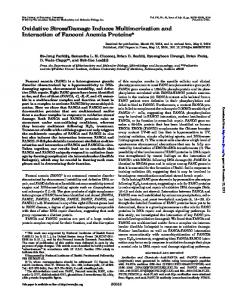
sorter (FACS) analysis using monoclonal antibodies (MoAbs) recognizing H-2D d, H-2K d and H-2L d (Figure 1). A representative example of 8-10 experiments is shown. 0.125mM t-BOOH induces the expression of H-2 class I antigens in both, G2 and B9 clones with differences worthwhile pointing out. As can be observed, in G2 clone treated with the xenobiotic, the percentage of fluorescent cells exposed to anti-H-2K d and anti-H-2D d antibodies is slightly higher than in control cells. However, the same clone increases the expression of the H-2L d molecule from a basal value of 3-40% (92% increase) after 4h incubation in the presence of t-BOOH. In B9 clone cells, presenting under basal conditions, a lack in the expression of H-2K a H-2D a and H-2L d class I molecules, the effect of t-BOOH is more evident and significant. In the presence of the oxidant, the increases of H-2Kd, H-2D d and H-2Ld antigens were from 5% to 30%, from 15% to 46% and from 9% to 43%, representing 83%, 67% and 79% increase percentages respectively. It should be emphasized that the observed increase in the expression of H-2 class I antigens by t-BOOH runs and developes in the absence of apparent signs of cytotoxicity.
Effect of t-BOOH on the Expression of MHC Class I Antigens in GR9 Fibrosarcoma Clones
Effect of t-BOOH on the Activation of NFIcB in B9 and G2 Clones
The effect of t-BOOH on the expression of cell surface H-2 class I antigens on B9 and G2 clones was measured by fluorescence activated cell
As shown in Figure 2, t-BOOH activates the retardation in native gels of the 32p-labeled DNA probe encompassing the decameric NF~B
TABLE II Comparative study of GSH status and the levels of 8-OHdG and MDA in BALB/c3T3, G2 and B9 cells Cell line GSH (nmoles/gProt) GSSG (nmoles/gProt) (GSSG/GSH) x 100 8-0HdG/105 dG MDA nmol/gProt.
3T3 20.7 + 0.23 ± 1.16 ± 2.14 ± 0.07 +
5.33 (21) 0.08 (21) 0.40 (21) 0.61 (7) 0.006 (15)
G2 46.2 • 0.33 + 0.71 ± 3.82 + 0.25 +
12.8 0.09 0.20 1.63 0.10
B9 (7)*** (7)*** (7)* (8)* (16)***
19.4 + 0.25 + 1.27 ± 3.13 + 0.06 +
5.40 0.12 0.49 0.75 0.02
(10)### (10) ~ (10) #~ (8)# (21) ~u~
Results are mean i S.D. with the number of experiments in parentheses. Assays were performed on individual cell culture plates and the mean of duplicate analysis were used for statistical calculations. For 8-OHdG, each DNA sample was assayed twice by HPLC-EC detection as described in the Methods section.*p < .05, **p < .01 and ***p< .005 comparing values in BALB/c3T3 cells with G2 and B9 cells. #p < .05, ##p < .01 and ~ p < .005 comparing values in G2 with B9 cells.
124
M.R. OLIVA et al. It-
[t
Free Radic Res Downloaded from informahealthcare.com by Radboud Universiteit Nijmegen on 10/27/14 For personal use only.
....
$-
m,
K~ B9 Basal: 5% B9 IBOOH: 30%
D~ B9 Basal: 15% B9 tBOOH: 46%
~/. Ka G2 Basal: 65% G2 tBOOH: 78%
Lj B9 Basal: 9% B9 IBOOH: 43%
A
""iiI ..... i~ ..... ~ ..... i'¢t De G2 Basal: 66% G2 tBOOH: 81%
Le G2 Basal: 3% G2 tBOOH: 40%
FIGURE 1 Effect of t-gOOH on MHC class I antigen expression in G2 and B9 murine fibrosarcoma clones. Fluorescence of cells was measured on a FACSort flow cytometer. Representative flow cytometry profiles show the effect of t-BOOH 0.125 m M (dark) by indirect i m m u n o fluorescence of B9 (upper panel) and G2 (lower panel) cell clones with the following MoAbs, 34.1.2 (Anti H-2K d D d ), 34.5.8 (Anti H-2D d ) and 28.14.8 (Anti H-2L d ). 10 3 events were collected and analyzed.
motif in both cell clones. The activation of NFnB was also shown by -y-INF in both cell clones although the incubation time required for this effect was 72 h which is in agreement with the previous observed lack time for H-2 antigen expression as will be discussed later.
DISCUSSION The purpose of the present study was to evaluate the role of oxygen reactive species in the regulation of H-2 class I expression. We used t-BOOH because of its considerable propensity for generating free radicals and electrophilic reactive intermediates. Its intracelullar decomposition by cytochrome P450 leads to the generation of free radicals including O~-, °OH, peroxyl
(LOO*) and alkoxyl (LO°) radicals. These latter radicals, peroxyl and alkoxyl, are common intermediates released from membrane phospholipids under oxidative stress conditions. [3sl The interaction of these reactive products with other cell macromolecules such as protein receptors, transcription factors or nucleic acids has been established and in so doing signal transducing pathways may be modulated in both directions, enhancement or inhibition. I1"2"391 Experiments were carried out using two cell clones (B9 and G2) derived from a well characterized fibrosarcoma. 121[ The clones were selected based on their different biological properties, H-2 class I antigen expression under basal conditions, and tumorigenic potential. I221 Analysis of oxidative stress parameters in both fibrosarcoma cell clones was performed
EFFECTOF t-BOOHON MHC ANTIGENS m
w ,e~ lID ~ 4~ ~ Ip4 g D OD CO M ; qm ~ OB
Free Radic Res Downloaded from informahealthcare.com by Radboud Universiteit Nijmegen on 10/27/14 For personal use only.
7 ~
~
gll Oil all
. . . .
!i!i!
FIGURE2 Effectof v-INF and t-BOOHon the inductionof ~-Bbindingactivityin G2 and B9 cells.Nuclearextractswere prepared from untreated (control)or after 4 h and 72h incubation in the presence of t-BOOH (0.125mM) and "¢-INF (1,000U/ml) respectively. Electrophoretic mobility shift assays were done with 0.05ng 32p-labelledprobe containing the NF~B consensus sequence. Competition experiments using50-100ng coldcompetitortogetherwithothermethodological details are specifiedin the correspondingepigraph.
and compared with those of BALB/c3T3 cells. There was a significant difference between the amount of antioxidant enzymes, especially between normal fibroblasts, where the respective activities were lower, and the G2 fibrosarcoma cell clone, presenting higher values of GSH, Cat, SOD and GPx. These differences may be attributed to a dispairment of oxygen metabolism in transformed cells [4°'411 or to the special characteristics of the tumor cell line tested.[4°,42] The antioxidant efficiency of B9 and G2 were also different. Thus, G2-clone cells presented significant higher values of GSH, Cat., SOD
125
and GPx when compared with B9 cells. In addition, GSH concentration in G2 cells was significantly (2-fold) higher when compared with the tripeptide levels in 3T3 and B9 cells. GSSG concentration in G2 cells was only 1.4-fold higher than that found in 3T3 or B9 cells. Therefore the redox status of G2 cells was more reduced showing a GSSG/GSH ratio of 0.71 in contrast with 1.16 and 1.27 obtained from 3T3 and B9 cells respectively. However, in spite of the higher GSH values in G2 cells which should represent a protection mechanisms against DNA damage and lipid peroxidation, as previously observed [321these cells showed higher levels of 8-OHdG and MDA, which are considered parameters of choice in oxidative stress processes.[ 1,43,44] We have shown that t-BOOH, increases the expression of H-2 class I antigens in both G2 and B9 fibrosarcoma cells, the effect being mediated by the activation of the transcription factor NF~B. Under basal conditions the expression of H-2K d and H-2D a is positive in G2 cells whereas in B9 cells it is negative. I21"22] These differences are difficult to explain through an oxidative stress mechanism. However, other molecular events such as differences in antigen processing and translocation, could explain the distinctive pattern of antigen expression observed between G2 and B9 cells under basal conditions. H-2 increased expression by t-BOOH was especially observed in those antigens which were low or not expressed under basal conditions i.e. H-2L a in G2 cells and H-2K d, H-2D d and H2-Ld in B9 cells. NF~B is a transcription factor which has specialized in the organism to induce the synthesis of defense and signalling proteins rapidly upon exposure of cells to a wide variety of most pathogenic agents, one of which is the xenobiotic used in this study. [4sl Here we have shown that t-BOOH induces the activation of NF~B shown by electrophoretic mobility shift assay. However, it is likely that t-BOOH derived metabolites including "OH which easily diffuses
Free Radic Res Downloaded from informahealthcare.com by Radboud Universiteit Nijmegen on 10/27/14 For personal use only.
126
M.R. OLIVA et al.
through the cell membranes, could directly activate NFnB by degrading or modifying InB in the cytoplasmic P50-p65-InB complex, a molecular mechanism allowing DNA-binding of NFnB and its translocation to the nucleus, lasl This hypothesis has been previously proposed and disccussed in the activation of NFe;B by H2021451 or TNF-derived oxygen species. [46! One of the most common characteristics of ROS effects is their rapid interactions with and modification of organic cell molecules. Consistant with this property, are also the short-timing effects of t-BOOH when compared with that of 7-INF, a well known stimulator of H-2 antigen expression and NFe;B activation. [471After 24 h of treatment "7-1NFsignificantly induces H-2 class I antigen expression at the cell surface, with expression reaching a maximun after 72h. [4s! Using the same fibrosarcoma clones, grown and incubated under the incubation conditions reported previously, we have shown that tBOOH enhances H-2 class I antigen expression and NFnB activation 4 h after treatment. Jointly considering these observations, it is concluded that t-BOOH is an efficient substrate in the induction of H-2 class I antigens. In agreement with previous reports [1"2'4s'461it is suggested that oxygen radicals acting as signal transducers or second messengers may integrate the diverse variety of NF~B-inducing effects, thus probably representing a common mechanism in the molecular machinery of gene expression. Recently the expression of MHC class II antigens by reactive oxygen species in peripheral blood dendritic cells has been reported I49] indicating that MHC antigens induction may respond to a common mechanism inplicating oxygen reactive species,
Acknowledgements We thank Miss Maria del Carmen Tormos Mufioz for her technical assistance. This work was supported by a Grant of Ftmdaci6n Ram6n
Areces (X Concurso Nacional de Ayudas a la Investigaci6n Cientifica y T6cnica 1997).
References [1] H. Sies (1991) Oxidative Stress. Oxidants and antioxidants, Academic Press, London, p. 650. [2] C.K. Sen and L. Packer (1996) Antioxidant and redox regulation of gene transcription. FASEB Journal, 10, 709-720. [3] Y. Sun and L.W. Oberly (1996) Redox regulation of transcriptional activators. Free Radical Biology and Medicine, 21, 335-348. [4] H.M. Hassan and I. Fridovich (1977) Regulation of the synthesis of superoxide dismutase in Escherichia coli. Journal of Biological Chemistry, 252, 7667-7672. [5] G.J. Finn and S. Condon (1975) Regulation of catalase synthesis in Salmonella typhimurium. Journal of Bacteriology, 123, 570--579. [6] M.F. Christaman, R.W. Morgan, F.S. lacobson and B. Ames (1985) Positive control of a regulon for defenses against oxidative stress and some heat-shock proteins in Salmonella typhimurium. Cell, 41, 753-762. [7] P. Cerutti (1985) Prooxidant states and tumor promotion. Science, 227, 375-381. [8] K. Crawford, I. Zbinden, P. Amstad and P. Cerutti (1988) Oxidant stress induces the protooncogenes c-fos and c-myc in mouse epidermal cells. Oncogene, 3, 27-32. [9] G.N. Rao, B. Lassegue, K.K. Griendling and R.W. Alexander (1993) Hydrogen peroxide stimulates transcription of c-jun in vascular smooth muscle ceils: role of arachidonic acid. Oncogene, 8, 2759-2764. [10] F.R. Collart, H. Horio and F. Huberman (1995) Heterogeneity in c-jun gene expression in normal and malignant cells exposed to either ionizing radiation or hydrogen peroxide. Radiation Research, 142, 188-196. [11] S.M. Keyse and R.M. Tyrrell (1989) Heine oxygenase is the major 32ka stress protein induced in human skin fibroblasts by UVA radiation, hydrogen peroxide, and sodium arsenite. Proceedings of the National Academy of Science of the USA, 85, 99-103. [12] S.J. Korsmeyer, J.R. Shutter, K.J. Veis, D.E. Merry and Z.N. Oltvai (1993) Bcl-2/Bax: a rheostat that regulates anti-oxidant pathway and cell death. Semin. Cancer Biology, 4, 327-332. [13] A.J. Fornace, I. Alamo and M.C. Hollander (1988) DNA damage-inducible transcripts in mammalian cells. Proceedings of the National Academy of Science of the USA, 85, 8800-8804. [14] D.R. Crawford, K.P. Leahy, Y. Wang and K.J.A. Davis (1996) Oxidative stress induces the levels of a MAFG homolog in hamster HA-1 cells. Free Radical Biology and Medicine, 21, 521-525. [15] P.A. Bauerle and D. Baltimore (1991) In Molecular aspects
of cellular regulation, hormonal control regulation of gene transcription (eds. P. Cohen and J.G. Foulkes), Elsevier/North Holland Biomedical Press, Amsterdam, pp. 409-432. [16] P.J. Bjorkman, M.A. Saper, B. Samraqui et al. (1987) Structure of human class I histocompatibility antigen, HLA-A2. Nature (London), 32% 506-512.
Free Radic Res Downloaded from informahealthcare.com by Radboud Universiteit Nijmegen on 10/27/14 For personal use only.
EFFECT OF t-BOOH ON MHC ANTIGENS [17] A. Townsend and H. Bodmer (1989) Antigen recognition by class I restricted T lymphocytes. Annual Reviews of Immunology, 7, 601-624. [18] M.A. Lopez-Nevot, F. Esteban, A. Ferron, J. Gutierrez, M.R. Oliva, C. Romero, C. Huelin, F. Ruiz-CabeUo and F. Garrido (1989) HLA class I gene expression on human primary tumors and autologous metastases: demonstration of selective losses of HLA antigens on colorectal, gastric and laryngeal carcinomas. British Journal of Cancer, 59, 221-226. [19] L.M. Torres, T. Cabrera, A. Concha, M.R. Oliva, F. RuizCabello and F. Garrido (1993) HLA class I expression and HLA class I expression and HPV-16 sequences in premalignant and malignant lesions of the cervix. Tissue Antigens, 41, 65-71. [20] F. Garrido, F. Rniz-Cabello, T. Cabrera, J.J. Perez-Villar, M. Lopez Botet, M. Duggan-Keen and P.L. Stern (1997) Implications for immunosurveillance of altered HLA class I phenotypes in human tumors. Immunology Today, 18, 89-95. [21] M. Perez, A. Garrido, I. Algarra and F. Garrido (1985) Different H-2 phenotypes in clones derived from a new BALB/c solid tumor: its relevance for local tumor growth. Inmunologia, 4, 60-69. [22] I. Algarra, J.J. Gaforio, A. Garrido, M.J. Mialdea, M. Perez and F. Garrido (1991) Heterogeneity of MHCclass I antigens in clones of methylcholanthrene-induced tumors. Implications for local growth and metastasis. International Journal of Cancer, 6, 73-81. [23] J.M. McCord and I. Fridovich (1968) The reduction of cytochrome c by milk xanthine oxidase. Journal of Biological Chemistry, 243, 5753-5760. [24] A. Clairbone (1986) In Handbook of Methods for Oxygen Radical Research (ed. R.A. Greenwald), CRC Press Inc, Florida, p. 283. [25] W.A. Gunzler, L. Flohe and A. Clairbone (1986) In Handbook of Methods for Oxygen Radical Research (ed. R.A. Greenwald), CRC Press Inc, Florida, p. 285. [26] R. Brigelius, C. Muckel, T.P.M. Akerboom and H. Sies (1983) Identification and quantitation of glutathione in hepatic protein mixed disulfides and its relation to glutathione disulfides. Biochemical Pharmacology, 32, 2529-2534. [27] J. Navarro, E. Obrador, J.A. Pellicer, M. Asensi, J. Vifia and J. Estrela (1997) Blood glutathione as an index of radiation-induced oxidative stress in mice and humans. Free Radical Biology and Medicine, 22, 1203-1209. [28] M. Asensi, J. Sastre, F.V. Pallard6, J. Garcia de la Asunci6n, J.M. Estrela and J. Vi~a (1994) A high-performance liquid chromatography method for measurements of oxidized glutathione in biological samples. Analytical Biochemistry, 217, 323-328. [29] S.H.Y. Wong, J.A. Knight, S.M. Hopfer, O. Zaharia, C.N. Leach Jr. and F.W. Sunderman Jr. (1987) Lipoperoxides in plasma as measured by liquid-chromatographic separation of malondialdehyde-thiobarbituric acid adduct. Clinical Chemistry, 33, 214-220. [30] M.M. Bradford (1976) A rapid and sensitive method for the quantitation of microgram quantities of protein utllising the principle of protein-dye binding. Analytical Biochemistry, 72, 248-254. [31] R.C. Gupta (1984) Non random binding of the carcinogen end-hydroxy-2-acetylaminofluorene to repetitive sequences of rat liver DNA in vivo. Proceed-
127
ings of the National Academy of Science of the USA, 81, 6943--6947. [32] P. Mufiiz, V. Valls, C. Perez-Broseta, A. Iradi, J.V. Climent, M.R. Oliva and G.T. SSez (1995) The role of 8-hydroxy-2'-deoxyguanosinein rffamycin-inducedDNA damage. Free Radical Biology and Medicine, 18, 747-755. [33] K. Frenkel, Z. Zong, H. Wei, J. Karkoszka, U. Patel, K. Rashid, M. Georgescu and ].J. Solomon (1991) Quantitative high performance liquid chromatography analysis of DNA oxidased in vitro and in vivo. Analytical Biochemistry, 196, 126-136. [34] H. Wei and K. Frenkel (1992) Suppression of tumor promoter-induced oxidative events and DNA damage in vivo by sarcophytol A: A possible mechanism of antipromotion. Cancer Research, 52, 2298-2303. [35] O. Blancher, J.F. Bourge, H. Zinszner, Z. Tatari, L. Degos and P. Paul (1991) DNA binding of regulatory factors interacting with MHC-class-I gene enhancer correlates with MHC-class-I transcriptional level in class-I-defective cell lines. International Journal of Cancer, 6, 138-145. [36] J.-D. Diguam, R.M. Lebovitz and R.G. Roeder (1983) Accurate transcription iniciation by RNA polymerase II in a soluble extract from isolated mammalian nuclei. Nucleic Acids Research, 11, 1475-1489. [37] F. Rodriguez, F. Peran, F. Garrido and F. Ruiz-Cabello (1994) Up modulation by estrogen of HLA class I expression in breast tumor cell lines. Immunogenetics, 39, 161-167. [38] E. Cadenas and H. Sies (1982) Low level of chemiluminescence of liver microsomal fractions initiated by tertbutyl hydroperoxide. European Journal of Biochemistry, 124, 349-356. [39] H. Monteiro and A. Stern (1996) Redox modulation of tyrosine phosphorylation-dependent signal transduction pathways. Free Radical Biology and Medicine, 21, 323-333. [40] M.R. Oliva, F. Ripoll, P. Mufiiz, A. Iradi, R. Trullenque, V. Vails, E. Drehmer and G.T. S~iez (1997) Genetic alterations and oxidative metabolism in sporadic colorectal tumors from a Spanish community. Molecular Carcinogenesis, 18, 232-243. [41] P. Vaupel, F. Kallinowski and P. Okunieff (1989) Blood flow, oxygen and nutrient supply, a metabolic microenvironment of human tumors: a review. CancerResearch, 49, 6449-6465. [42] Y. Sun (1990) Free radicals, antioxidant enzymes and carcinogenesis. Free Radical Biology and Medicine, 8, 583-599. [43] R.A. Floyd (1990) Role of 8-hydroxyguanine in carcinogenesis. Carcinogenesis, 11, 1447-1450. [44] G.T. S~ez, W.H. Bannister and J.V. Bannister (1990) Free radicals and thiol compounds: The role of glutathione against free radical toxicity. In CRC handbook of physiological functions ofglutathione (ed. J. Vina), CRC Press Inc, Florida, pp. 237-254. [45] R. Schreck, P. Rieber and P. Bauerly (1991) Reactive oxygen intermediates as apparently widely used messengers in the activation of the NF~B transcription factor and HIV-1. EMBO Journal, 10, 2247-2258. [46] K. Schulze-Osthoff, R. Beyaert, V. Vandevoorde, G. Haegeman and W. Fiefs Depletion of the mitochondrial electron transport abrogats the cytotoxic and gene-inductive effects of TNF. EMBO Journal, 12, 3095-3104.
128
M.R. OLIVA et al.
Free Radic Res Downloaded from informahealthcare.com by Radboud Universiteit Nijmegen on 10/27/14 For personal use only.
[47] B. David-Watine, A. Israel and P. Kourilsky (1990) The regulation and expression of MHC class I genes. Immunology Today, 8, 286-291. [48] J.J. Gaforio, M. P6rez, I. Algarra, M.J. Mialdea, H.G. Ljunggren and F. Garrido (1991) Differential mRNA levels of c-myc, c-fos and MHC class I in several clones
of a murine fibrosarcoma. International Journal of Cancer, 49, 906-910. [49] K. Rutault, C. Alderman, B.M. Chain and D.R. Katz (1999) Reactive oxygen species activate human peripheral blood dendritic cells. Free Radical Biology and Medicine, 26, 232-238.