RESEARCH ARTICLE 1833
Development 137, 1833-1842 (2010) doi:10.1242/dev.047647 © 2010. Published by The Company of Biologists Ltd
PAR-3 mediates the initial clustering and apical localization of junction and polarity proteins during C. elegans intestinal epithelial cell polarization Annita Achilleos1, Ann M. Wehman1 and Jeremy Nance1,2,* SUMMARY The apicobasal polarity of epithelial cells is critical for organ morphogenesis and function, and loss of polarity can promote tumorigenesis. Most epithelial cells form when precursor cells receive a polarization cue, develop distinct apical and basolateral domains and assemble junctions near their apical surface. The scaffolding protein PAR-3 regulates epithelial cell polarity, but its cellular role in the transition from precursor cell to polarized epithelial cell has not been determined in vivo. Here, we use a targeted protein-degradation strategy to remove PAR-3 from C. elegans embryos and examine its cellular role as intestinal precursor cells become polarized epithelial cells. At initial stages of polarization, PAR-3 accumulates in cortical foci that contain Ecadherin, other adherens junction proteins, and the polarity proteins PAR-6 and PKC-3. Using live imaging, we show that PAR-3 foci move apically and cluster, and that PAR-3 is required to assemble E-cadherin into foci and for foci to accumulate at the apical surface. We propose that PAR-3 facilitates polarization by promoting the initial clustering of junction and polarity proteins that then travel and accumulate apically. Unexpectedly, superficial epidermal cells form apical junctions in the absence of PAR-3, and we show that PAR-6 has a PAR-3-independent role in these cells to promote apical junction maturation. These findings indicate that PAR-3 and PAR-6 function sequentially to position and mature apical junctions, and that the requirement for PAR-3 can vary in different types of epithelial cells.
INTRODUCTION Polarized epithelial cells perform vital roles in organ morphogenesis and function. Most types of epithelial cells form when precursor cells respond to a polarizing cue, develop distinct apical and basolateral surfaces and assemble apical junctions with neighboring cells (Giepmans and van Ijzendoorn, 2009; Shin et al., 2006). Mutations that disrupt the apicobasal polarity of epithelial cells cause defects in tissue morphogenesis and integrity, impair organ function and can lead to unchecked proliferation and tumorigenesis (Bilder, 2004; Dow and Humbert, 2007; Wodarz and Nathke, 2007). Therefore, there is considerable interest in learning how cells polarize to form epithelial cells and how polarity is lost in certain types of cancer. Many proteins that regulate epithelial cell polarity have been identified through genetic studies in model organisms. One important polarity regulator is the multi-PDZ domain protein PAR3. First identified for its role in polarizing the C. elegans one-cell embryo (Etemad-Moghadam et al., 1995; Kemphues et al., 1988), PAR-3 is now known to help polarize a wide variety of animal cell types, including epithelial cells (Goldstein and Macara, 2007). PAR-3 localizes asymmetrically at the cortex of polarized cells and can interact with a variety of polarity proteins. Within many polarized cells, PAR-3 associates with PAR-6 (a PDZ and CRIB domain protein) and its binding partner aPKC (atypical protein kinase C), which regulates downstream effectors by 1
Helen L. and Martin S. Kimmel Center for Biology and Medicine at the Skirball Institute for Biomolecular Medicine and 2Department of Cell Biology, NYU School of Medicine, New York, NY 10016, USA. *Author for correspondence (
[email protected])
Accepted 29 March 2010
phosphorylation (Izumi et al., 1998; Joberty et al., 2000; Lin et al., 2000). These findings suggest that PAR-3 serves as a scaffold that recruits other polarity proteins to set up an asymmetric cortical signaling center. The role of PAR-3 in polarizing epithelial cells has been investigated most extensively in the Drosophila blastoderm. In contrast to most epithelial cell types, blastoderm epithelial cells polarize as they form by cellularization, which occurs when membrane furrows invaginate from the embryo surface to separate cortical nuclei. As the cellularization furrows grow inward, Bazooka (Baz), the PAR-3 homolog in Drosophila, accumulates within spot-like lateral clusters just below the apical surface (Harris and Peifer, 2004; Harris and Peifer, 2005; McGill et al., 2009). Clusters of E-cadherin (Shotgun) form independently at the apical surface and travel to the apicolateral region, where they are trapped by Baz and eventually form adherens junctions (McGill et al., 2009). Although both Baz and E-cadherin are required for the polarization of blastoderm epithelial cells, Baz appears to act upstream and is required for E-cadherin localization (Harris and Peifer, 2004). The role of PAR-3 in epithelial cells that form from unpolarized precursor cells is less clear. PAR-3 has been studied in cultured MDCK cells, which are derived from canine kidney cells that polarize by mesenchymal-to-epithelial transition in vivo. MDCK cells can be depolarized by removing calcium and subsequently repolarized by returning calcium to the medium (a calcium switch). PAR-3 localizes to tight junctions of MDCK cells (Izumi et al., 1998), and when its levels are reduced by siRNA treatment, the relocalization of tight junction and other apical proteins is severely delayed following a calcium switch (Chen and Macara, 2005; Horikoshi et al., 2009; Ooshio et al., 2007). Whether PAR-3 functions to concentrate junction components, analogous to its role
DEVELOPMENT
KEY WORDS: PAR-3, PAR-6, Epithelial, Mesenchymal, Polarity, C. elegans
1834 RESEARCH ARTICLE
MATERIALS AND METHODS Strains
The following mutations and balancers were used. LGI: hmr-1(zu389) (Costa et al., 1998), unc-101(m1), par-6(zu222, tm1425) (Totong et al., 2007; Watts et al., 1996), hT2 [bli-4(e937) let-?(q782) qIs48]; LGIII: par3(it71) (Cheng et al., 1995), unc-32(e189), unc-119(ed3), qC1[dpy19(e1259) glp-1(q339)]; LGIV: him-8(e1489). The par-3(tm2010) and par3(tm2716) alleles were obtained from the National BioResource Project for the Nematode C. elegans (Japan), outcrossed six times, and sequenced to confirm deletion breakpoints. tm2010 removes nucleotides 5050 to 5457 of the par-3 genomic sequence (start codon, +1) and tm2716 removes
nucleotides 5773 to 6373. Both alleles failed to complement par-3(it71) for the maternal-effect lethal (Mel) phenotype (20/20 tm2010/it71 and 7/7 tm2716/it71 were Mel). The following transgenes were used: naIs8 [Plim-7::mCherry] (Voutev et al., 2009), xnIs96 [hmr-1::gfp, unc-119], xnIs123-127 [par-3s::gfp], xnIs199 [yfp::par-3s], zuIs20 [par-3::zf1::gfp, unc-119] (Nance et al., 2003), zuIs43 [Ppie-1::gfp::par-6::zf1, unc-119] (Totong et al., 2007), zuIs73 and zuIs74 [par-3::gfp, unc-119]. Unless otherwise indicated, endogenous promoters were used. Molecular biology
DNA was manipulated by standard techniques or by fosmid recombineering using galK selection in bacterial strains SW105 or SW106 (Tursun et al., 2009; Warming et al., 2005; Zhang et al., 2008). For analysis of par-3s transcripts, embryos were collected by alkaline hypochlorite treatment of adults and aged 4 hours before RNA was extracted with Trizol (Krause, 1995); poly(dT)-primed cDNA was amplified using par-3sspecific primers. Transgenes and worm transformation
par-3::gfp was created by subcloning the 16,526 bp SalI fragment of cosmid F54E7 into pBluescript KS+ and inserting gfp (amplified from pPD95.75) into a PstI site engineered at the 3⬘ end of the coding sequences; unc-119 was inserted into the vector NotI site (Nance et al., 2003). par3s::gfp was constructed by modifying par-3::gfp to remove sequence upstream of the StuI site within the third intron, replacing par-3 exons and introns with an AvrII site and inserting par-3s cDNA. yfp::par-3s was created by recombineering fosmid WRM0616cG01; yfp from plasmid pBALU2 (Tursun et al., 2009) was inserted at the par-3s start codon and unc-119 was recombined into the fosmid backbone using pLoxp unc-119 (Zhang et al., 2008). hmr-1::gfp was constructed by replacing the hmr-1 genomic sequence in plasmid pW02-21 (Broadbent and Pettitt, 2002) with hmr-1 cDNA containing introns 2-4, inserting an ApaI site before the stop codon, and cloning gfp plus the unc-54 3⬘ UTR into this site. HMR-1GFP produced from the hmr-1::gfp transgene xnIs96 is localized similarly to endogenous HMR-1 as detected by immunostaining, and rescues the strict embryonic lethality of hmr-1(zu389) mutants [1064 of 1485 (72%) were viable]. Transgene insertions were created by biolistic transformation of unc-119 mutants (Praitis et al., 2001). RNAi
RNAi was performed by the feeding method using HT115 bacteria containing empty vector (pPD129.36) or gfp plasmids (Timmons and Fire, 1998). Feeding was performed at room temperature as described (Kamath et al., 2001), but substituting -lactose (0.2%) for IPTG. par-3(MZ) and par-6(MZ); par-3(MZ) embryos
par-3(MZ) embryos were obtained by crossing par-3(tm2716 or tm2010) unc-32/qC1; him-8; naIs8 [Plim-7::mCherry] males with par-3(tm2716 or tm2010) unc-32; zuIs20 [par-3::zf1::gfp] hermaphrodites and selfing the Unc outcross progeny. One quarter of the F2 embryos were ‘par-3(MZ)’, i.e. par-3(tm2716) unc-32 homozygotes that lack zuIs20 and inherit maternal PAR-3ZF1-GFP protein, which is degraded rapidly in early embryos. par-3(MZ) embryos were identified by a lack of GFP and arose at the expected frequency [40 of 173 (23%)]. par-3(MZ) embryos had normal anterior-posterior polarity (13 of 13 embryos localized PGL-1 to the germline precursor). In some experiments, par-3(MZ) embryos were obtained by crossing par-3(tm2716); zuIs20 hermaphrodites with par3(tm2716)/hT2; him-8 males. par-3(MZ) embryos expressing HMR-1GFP were obtained by using par-3(tm2716) unc-32/qC1; him-8; xnIs96 [hmr1::gfp] males. In experiments with par-3(MZ) embryos, controls were siblings of par-3(MZ) embryos that expressed PAR-3ZF1-GFP zygotically. par-6(MZ); par-3(MZ) embryos were obtained by crossing par6(tm1425)/hT2; par-3(tm2716)/hT2; him-8 males with unc-101 par6(zu222); zuIs20; par-3(tm2716) unc-32; zuIs43 [Ppie-1::gfp::par-6::zf1] hermaphrodites and allowing non-Unc F1 lacking hT2 to self. Because the pie-1 promoter in zuIs43 is active only maternally (Totong et al., 2007), one quarter of embryos lacking zygotic PAR-3ZF1-GFP should be ‘par6(MZ); par-3(MZ)’ double mutants, i.e. par-6(tm1425); par-3(tm2716)
DEVELOPMENT
in trapping clusters of adherens junction proteins in polarizing blastoderm epithelial cells, is not known. Because RNAi-mediated knockdown of PAR-3 in MDCK cells is incomplete, it is also unclear whether the ability of junctions to reform in PAR-3depleted cells is due to residual PAR-3 or to the presence of a PAR3-independent polarization mechanism. In vivo, PAR-3 has been shown to regulate the polarity of some mammalian epithelial cell types, although its cellular role during polarization is not known (Hirose et al., 2006). In this study, we use a combination of live imaging and loss-offunction genetics to establish the in vivo cellular role of PAR-3 in C. elegans embryonic epithelial cells. C. elegans epithelial cells form during organogenesis, which begins during the middle stages of embryogenesis, and zygotically expressed PAR-3 localizes to apical junctions in these cells. Examining the function of PAR-3 in C. elegans epithelial cells has been hindered by the essential requirement of maternal PAR-3 in polarizing the one-cell embryo. Existing mutations in the single par-3 gene eliminate maternal but not zygotic PAR-3 protein and cause a maternal-effect lethal phenotype (Aono et al., 2004; Etemad-Moghadam et al., 1995; Kemphues et al., 1988); mutant embryos have scrambled cell fates and fail to form organized epithelia. The function of PAR-3 in epithelial cells that form in larvae has been examined by feeding par-3 double-stranded (ds) RNA to freshly hatched worms, reducing levels of zygotically expressed PAR-3. par-3(RNAi) larvae develop defects in the localization of several apical proteins within a subset of late-forming epithelial cells (Aono et al., 2004). This finding suggests that PAR-3 might have a broad role in epithelial polarity establishment or maintenance that could be studied if it were possible to remove both maternal and zygotic PAR-3 before epithelial cells form. Here, we combine a targeted protein-degradation strategy and newly isolated par-3 deletion alleles to eliminate PAR-3 from epithelial cells without disrupting polarization of the one-cell embryo. We show that at the initial stages of intestinal epithelial cell polarization, PAR-3 colocalizes with HMR-1 (E-cadherin), other adherens junction proteins, and with the polarity proteins PAR-6 and PKC-3 (aPKC) within cortical foci, which travel apically and cluster as polarization proceeds. Intestinal epithelial cells lacking PAR-3 mislocalize apical and junction proteins. Using live imaging, we establish that PAR-3 is required for the formation of HMR-1GFP foci as intestinal epithelial cells polarize. Our results suggest that PAR-3 facilitates polarization by clustering polarity and junction proteins at the cell surface, which then accumulate at apical regions of the cell. Finally, we show that superficial epidermal cells can form apical junctions in the absence of PAR-3, and that PAR-6 has a PAR-3-independent role in these cells to promote junction maturation. These findings indicate that PAR-3 and PAR-6 have sequential roles in the positioning and maturation of apical junctions, and that some epithelial cell types can utilize PAR-3-independent mechanisms to form apical junctions.
Development 137 (11)
PAR-3 in C. elegans epithelia homozygotes that express maternal PAR-3ZF1-GFP and PAR-6ZF1-GFP protein only during early embryonic stages. Double-mutant embryos were triple stained for GFP, PAR-6 and DLG-1 and arose at the expected frequency [21/68 (32%) of the embryos lacking zygotic PAR-3ZF1-GFP]. Control crosses were performed using par-3(tm2716)/hT2; him-8 males; 34/35 control par-3(MZ) embryos showed normal junction maturation. par6(MZ); par-3(MZ) embryos and their siblings had normal anterior-posterior polarity (29/29 embryos had asymmetric first and second embryonic cleavages).
RESEARCH ARTICLE 1835 1-m intervals were captured every 1-2 minutes. Fluorescence movies were acquired using a Leica SP5 confocal microscope, 63⫻ 1.3 NA waterimmersion objective, 488 or 514 nm laser and a 3⫻ zoom. Laser intensity and scan speed were adjusted so that embryos hatched after imaging. Maximum intensity projections of several planes at 800-nm intervals were compiled in ImageJ (NIH) and processed using Photoshop (Adobe). For movies of par-3(MZ) embryos, mutant and control sibling embryos were imaged together. Mutant embryos were distinguished as those that arrested at the 2-fold stage and developed cell adhesion defects.
Immunostaining
Live imaging
Differential interference contrast (DIC) movies were acquired using a Zeiss AxioImager, 63⫻ 1.4 NA or 40⫻ 1.3 NA objective, Nomarski optics, an Axiocam MRM camera and AxioVision software. z-stacks of sections at
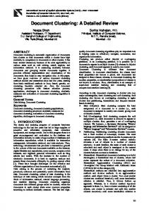
RESULTS PAR-3 colocalizes with junction and polarity proteins in apically targeted foci PAR-3 localizes to apical regions of polarized embryonic epithelial cells, including those of the intestine, pharynx and epidermis (Fig. 1A,G) (Bossinger et al., 2001; McMahon et al., 2001; Nance et al., 2003). We used two different anti-PAR-3 monoclonal antibodies to determine whether PAR-3 is present within embryonic epithelial cells as apicobasal polarity first appears, and where within the cell it localizes. Initially, we focused our analysis on intestinal epithelial cells, which are among the first epithelial cells to form during embryogenesis and have a simple organization. The intestinal epithelium differentiates from intestinal precursor cells (IPCs) that are arranged in contacting left and right rows aligned with the anterior-posterior axis (Fig. 1C). Each IPC polarizes such that its nascent apical surface forms at the midline where the left and right rows of IPCs meet; apical surfaces of cells on opposite sides of the midline eventually separate to form the intestinal lumen (Leung et al., 1999). Before the IPCs showed other signs of polarity, we observed PAR-3 immunostaining in small foci that formed at regions of contact between these cells (Fig. 1B; see Fig. S1A and Movie 1 in the supplementary material); PAR-3 foci gradually accumulated at the nascent apical surface (Fig. 1C; see Fig. S1B in the supplementary material). Similar spot-like formations within polarizing epithelial cells have been described for other PAR polarity proteins and adherens junction proteins (Bossinger et al., 2001; Leung et al., 1999; McMahon et al., 2001; Totong et al., Fig. 1. PAR-3 localization within polarizing epithelial cells. DNA is blue (in this and all subsequent figures). Anterior is left. C. elegans embryos are ~50m. (A-C⬘) PAR-3 in polarized intestinal and pharyngeal cells (A), in intestinal precursor cells (IPCs) that are beginning to polarize (B), or during polarization (C). The boxed regions in B,C are shown in B⬘,C⬘ and show colocalization of PAR-3 with HMR-1. (D,E)PAR-3 colocalization with HMP-1 (D) and PAR-6 (E) in IPCs at the onset of polarization. (F)Stills from Movie 2 of IPCs expressing PAR3YFP (see Movie 2 in the supplementary material). Dashed line indicates the future apical surface. A focus of PAR-3YFP (arrowhead) is shown over time (minutes). (G)Lateral view of polarizing epidermis (bracketed region) showing apical PAR-3 (arrow) and PAR-6. Scale bars: 2.5m.
DEVELOPMENT
Embryos were fixed in methanol and paraformaldehyde and stained as described (Anderson et al., 2008). The following antibodies and dilutions were used: mouse anti-AJM-1 ‘MH27’, 1:10 (Francis and Waterston, 1991); guinea pig anti-EAT-20, 1:100 (Shibata et al., 2000); rabbit antiERM-1, 1:200 (van Furden et al., 2004); rabbit anti-GFP, 1:2000 (Abcam); rat anti-GFP, 1:250 (Nacalai Tesque); rabbit anti-HMR-1, 1:50 (Costa et al., 1998); mouse anti-HMP-1, 1:10 (Costa et al., 1998); rabbit anti-HMP2, 1:5 (Costa et al., 1998); mouse anti-IFB-2 ‘MH33’, 1:150 (Francis and Waterston, 1991); mouse anti-PAR-3 P4A1 (IgG1) and P1A5 (IgG2a), both 1:10 (Nance et al., 2003); rabbit anti-PAR-6, 1:40,000 (Schonegg et al., 2007) and 1:30 (Hung and Kemphues, 1999); rabbit anti-PGL-1, 1:10,000 (Kawasaki et al., 1998); rat anti-PKC-3, 1:30 (Tabuse et al., 1998); mouse anti-PSD-95 (recognizes DLG-1), 1:200 (Affinity BioReagents) (Firestein and Rongo, 2001); and rat anti--tubulin, 1:2000 (Harlan). Mouse anti-PAR-3 monoclonal antibody P1A5 was isolated as described previously (Nance et al., 2003). In co-staining experiments, anti-PAR-3 monoclonal antibodies P1A5 and P4A1 showed a largely overlapping pattern that was absent in par-3(MZ) embryos; each antibody also showed non-specific staining that persisted in par-3(MZ) embryos and par-3(RNAi) embryos (P1A5 stained nuclei, and P4A1 stained the epidermis or cuticle of late-elongation stage embryos). z-stacks of embryos were acquired and processed as described (Anderson et al., 2008). Unless stated otherwise, a minimum of 50 control and 40 par-3(MZ) embryos at the indicated stages were examined in each staining.
2007). In co-staining experiments, we observed that PAR-3 foci contained the adherens junction proteins HMR-1, HMP-1 (catenin), HMP-2 (-catenin) (Fig. 1B⬘,D; data not shown) and the PAR proteins PAR-6 and PKC-3 (Fig. 1E; data not shown). By contrast, we could not detect the junction proteins DLG-1 and AJM-1 within PAR-3 foci. DLG-1 and AJM-1, which localize to a distinct basal region of mature junctions (Koppen et al., 2001; McMahon et al., 2001), first appeared within intestinal epithelial cells after the apical accumulation of PAR and adherens junction proteins was already evident. To determine whether the asymmetric localization of PAR-3 within mature epithelial cells arises from apical movement of PAR3 foci, we created a transgene expressing functional YFP-tagged PAR-3 (PAR-3YFP) from the par-3 promoter (see below) and captured time-lapse movies. In polarizing IPCs, PAR-3YFP formed foci similar to those observed in immunostained embryos (7 of 7 embryos) (Fig. 1F; see Movie 2 in the supplementary material). Individual PAR-3YFP foci were dynamic and moved erratically along the surfaces of IPCs until they adopted a more directed motion, concentrated apically and aggregated. These results, taken together with the co-staining experiments described above, suggest that foci containing PAR-3, adherens junction proteins and the polarity proteins PAR-6 and PKC-3 travel apically and condense as IPCs begin to polarize. Within fully polarized intestinal epithelial cells, PAR-3 segregated away from apical PAR-6 and PKC-3 and colocalized with HMR-1, HMP-1 and HMP-2 at adherens junctions, which form where apical and lateral surfaces meet (data not shown) (Totong et al., 2007). In contrast to adherens junction proteins, which maintained high levels of expression within epithelial cells throughout embryogenesis, PAR-3 immunostaining peaked during and after epithelial polarity establishment and diminished during subsequent stages. PAR-3 showed a similar localization within polarizing pharyngeal epithelial cells (Fig. 1A). In polarizing epidermal epithelial cells, we also detected foci of apically directed PAR-3YFP (see Movie 2 in the supplementary material). However, foci did not persist upon reaching the apical surface. Rather, PAR-
Development 137 (11)
3 developed a smooth cortical enrichment at the contact-free apical surface, where it colocalized transiently with PAR-6 and PKC-3 before becoming enriched at apical junctions (Fig. 1G; see Fig. S6 in the supplementary material; data not shown). Combined, these observations show that PAR-3 is present within epithelial precursor cells at the initial stages of polarity establishment, is among the earliest proteins to develop apicobasal asymmetry, colocalizes with other polarity proteins and junction proteins within apically targeted foci, and appears to peak in intensity as epithelial polarization is established and elaborated. Zygotic par-3 expression is required for viability To determine whether par-3 is required to establish polarity in epithelial cells, we first searched for par-3 null mutants. We obtained two uncharacterized par-3 deletion mutants from the National BioResource Project, Japan (Fig. 2A). tm2010 is a 408 bp deletion that removes sequences just prior to the first PDZ domain and is predicted to alter splicing. The 601 bp tm2716 deletion removes most of the first PDZ domain and causes a frameshift that is predicted to terminate translation just after the deletion; this allele is likely to be a genetic null, as the expected mutant gene product is severely truncated and lacks most of the functional domains of PAR-3. tm2010 and tm2716 failed to complement the maternal-effect lethal allele par-3(it71), but each mutation on its own caused a more severe L1 larval lethal phenotype (Table 1; see Materials and methods). We reasoned that the deletions disrupt an essential par-3 isoform that is unaffected by existing par-3 mutations, all of which cause maternal-effect lethal phenotypes. We searched for alternative par-3 transcripts by sequence comparison and RT-PCR. By aligning genomic sequences from C. elegans and C. briggsae, we identified a highly conserved region near the end of the large third intron. The conserved region contains an SL1 trans-splice acceptor, which indicates the 5⬘ end of a transcript (Hwang et al., 2004), and forms an open reading frame that extends in-frame into the fourth exon. Using RT-PCR, we verified that the conserved region is the beginning of an alternative par-3 transcript (which we named par-3s) that contains Fig. 2. Isoforms of C. elegans par-3. (A)par-3l, par-3s and yfp::par-3s. Exons (rectangles), introns (chevrons) and mutations are indicated. (B)Predicted PAR-3L and PAR-3S products showing the oligomerization domain (magenta), PDZ domains (yellow) and aPKC-binding domain (cyan). (C)Expression and rescuing activity of par-3 transgenes. (D,E)Expression of par-3::gfp in one-cell (D) and 1.5-fold stage (E) embryos. (F)Expression of yfp::par-3s in 1.5-fold stage embryo. Scale bar: 2.5m.
DEVELOPMENT
1836 RESEARCH ARTICLE
PAR-3 in C. elegans epithelia
RESEARCH ARTICLE 1837
Table 1. Phenotypes of par-3(tm2716) and par-3(MZ) mutants Self-progeny phenotype Maternal genotype
Wild type par-3(tm2716)/+ par-3(tm2716); par-3::zf1::gfp par-3(tm2716); par-3::zf1::gfp/+
Wild type
Embryonic lethal
Larval lethal
356 (99.5) 309 (78) 697 (97.5) 490 (61)
2 (0.5) 7 (1.7) 18 (2.5) 273 (34)
0 82 (20.3) 0 45 (5)
The number of embryos or larvae is shown, with the percentage in parentheses.
each of the previously annotated downstream par-3 exons (Fig. 2A; see Fig. S2 in the supplementary material). The predicted PAR-3S protein includes all of the known functional domains of full-length PAR-3 (hereafter PAR-3L), although the putative N-terminal oligomerization domain is truncated (Fig. 2B). par-3s mRNA is disrupted by the tm2010 and tm2716 deletions, but is unlikely to be affected by the two molecularly characterized par-3 maternaleffect lethal mutations; these nonsense mutations occur in upstream exons that are not included in the par-3s transcript (Aono et al., 2004) (Fig. 2A). To determine where the different PAR-3 isoforms are expressed, we created translational reporters by fusing gfp to par-3 within large genomic clones. First, we created a C-terminal GFP fusion (par-3::gfp) that reports on both PAR-3L and PAR-3S expression. par-3::gfp expressed PAR-3GFP in the early embryo and in epithelial cells (Fig. 2C-E), in a pattern similar to that of endogenous PAR-3 (2/2 lines). Reasoning that the large third intron of par-3 contains an internal promoter driving par-3s transcription, we created a par-3::gfp derivative lacking all sequences upstream of the third intron (par-3s::gfp). Embryos expressing par-3s::gfp showed zygotic PAR-3SGFP expression in epithelial cells (6/6 lines) but no maternal expression (Fig. 2C; data not shown). Our inability to detect maternal PAR-3SGFP suggests that sequences upstream of the third intron might be required for germline PAR-3 expression; alternatively, par-3s::gfp transgenic lines could be silenced within the germ line (Kelly et al., 1997). To distinguish between these possibilities, we fused yfp to the beginning of par-3s within a genomic clone containing the entire par-3 gene (Fig. 2A); since the yfp insertion site is within coding sequences of par-3s but within intronic sequences of par-3l, the yfp::par-3s transgene should express PAR-3SYFP as well as untagged PAR-3L. To detect both transgenic PAR-3L and PAR-3SYFP we crossed the yfp::par-3s transgene into par-3(tm2716) mutants, which lack endogenous PAR-3 immunostaining (see below), and co-stained embryos with PAR-3 and GFP antibodies. In a line of yfp::par-3s that did not show germline silencing, we detected PAR-3SYFP only in epithelial cells, and PAR-3L was present weakly in early embryos (Fig. 2C,F; data not shown). These results indicate that PAR-3L is expressed maternally, and that PAR-3S is expressed zygotically but not maternally. Consistent with these expression patterns, transgenes
expressing both PAR-3L and PAR-3S (par-3::gfp and yfp::par-3s) rescued larval and maternal-effect lethality of par-3(tm2716) mutants (par-3::gfp, 2/2 lines; yfp::par-3s, 1/1 line) (Table 2); a transgene expressing only PAR-3S (par-3s::gfp) rescued larval lethality but not maternal-effect lethality [1 (the highest-expressing) of 4 lines rescued] (Fig. 2C). To establish whether PAR-3S has an important role during development, we treated par-3(tm2716); yfp::par-3s worms with gfp dsRNA, which targets transgenic par-3 tagged with gfp or yfp (Table 2). When fed to par-3(tm2716); yfp::par-3s worms, gfp dsRNA targets expression of zygotic PAR-3SYFP but not maternal PAR-3L, which is untagged. par-3(tm2716); yfp::par-3s worms fed at the L4 stage produced embryos that hatched but frequently developed into sterile adults. This phenotype is observed in wildtype larvae treated with par-3 dsRNA and is due to a reduction in zygotic PAR-3 protein (Aono et al., 2004), indicating that PAR-3S has an essential zygotic function. gfp dsRNA fed to control par3(tm2716); par-3::gfp worms targets expression of both PAR3LGFP and PAR-3SGFP. As expected, par-3(tm2716); par-3::gfp worms fed gfp dsRNA at the L4 stage produced dead eggs owing to the loss of maternal PAR-3. When fed at the L1 stage to bypass the requirement for maternal PAR-3, some par-3(tm2716); par3::gfp worms became sterile, as we observed for par-3(tm2716); yfp::par-3s worms. Taken together, these findings indicate that maternal PAR-3L has an essential function in the early embryo, whereas zygotic PAR-3S, which is found in epithelial cells, has an essential function during embryonic or larval stages. par-3 is required for morphogenesis of epithelial organs To determine whether PAR-3 is required for the initial polarization of epithelial cells, we designed a strategy to eliminate PAR-3 protein from the embryo after its essential role in polarizing the one-cell embryo has been completed but before epithelial cells are formed. We previously described the par-3::zf1::gfp transgene, which transiently expresses maternal PAR-3ZF1-GFP (Nance et al., 2003). par-3::zf1::gfp is a derivative of par-3::gfp that includes sequences encoding the PIE-1 protein ZF1 (zinc-finger) domain fused to par-3; proteins tagged with the ZF1 domain degrade rapidly in all early embryonic somatic cells (Reese et al., 2000).
L4 feeding Embryo phenotype
L1 feeding Adult phenotype*
Genotype and treatment†
Hatched
Dead
Fertile
Sub-fertile
Sterile
par-3(tm2716); par-3::gfp, vector RNAi par-3(tm2716); par-3::gfp, gfp RNAi par-3(tm2716); yfp::par-3s, vector RNAi par-3(tm2716); yfp::par-3s, gfp RNAi
389 (94) 0 385 (99) 341 (98)
27 (6) 450 2 (1) 9 (2)
68 63 (88) 62 (91) 3 (4)
0 0 5 (7) 42 (61)
0 9 (12) 1 (1) 24 (35)
The number of embryos or adults is shown, with the percentage in parentheses. *Sterile worms laid no eggs; sub-fertile worms laid 1-20 eggs; fertile worms laid more than 100 eggs. † Worms were also homozygous for unc-32. Transgenes used were zuIs73 [par-3::gfp] and xnIs199 [yfp::par-3s].
DEVELOPMENT
Table 2. Function of par-3 isoforms
par-3::zf1::gfp rescues the polarity defects of par-3 mutant one-cell embryos before the fusion protein starts to rapidly degrade at the four-cell stage (Nance et al., 2003). Zygotic par-3::zf1::gfp expression begins later in epithelial cell precursors and is stable because ZF1-mediated degradation appears to be limited to early embryos. Therefore, par-3(tm2716) mutants expressing par3::zf1::gfp were viable and produced embryos that had normal anterior-posterior polarity (Table 1; see Materials and methods). To obtain embryos with epithelial cells lacking both maternal and zygotic PAR-3, we selfed par-3(tm2716); par-3::zf1::gfp/+ worms and analyzed progeny lacking the par-3::zf1::gfp transgene. Hereafter, we refer to these embryos as par-3(MZ) embryos [par3(MZ) embryos differ from the previously described par-3(ZF1) embryos (Nance et al., 2003), which still express PAR-3ZF1-GFP zygotically]. Using antibodies that recognize sequences downstream of the tm2716 deletion, we could not detect PAR-3 in par-3(MZ) embryos after PAR-3ZF1-GFP had degraded from early blastomeres (Fig. 4B; see Fig. S6B in the supplementary material). Therefore, PAR-3 is depleted from par-3(MZ) embryos well before epithelial cells form. One quarter of the progeny from par-3(tm2716); par3::zf1::gfp/+ mothers died during embryogenesis, suggesting that par-3(MZ) embryos arrest at an earlier stage than par-3 deletion mutants, which inherit maternal PAR-3 (Table 1). We acquired time-lapse DIC movies of developing embryos to determine why par-3(MZ) embryos arrested. In wild-type embryos, epidermal epithelial cells migrate ventrally to encase the embryo in skin then contract circumferentially to squeeze the embryo and elongate the body axis; simultaneously, the pharynx and intestine form a linked tube that connects to the mouth and rectum. par-3(MZ) embryos elongated much more slowly than wild-type embryos and arrested before reaching 2-fold elongation, as compared with the 4-fold elongation of wild-type embryos at this stage (Fig. 3A-C; see Movies 3, 4 in the supplementary material). We observed defects in morphogenesis of the pharynx and intestine, such as mispositioned intestinal cells and an improperly elongated pharynx (Fig. 3A,B, outlined). Mutant embryos also frequently formed epidermal lesions, although these were restricted to the mouth and rectum, where internal and external epithelial cells connect (Fig. 3B, arrowheads). This phenotype contrasts with that of par-6(MZ) embryos, which form only immature junctions and develop lesions throughout the epidermis (Totong et al., 2007). The phenotype of par-3(MZ) mutants is consistent with a defect in epithelial cell polarization or junction formation that interferes with the morphogenesis of internal epithelial organs. Additionally, the different phenotypes of par-3(MZ) and par-6(MZ) embryos suggest that PAR-3 and PAR-6 have distinct roles in epithelial cells.
Development 137 (11)
PAR-3 mediates intestinal epithelial cell polarization To investigate the cellular basis of the morphogenesis defects in par-3(MZ) embryos, we immunostained for proteins that develop asymmetric localizations within polarized epithelial cells. PAR-6 and PKC-3 localize to the apical cortex of wild-type intestinal epithelial cells (Totong et al., 2007), but in par-3(MZ) embryos both proteins were present diffusely within the cytoplasm and failed to enrich apically (Fig. 4C,D; data not shown). The intermediate filament protein IFB-2 is found just beneath apical microvilli in wild-type intestinal cells (Bossinger et al., 2004). In par-3(MZ) embryos, IFB-2 formed large discontinuous aggregates, rather than the continuous apical band observed in wild-type embryos (see Fig. S4C,D in the supplementary material). HMP-1 localizes to adherens junctions, which form at the interface of apical and lateral surfaces. In par-3(MZ) embryos, HMP-1 accumulated within irregular aggregates, similar to those seen in embryos stained for IFB-2 (see Fig. S4A,B in the supplementary material). The junction proteins DLG-1 and AJM-1 also formed irregular aggregates in par-3(MZ) embryos (Fig. 4E,F; data not shown). We co-stained embryos for DLG-1 and either HMP-1 or IFB-2 to determine the composition of aggregates (see Fig. S4 in the supplementary material). In most aggregates, DLG-1 showed considerable colocalization with HMP-1 or IFB-2. Therefore, although PAR-3 is required to properly position these apically enriched proteins, they retain the ability to aggregate when PAR-3 is absent. In addition to these cortical proteins, we examined two apically enriched transmembrane proteins: the apical Crumbs-like protein EAT-20 and the adherens junction protein HMR-1 (Shibata et al., 2000). Both EAT-20 and HMR-1 were mislocalized in par-3(MZ) embryos but were also more dispersed, unlike DLG-1 and HMP-1, and did not form large aggregates (Fig. 4G-J; see Fig. S4E,F in the supplementary material). The irregular organization of apical surfaces in par-3(MZ) embryos made it difficult to assess the localization of two other markers: LET-413 (the homolog of Drosophila Scribble) and microtubules. LET-413 normally localizes to basolateral surfaces and is excluded from apical surfaces (Legouis et al., 2000). par-3(MZ) embryos showed normal basolateral LET-413 localization but apical exclusion was difficult to ascertain given the lack of a defined apical surface in mutant embryos (Fig. 4K,L, arrowheads). Microtubules are normally enriched just below the apical cortex as the IPCs polarize (see Fig. S3A in the supplementary material). In par-3(MZ) embryos, microtubules in the IPCs were concentrated in patches, rather than uniformly, beneath the apical surface (see Fig. S3B in the supplementary material). In summary, PAR-3 is required to
Fig. 3. Morphogenesis in par-3(MZ) embryos. (A,B)DIC time-lapse stills of wild-type and par-3(MZ) mutant C. elegans embryos at 1.5-fold stage. The pharynx (outlined) does not extend anteriorly in the par-3(MZ) embryo and extruded cells (arrowheads) are visible. (C)Elongation rate to 1.5-fold stage. Time is minutes from completion of ventral enclosure to 1.5-fold stage. Error bars indicate s.d.; *, P