The Journal of Immunology
Monocyte Surface-Bound IL-15 Can Function as an Activating Receptor and Participate in Reverse Signaling1 Graham G. Neely,* Slava Epelman,† Ling Ling Ma,* Pina Colarusso,‡ Christopher J. Howlett,§ Ernest K. Amankwah,† Amanda C. McIntyre,† Stephen M. Robbins,§ and Christopher H. Mody2†¶ IL-15 is a short chain, four-␣ helix cytokine that shares some biological function with IL-2. One striking difference between IL-2 and IL-15 is the ability of monocytes to express IL-15 on their cell surface after activation. In the current study we have investigated the ability of human monocyte cell surface IL-15 to participate in reverse signaling. Cross-linking anti-IL-15 Abs were used as a surrogate ligand for surface IL-15 engagement. Ligation of cell surface-expressed IL-15 induced monocyte adhesion that required the activity of small m.w. GTPases. Reverse signals through surface IL-15 activated the Rho-GTPase Rac3. In addition, engagement of cell surface IL-15 was found to activate a number of signaling pathways, including both extracellular signalregulated kinase 1/2 and p38, and resulted in the secretion of IL-8. IL-8 production required mitogen-activated protein kinase activity. Thus, the current study has established that cell surface IL-15 is more than just a ligand; it can function as a receptor and participate in reverse signaling that results in cellular adhesion and production of inflammatory cytokines. The Journal of Immunology, 2004, 172: 4225– 4234.
T
raditionally, the interaction between a receptor and a ligand is considered in terms of the unidirectional effect a soluble mediator has on its corresponding receptor. It is now becoming clear that membrane-bound ligands can participate in reverse and bidirectional signaling. In some circumstances, bidirectional signaling involves the cognate juxtacrine interaction of transmembrane receptor pairs on adjacent cells. MHC/TCR interactions typify this process (1–3). In other systems the bidirectional responses involve receptor-ligand interactions, where one partner is a transmembrane receptor and the other is a tethered ligand. Such is the case for the well-characterized bidirectional ephrin/eph system directing axonal guidance (4) as well as the bidirectional nature of some TNF family members contributing to multiple stages of immune regulation, including T cell costimulation, cytokine production, and germinal center formation (5– 8). IL-15 is a four-␣ helix, short chain, type I T cell growth factor that shares some structural and biological similarities with IL-2. Studies using knockout mice have revealed that IL-15 is indispensable for innate lymphoid ontogeny (9, 10) and for the generation and maintenance of a population of CD8 memory cells (11, 12). In addition to these lymphoid functions, IL-15 can directly regulate monocyte cytokine production (13, 14), lineage commit*Departments of Medical Sciences, †Microbiology Infectious Disease, ‡Physics and Biophysics, §Oncology, and ¶Internal Medicine, University of Calgary, Calgary, Alberta, Canada Received for publication August 11, 2003. Accepted for publication January 20, 2004. The costs of publication of this article were defrayed in part by the payment of page charges. This article must therefore be hereby marked advertisement in accordance with 18 U.S.C. Section 1734 solely to indicate this fact. 1 This work was supported by grants from the Canadian Institutes for Health Research and the Canadian Foundation for AIDS Research. C.H.M. and S.M.R. are Senior Scholars of the Alberta Heritage Foundation for Medical Research. S.M.R. holds a Canada Research Chair in cancer biology. C.H.M. is the Jessie Bowden Lloyd Professor of Immunology. G.G.N. is supported by a studentship from the Alberta Lung Association. This work conforms to the guidelines established by the Conjoint Committee on Medical Ethics at the University of Calgary. 2
Address correspondence and reprint requests to Dr. Christopher H. Mody, Room 273, Heritage Medical Research Building, University of Calgary, Calgary, Alberta, Canada T2N 4N1. E-mail address:
[email protected] Copyright © 2004 by The American Association of Immunologists, Inc.
ment (14, 15), and microbiocidal activity (16 –18). In addition, there are many subtle roles for IL-15 in the generation and regulation of a productive immune response (reviewed by Fehniger et al. (19)). IL-15 can be found in a membrane-tethered form in some diseases and after stimulation (20 –22). For IL-15, the capacity for stable membrane expression may depend on association with its unique high affinity receptor, IL-15R␣ (23). Alternatively, this cell surface expression could result from the abnormal IL-15 pro pieces or signal peptides generated through differential splicing. Regardless, cell surface IL-15 is expressed in a biologically active form where it can mediate CD8 T cell proliferation (23, 24) and NK cell development (25). Our current understanding of the mechanism of IL-15 function does not provide an explanation for what advantage, if any, is conferred by tethering IL-15 to the cell surface. Potentially, surface IL-15 could function as a ligand and, through increased avidity, promote receptor clustering, thereby amplifying IL-15 responses. Indeed, we have previously reported that cell surface IL-15 was roughly 5 times more effective at supporting T cell proliferation than its soluble counterpart (24). Moreover, cell surface retention of IL-15 may, as is the case for both the TNF/TNF receptor (TNFR)3 and the ephrin/eph system, confer the ability to participate in reverse or bidirectional responses. In the current study we addressed the potential for cell surface IL-15 to function as a cellular activation receptor. We reasoned that, through retention on the plasma membrane, cell surface IL-15 might possess distinct biological activities that are lacking in its soluble counterpart. It was our hypothesis that cell surface retention may confer the capacity to function as a receptor and allow cell surface IL-15 to participate in reverse signaling events. To address this, hypercross-linking of cell surface IL-15 on monocytic cells was used. Cells stimulated in this manner were analyzed for
3 Abbreviations used in this paper: TNFR, TNF receptor; CDC, cell division cycle; ERK, extracellular signal-regulated kinase; JAK, Janus kinase; MAPK, mitogen-activated protein kinase; Rac, Ras-related C3 botulinum toxin substrate; rh, recombinant human; Rho, Ras homology; Syk, spleen tyrosine kinase; Tyr, tyrosine.
0022-1767/04/$02.00
4226 their ability to activate a number of downstream signaling effectors. The activity of the Rho family members, Rac and CDC42, was measured by a Rac activity assay. Activation of mitogen-activated protein kinase (MAPK) family members, extracellular signal-regulated kinase 1/2 (ERK1/2) and p38, was assessed by phosphospecific immunoblots. The roles of these signaling molecules were assessed by the use of inhibitors, and the biological consequences of activation were assessed by monocyte adhesion to plastic and VCAM-1, and the production of IL-8 protein.
Materials and Methods Cells and cell lines THP-1 cells (American Type Culture Collection, Manassas, VA) were maintained in RPMI 1640 supplemented with 10% heat-inactivated FBS, 1% penicillin/streptomycin, and 1 mM sodium pyruvate (all from Life Technologies, Burlington, Canada) at 37°C in a 5% CO2 atmosphere. PBMC were obtained by venipuncture from healthy adults. Blood was anticoagulated by adding 10 U/ml heparin (Organon Teknika-Cappel, Scarborough, Canada). PBMC were purified by centrifugation (800 ⫻ g, 20 min) on a Ficoll-Hypaque density gradient (C-six Diagnostics, Mequon, WI). The cells were washed three times in HBSS (Life Technologies), counted in a hemocytometer, and resuspended in complete medium containing RPMI 1640 (Life Technologies), 5% heat-inactivated human AB serum (BioWhittaker, Walkersville, MD), 2 mM L-glutamine (Life Technologies), 100 U of penicillin per ml, 100 g of streptomycin per ml, 0.2 g of amphotericin B/ml (Life Technologies), 1 mM sodium pyruvate (Life Technologies), and 0.1 mM nonessential amino acids (Life Technologies).
Flow cytometry Resting THP-1 cells (1.0 ⫻ 106/ml) were cultured in low adherence plates (Costar, Cambridge, MA) with complete medium (as described above) or medium containing GM-CSF (30 ng/ml; R&D Systems, Minneapolis MN), Escherichia coli LPS (10 ng to 1 g/ml; Sigma-Aldrich, Oakville, Canada), PMA (10 ng/ml; Calbiochem, San Diego, CA), PHA (10 g/ml; Sigma-Aldrich), or IFN- (100 ng/ml; R&D Systems). Cell surface IL-15 was detected as previously described (24). Briefly, cells were washed three times with PBS (1% FBS and 0.1% NaN3) and incubated in the dark at 4°C with anti-IL-15 Ab (Mab247; R&D Systems) or isotype-matched control Ab (R&D Systems). Cells were washed twice and incubated with goat anti-mouse IgG1-PE (Molecular Probes, Eugene, OR). Cells were fixed with 1% p-formaldehyde for 20 min before fluorescent analysis using LYSIS II software on a FACScan fluorocytometer (BD Biosciences, Mountain View, CA).
Real-time adhesion assay One million THP-1 cells were incubated for 30 min at 4°C with 1 g/ml of anti-IL-15 mAbs (MAB247 and MAB647; R&D Systems) or an equivalent amount of isotype-matched control Ab. Cells were then added to 35-mm glass-bottom culture dishes (MatTek, Ashland, MA) and allowed to settle at 4°C for 30 min. A secondary Ab was then added (1 g of antimouse F(ab)2; Pierce, Rockford, IL) to hypercross-link surface IL-15. Cells were transferred to 37°C where images were captured with a Retiga EX camera (QImaging, Burnaby, Canada) monochrome 12-bit peltier cooled to 25°C below ambient over 60 min using the ⫻40 phase contrast objective of a wide-field Olympus 1X-70 microscope (Olympus, Melville, NY) equipped with a temperature-controlled box kept at 37°C.
Plastic adhesion assay THP-1 cells were preincubated with inhibitor (Clostridium difficile toxin B, 3 ng/ml; AG490, 100 nm; Calbiochem), incubated for 30 min at 4°C with 1 g/ml of two anti-IL-15 mAbs (MAB247 and MAB647; R&D Systems), 50 ng recombinant human IL-15 (rhIL-15; R&D Systems), or an equivalent amount of isotype-matched control Ab (R&D Systems). Cells were washed with cold medium and then cultured in 24-well, flat-bottom plates (Falcon; BD Biosciences, Franklin Lakes, NJ) for 4 h in the presence or the absence of 1 g of a hypercross-linking anti-mouse F(ab)2 (Pierce). Nonadherent cells were washed away with cold RPMI 1640, and adherent cells were counted. In addition to visual counting, an MTT assay was performed to enumerate adherent cells. In this case, 10 l of a 5-mg/ml MTT solution (Sigma-Aldrich) was added to each washed well, and adherent cells were incubated for an additional 4 h, at which time 100 l of isopropanol/0.04 N HCl solution was added to each well and read spectrophotometrically at 570 nm.
REVERSE SIGNALING BY SURFACE IL-15 Rac activation assay IL-15 was hypercross-linked on the surface of THP-1 cells as described above. Instead of hypercross-linking, some THP-1 cells were treated with either rhIL-15 (50 ng/ml; R&D) or LPS (10 ng/ml; Sigma-Aldrich). After a 30-min incubation, cells were lysed and a P-21 binding domain pulldown assay was performed according to the manufacturer’s instructions (Upstate Biotechnology, Lake Placid, NY). Samples were resolved using SDSPAGE, and immunoblots were performed for RAC1, RAC3, and CDC42 (Santa Cruz Biotechnology, Santa Cruz, CA).
Phosphospecific Western blotting IL-15 was hypercross-linked on the surface of THP-1 cells as described above. Instead of hypercross-linking, some THP-1 cells were treated with rhIL-15 (10 ng/ml; R&D Systems). Cells were lysed in a Nonidet P-40 lysing buffer (50 mM HEPES, 150 mM NaCl, 1.5 mM MgCl2, 1 mM EGTA, 100 mM NaF, 1 mM Na3VO4, and 1% Nonidet P-40). Debris and unlysed cells were removed through low speed centrifugation (3000 rpm). A Western blot was performed on the soluble lysate using MAPK-phosphospecific Abs according to the manufacturer’s instructions (Cell Signaling Technology, Mississauga, Canada).
VCAM-1 adhesion assay An in vitro adhesion assay was performed as previously described (26). Briefly, 96-well Maxisorp Immuno Module flat-bottom plates (Nunc, Naperville, IL) were coated with 10 g/ml BSA (Sigma-Aldrich), or recombinant VCAM-1 (R&D Systems). After hypercross-linking or treatment with LPS, 2.0 ⫻ 105 THP-1 cells were plated in triplicate and allowed to settle with treatment for 1 h at 4°C. Samples were incubated at 37°C for 30 min and washed with 2.5% BSA in HBSS (Life Technologies). Wells were washed with cold RPMI 1640. The resultant adherent cells were detached by vigorous pipetting using cold HBSS without Ca2⫹ or Mg2⫹ and 10 mM EDTA and were counted using a Coulter counter (Beckman Coulter, Miami, FL). Photographs of cells were obtained before detachment using the ⫻40 objective of an Axiovert inverted microscope (Zeiss, Thornwood, NY).
Intracellular cytokine detection After IL-15 hypercross-linking, THP-1 cells were cultured for 4 h in the presence of monensin (2 M; Golgi-Stop; BD PharMingen, San Diego, CA). Cells were washed three times in cold PBS (1% FCS and 0.1% sodium azide) and permeabilized using the Cytofix/Cytoperm reagent (BD PharMingen) according to the manufacturer’s instructions. Cells were washed twice in Perm Wash (BD PharMingen) and incubated for 30 min at 4°C with mouse monoclonal anti-human IL-8 Ab (BD PharMingen) or an equal amount of mouse isotype-matched control Ab (Caltag, Burlingame, CA). Cells were washed twice with Perm Wash and then washed twice with PBS (1% FCS and 0.1% NaN3) before fluorescent analysis using LYSIS II software on a FACScan fluorocytometer (BD Biosciences). In some cases, 10 M of the MAPK kinase 1/2 inhibitor U 0126 (Calbiochem), the p38 inhibitor SB203580 (A.G. Scientific, San Diego, CA), or empty vehicle control was added to the cells before IL-15 hypercross-linking.
IL-8 ELISA After IL-15 hypercross-linking, THP-1 cells were cultured in AIM V medium (Life Technologies), and culture supernatants were collected at 18 h. An IL-8 sandwich ELISA was performed (R&D Systems). After addition of goat anti-mouse-HRP (Amersham Pharmacia Biotech, Little Chalfont, U.K.), wells were washed, and 50 l of a 1/1 mixture of H2O2 and tetramethylbenzidine solutions (Genzyme, San Carlos, CA) was added. The reaction was stopped with 50 l of 1 M H2SO4 and was read spectrophotometrically at 450 nm. IL-8 concentrations were determined by comparison with a standard curve generated with rhIL-8 (R&D Systems).
Hypercross-linking IL-15 on activated PBMC PBMC (1.0 ⫻ 106/ml) were activated with 1 g/ml LPS (or medium alone as a control) for 1 h in low adherence plates containing complete RPMI 1640 (Life Technologies). After the initial stimulation, cells were washed, surface IL-15 was hypercross-linked, and then cells were incubated in the presence of monensin plus inhibitors to p38 or MAPK kinase 1/2. Eighteen hours after hypercross-linking, cells were harvested and prepared for the detection of intracellular IL-8 by flow cytometry, as described above.
The Journal of Immunology Statistics When applicable, data are presented as the mean ⫾ SEM. Statistical analysis was performed using Student’s t test with the Bonferroni correction for comparison of multiple groups.
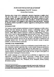
Results THP-1 monocytes constitutively express cell surface IL-15 and do not alter IL-15 levels after stimulation To test the hypothesis that cell surface IL-15 participates in reverse signaling by functioning as a receptor, we chose to use the monocytic cell line THP-1. THP-1 cells were chosen because they have previously been reported to exhibit constitutive IL-15 surface expression (27). Indeed, our data demonstrate that the expression of cell surface IL-15 was stable in culture (Fig. 1a). In separate experiments, a variety of stimuli known to modulate IL-15 expression on primary human monocytes (LPS, GM-CSF, IFN-, PMA, and PHA) (G. G. Neely, E. Amankwah, and C. H. Mody, unpublished observations) (24) had no clear effect on the levels of membrane IL-15 observed (Fig. 1b). Thus, THP-1 cells presented an opportunity to examine the function of surface-expressed IL-15 without having to activate the cells, which might elicit a superimposed downstream consequence.
4227 Cell surface IL-15 can function as a receptor to promote monocyte adhesion Although resting THP-1 cells are nonadherent in culture, activated THP-1 cells are adherent (28). We thought that if cell surface IL-15 was a receptor capable of promoting monocyte activation, then engagement of cell surface IL-15 might also promote monocyte adhesion. To test this hypothesis, THP-1 cells were pretreated with mAbs directed to human IL-15. Cells were then placed in glass-bottom culture dishes, and surface IL-15 was hypercrosslinked with the F(ab⬘)2 portion of an anti-mouse secondary Ab (Fig. 2a). In both the control and cross-linking conditions, a basal level of cells settled and remained in the plane of view over the course of the experiment. After the addition of hypercross-linking secondary Ab (at 2 min), inducible adhesion was observed in the IL-15 hypercross-linked group (10 and 60 min), but not in the control group. By visual inspection, no morphological differences were evident between cells that had initially adhered spontaneously compared with cells that had undergone hypercross-linking IL-15-induced adhesion. Cell surface, but not soluble, IL-15 can induce monocyte adhesion To differentiate firm adhesion from settling, this assay was modified to quantify cells tightly associated with the culture substrate. After a 4-h incubation, nonadherent cells were removed by washing, and the remaining adherent cells were counted. Although no treatment, treatment with isotype control primary Abs and secondary cross-linking Ab treatment, or exogenous rhIL-15, which would engage the IL-15R␣, were all incapable of inducing adhesion, hypercross-linking surface IL-15 induced a significant (⬎7fold) increase in monocyte adhesion (Fig. 2b). This increased adhesion required hypercross-linking and was not a phenomenon of Ab treatment, as adhesion was not observed when soluble IL-15 Abs were used without a secondary Ab, or when both primary and cross-linking Abs were added simultaneously (data not shown). This adhesion was not a result of LPS contamination, as adhesion occurred in the absence of serum, and polymyxin B did not abrogate this response (data not shown). To enumerate cell number objectively and to assess the viability of adherent cells, an MTT assay was performed in parallel (Fig. 2c). From these independent approaches, it follows that cell surface IL-15 acts as a monocyteactivating receptor. The observation that cell surface IL-15 can trigger monocyte activation, whereas its soluble isoform cannot, supports the hypothesis that cell surface and soluble IL-15 have the capacity to exhibit distinct biological functions. Rho family GTPases are essential for surface IL-15-induced monocyte activation
FIGURE 1. THP-1 cells express IL-15 on their cell surface and do not up-regulate IL-15 in response to various stimuli. a, THP-1 cells were cultured for the indicated times, and then flow cytometry was performed to assess levels of cell surface IL-15. b, THP-1 cells were stimulated for 24 h with various agents known to modulate monocyte IL-15 cell surface levels, and flow cytometry was performed to detect IL-15 surface expression. The experiment was repeated three times with the same results.
Rho family GTPases are essential for actin rearrangement and subsequent cellular adhesion. To determine whether Rho family GTPases were necessary for surface IL-15-induced reverse signals resulting in monocyte activation/adhesion, the natural Rho family inhibitor C. difficile toxin B was used (Fig. 3, a and b). In the presence of the Rho family inhibitor, the adhesion of THP-1 cells was abrogated, implicating Rho family members as downstream effectors contributing to monocyte responses after surface IL-15 engagement. Trypan blue exclusion demonstrated that no toxicity was observed when monocytes were treated with C. difficile toxin B over the course of the experiment (data not shown). Soluble IL-15 activates cells primarily through the Janus kinase (JAK)/Stat signaling pathway (29). To explore the possible role of this pathway in IL-15 reverse signals, cells were also treated with AG490, a JAK inhibitor (Fig. 3b). The presence of the JAK inhibitor did
4228
FIGURE 2. Cell surface IL-15 can function as a receptor to induce monocyte adhesion. a, THP-1 cells were incubated with isotype control or antiIL-15 Abs and then hypercross-linked (IL-15-XL) with anti-mouse F(ab)2. Images were taken every minute for 60 min. b and c, THP-1 cells were incubated with anti-IL-15 Abs or equal amounts of isotype-matched control Ab, washed, and then hypercross-linked with anti-mouse F(ab)2. b, After 4 h, the nonadherent cells were removed with RPMI 1640, and cell numbers were counted and presented as the mean of quadruplicate wells ⫾ SEM. c, An MTT assay was performed and presented as the mean of quadruplicate wells ⫾ SEM. The experiment was repeated three times with similar results. ⴱ, p ⬍ 0.05 compared with unstimulated. ⴱⴱ, p ⬍ 0.05 compared with IL-15 hypercross-linked. NS, Not significant compared with unstimulated.
REVERSE SIGNALING BY SURFACE IL-15
FIGURE 3. Surface IL-15 reverse signals can activate Rac3 and induce monocyte adhesion to plastic in a Rho family-dependent fashion. After toxin B treatment, surface IL-15 was hypercross-linked (IL-15-XL), and then cells were incubated for 4 h. Nonadherent cells were removed, and adherent cell numbers were counted and presented as the mean of quadruplicate wells ⫾ SEM (a), or an MTT assay was performed, and data were presented as the mean of quadruplicate wells ⫾ SEM (b). c, THP-1 cells were incubated with medium, isotype-matched control mAbs, or anti-IL-15 mAbs, then stimulated with cross-linking secondary or either 25 ng of rhIL-15 or 10 ng of LPS. After 15 min (Rac3) or 30 min (Rac1, CDC42), cells were lysed, and a Rac activity assay was performed by affinity precipitation. Precipitate was resolved on SDS-PAGE, followed by Western blots for Rac1, Rac3, or CDC42. The experiment was repeated three times with similar results. ⴱ, p ⬍ 0.05 compared with unstimulated. ⴱⴱ, p ⬍ 0.05 compared with IL-15 hypercross-linked. NS, Not significant compared with unstimulated. #, p ⬍ 0.05 compared with isotype plus DMSO.
The Journal of Immunology not block surface IL-15-induced adhesion, indicating that small GTPases of the Rho family, but not tyrosine kinases of the JAK family, are essential for surface IL-15 reverse signals resulting in monocyte adhesion. IL-15 functions as a receptor to activate Rho family GTPases To determine whether engagement of surface IL-15 could result in the activation of a small GTPase such as Rac or CDC42, a Rac activity assay was performed (Fig. 3c). As previously demonstrated, stimulation of monocytes with LPS promoted the activation of Rac1 (30). However, hypercross-linking cell surface IL-15 resulted in the selective activation of Rac3. Further, whereas 50 ng of rhIL-15 failed to activate Rac3, a low level of CDC42 activation could be detected. Neither the cells treated with isotype-matched control Ab, nor cells that had hypercross-linked surface IL-15 demonstrated Rac1 or CDC42 activation, known effectors of FcR signaling, confirming minimal contribution by FcRs to the observed surface IL-15-induced adhesion (31, 32). Thus, cell surface IL-15 can activate Rac3, an ability that its soluble counterpart lacks. These data reinforce the idea proposed by ourselves (24) and others (23, 33) that cell surface IL-15 may function differently than its soluble counterpart and provide some new insight into how this functional difference may manifest itself at the cellular level. Surface IL-15 can induce monocyte adhesion to VCAM-1 dependant on Rho family GTPases Although cellular adherence to plastic substrates may be of some medical importance, experiments were preformed to determine whether surface IL-15 resulted in adherence to a more physiological substrate. For this purpose, the ability of cell surface IL-15 to initiate monocyte adhesion to immobilized VCAM-1 was evaluated. In vivo, marginalized monocytes interact with the endothelium until monocyte activation occurs. After activation, monocytes will firmly adhere to the endothelium in a VCAM-1/␣4-dependent manner (34, 35). Adhesion through VCAM-1 also plays a role in subsequent monocyte diapedesis (36). To study regulated monocyte adhesion in vitro, 96-well plates were coated with either
4229 VCAM-1 or BSA (as a control), and an adhesion assay was performed (Fig. 4). Activated monocytes did not adhere to BSAcoated plastic wells. Hypercross-linking cell surface IL-15 resulted in a substantial increase in the number of cells adherent to VCAM1-coated plates without morphological differences between IL-15 hypercross-linked monocytes compared with spontaneously adherent control cells (Fig. 4a). When assessed quantitatively, hypercross-linking cell surface IL-15 was sufficient to promote a large (⬎30-fold) amount of monocyte adhesion to VCAM-1 (Fig. 4b). To determine whether Rho family GTPases were also involved in the adherence of surface IL-15-activated monocytes to VCAM-1, monocytes were treated with the Rho family inhibitor C. difficile toxin B. Toxin B abrogated adhesion to VCAM-1. Trypan blue exclusion demonstrated that no toxicity was observed when monocytes were treated with C. difficile toxin B over the course of the experiment (data not shown). These data support the previous observations that surface-IL-15 acts as a monocyte activation receptor that signals via Rho family GTPases for monocyte adhesion to plastic and extends the observation to adherence to VCAM-1. Cell surface IL-15 functions as a receptor to activate MAPK Reverse signaling through the TNF/TNFR receptor system has established a clear role for the MAPK family members ERK1/2 and p38 (6, 7, 37). In addition, MAPK activation has been observed both up- and downstream of activated Rac (38 – 40). To address the participation of MAPK family members in response to hypercross-linking surface IL-15, levels of MAPK phosphorylation were evaluated after surface IL-15 engagement (Fig. 5a). Hypercross-linking surface IL-15 was sufficient to promote both p38 and ERK1/2 activation. As expected, treatment with soluble IL-15 was also capable of initiating p38 and ERK1/2 activation, whereas low level stress-activated protein kinase phosphorylation was only observed after treatment with soluble IL-15. Hypercross-linking cell surface IL-15 triggers IL-8 production The MAPK family members ERK1/2 and p38 have been implicated in the production of IL-8 by monocytes (41– 43). Thus, we
FIGURE 4. Surface IL-15 reverse signals can induce monocyte adhesion to VCAM-1 in a Rho-dependant fashion. THP-1 cells were incubated with anti-IL-15 Abs or equal amounts of isotype-matched control Ab, washed, hypercross-linked with anti-mouse F(ab)2, and incubated in 96-well plates in the presence of the blocking agent BSA or rhVCAM-1 plus BSA. Cells were allowed to settle at 4°C and then were incubated at 37°C for 15 min. Nonadherent cells were removed with RPMI 1640. Adherent cells were detached with trypsin, and cells were counted with a Coulter counter and presented as the mean of quadruplicate wells ⫾ SEM. ⴱ, p ⬍ 0.05 compared with isotype plus VCAM-1. ⴱⴱ, p ⬍ 0.05 compared with IL-15 hypercross-linked. NS, Not significant compared with isotype plus VCAM-1. The experiments were performed three times with similar results.
4230
REVERSE SIGNALING BY SURFACE IL-15
FIGURE 5. Surface IL-15 reverse signals activate MAPK family members resulting in IL-8 production. a, THP-1 cells were incubated with anti-IL-15 Abs or equal amounts of isotype-matched control Ab, washed, and hypercross-linked with anti-mouse F(ab)2. In parallel, THP-1 cells were incubated with recombinant IL-15 (10 ng/ml). After 30 min, cells were lysed, and a Western blot was performed to detect the phosphorylated forms of ERK1/2, p38, or stress-activated protein kinase/c-Jun NH2-terminal kinase. An immunoblot for actin was performed as a loading control. b, THP-1 cells were pretreated with monensin, and isotype or IL-15 was hypercross-linked. Cells were incubated for 6 or 18 h, and then cells were washed, permeabilized, and probed for IL-8 production using intracellular FACS. Data are expressed as the mean fluorescence intensity of IL-8 detected. c, THP-1 cells were treated with IL-15 or isotype plus secondary cross-linking Ab, and monocytes were incubated for 18 h. Culture supernatants were collected, and an IL-8 ELISA was performed. Data are presented as the mean of quadruplicate wells ⫾ SEM. ⴱ, p ⬍ 0.05 compared with isotype plus secondary Ab. NS, Not significant compared with unstimulated. d, THP-1 cells were pretreated with monensin and DMSO, 5 mol of U0126, 5 mol of SB203580, or both inhibitors together, and IL-15 was hypercross-linked. Cells were incubated for 18 h, then washed, permeabilized, and probed for IL-8 production using intracellular FACS. Data are expressed as the percent inhibition of IL-8 production compared with the isotype control. The experiments were performed three times with similar results.
hypothesized that MAPK activation after IL-15 hypercross-linking might result in IL-8 production. To address this possibility, intracellular cytokine labeling and flow cytometric analysis were used (Fig. 5b). Although both unstimulated and isotype cross-linked monocytes expressed minimal IL-8 protein, monocytes in which cell surface IL-15 had been hypercross-linked exhibited an accumulation of intracellular IL-8 protein. As IL-8 has been known to reside within regulated secretory granules (44, 45), and production of IL-8 protein is not necessarily indicative of IL-8 secretion, the degree of IL-8 secretion by ELISA was quantified (Fig. 5c). Crosslinking surface IL-15 induced IL-8 secretion compared with either unstimulated or isotype control hypercross-linked cells. In our system IL-8 induction was the exclusive ability of cell surface IL-15, as the treatment of monocytes with a high dose (50 ng/ml) of soluble rhIL-15 failed to induce significant IL-8 secretion. Thus, in addition to identifying another biological function for cell surface IL-15 as a receptor, we have identified another divergent cellular
response that can be mediated by cell-associated, but not soluble, IL-15. MAPK activation is necessary for surface IL-15-induced IL-8 production To establish a causal relation between MAPK activation and IL-8 production after IL-15 reverse signals, the selective inhibitor of ERK activation, U0126, and the selective p38 inhibitor, SB203580, were used (Fig. 5d). In the presence of the ERK activation inhibitor, a consistent decrease in IL-8 protein production (⬃50% decrease) was observed. Although the p38 inhibitor alone did not reduce IL-8 production, when used in concert with the ERK activation inhibitor, we observed abrogation of IL-8 synthesis. Cells remained alive during the course of this experiment, as assessed by forward and side scatter flow cytometric characteristics. Thus, cell surface IL-15 functions as a receptor to activate
The Journal of Immunology MAPKs, which then cooperate to promote IL-8 synthesis and secretion. To verify that cell surface IL-15 can function as a receptor, IL-15 hypercross-linking experiments were performed with primary human monocytes. As primary monocytes do not constitutively express biologically active amounts of cell surface IL-15 (24), IL-15 surface expression was induced with LPS stimulation. After a 1-h stimulation, the LPS was washed out, and surface IL-15 was hypercross-linked as before. IL-8 production was quantified using flow cytometry (Fig. 6). Hypercross-linking cell surface IL-15 induced a consistent increase in IL-8 protein compared with control cross-linked cells. Of note, the isotype control was capable of inducing a modest increase in IL-8 accumulation (also seen in Fig. 5c), suggesting that this response includes a minimal contribution through the FcRs. As expected, blocking ERK activation decreased IL-8 accumulation, and blocking both ERK and p38 abrogated surface IL-15-induced IL-8 accumulation.
Discussion We have made four major observations. 1) Cell surface IL-15 can function as a receptor and can initiate the activation of the Rho family GTPase Rac3 and MAPK members, ERK1/2 and p38. 2) Through Rho GTPases, engagement of cell surface IL-15 can initiate adhesion to both plastic and VCAM-1. 3) Through MAPKs, engagement of cell surface IL-15 can promote IL-8 production. 4) Cell surface IL-15 can signal through different effectors than soluble IL-15, resulting in distinct biological responses. Our current view of how cell surface IL-15 contributes to immune responses is summarized in Fig. 7. Our laboratory and others have revealed that cell surface IL-15 can act in a juxtacrine fashion to promote T cell proliferation (23, 24) and NK cell development (25) (Fig. 7a). This cognate juxtacrine signal can occur even if cell surface IL-15 is tethered by its own high affinity receptor (23). It is not clear if the same protein complex that tethers IL-15 to the cell surface and promotes juxtacrine responses also mediates cell surface IL-15 reverse effects. It is possible that different cell-cell interactions and macromolecular receptor/ligand complexes mediate distinct forward and reverse effects, resulting from the engagement of cell surface IL-15. In the current study we have traced this receptor-like function of surface IL-15 back to the activation of Rac3, ERK1/2, p38, and subsequent monocyte adhesion and/or cytokine elaboration (Fig. 7, b and c). Further, we have clearly established that the biological function of cell surface IL-15 is distinct from that of soluble IL-15 in monocytes. These observa-
FIGURE 6. Cell surface IL-15 functions as a receptor in primary human monocytes to induce MAPK-dependant IL-8 production. PBMC were stimulated with LPS (1 g) for 1 h, then washed, and surface IL-15 was hypercross-linked in the presence of monensin and MAPK family inhibitors (p38 or ERK) or vehicle. Cells were incubated for 18 h, then prepared for flow cytometry. CD14 was used to identify monocytes, and the IL-8 detected is expressed as the mean fluorescence intensity. A similar experiment was performed three times with the same results.
4231 tions shed new light on the mechanism by which IL-15 function may be regulated and suggest that the divergence of function observed in vivo between IL-2 and IL-15 may partially be explained by the additional ability of cell surface IL-15 to modulate the function of those cells bearing this cytokine. Previously, we have reported that activated monocytes mobilize IL-15 to the cell surface. In this study we have shown that engagement of surface IL-15 promotes further monocyte activation. This type of positive feedback regulation may be important for achieving a threshold level of activation during the generation of a productive immune response. Recent evidence suggests that other tethered ligands can function as receptors to initiate reverse signaling. TNF-␣ has been shown to participate in reverse signaling events, and engagement of membrane-bound TNF-␣ family members can suppress the macrophage proinflammatory response after LPS exposure, provide T cell costimulation, and promote cytokine production in concert with TCR engagement (5, 6, 8, 37, 46). It is intriguing to consider the possibility that TNF or TNF family members can also transduce similar reverse signals that modulate monocyte adhesion and cytokine production. Although the phenomenon of cell-tethered IL-15 was observed shortly after its initial isolation (20 –22), it has been difficult to define, both conceptually and experimentally, the advantages of cell tethering in IL-15 function. Recently, we have shown that cell-tethered IL-15 can promote juxtacrine T cell proliferation, and that membrane retention appeared to amplify IL-15 activity (24). Further, this juxtacrine effect can occur even while membrane IL-15 is bound to its own high affinity receptor on the IL-15bearing cell, revealing that the IL-15 respondent cells require only the  and ␥ common subunits of the IL-15R (23). Initially the IL-15R␣ was assumed to possess no intrinsic signaling capacity, but, rather, to convey its effects through interactions with the IL-2R  and ␥ common chains (47). Recent evidence shows that IL-15R␣ plays a direct and active role in cell signaling. IL-15R␣ can modulate cell survival by interacting with TNFR-associated factor-2 and deflecting proapoptotic cascades (48, 49). Despite a small 37-aa cytoplasmic tail, IL-15R␣ has one site of tyrosine phosphorylation (Tyr227). This site becomes phosphorylated after IL-15 treatment, allowing the recruitment of the tyrosine kinase Syk and the subsequent activation of phospholipase C␥ (50). Mutation of this residue disrupts both IL-15R␣ phosphorylation and recruitment of Syk to the IL-15R␣ and abrogates subsequent phospholipase C␥ activation. The participation of IL15R␣ in cell surface IL-15 reverse signaling is an intriguing
4232
FIGURE 7. Proposed model for functional significance of cell surface IL-15. IL-15 is expressed in a cell surface form on activated monocytes and other cell types. a, Forward signals. Cell surface IL-15 can promote CD8 T cell proliferation and NK cell development in a juxtacrine fashion (23– 25). Surface IL-15 is depicted attached to a surface molecule labeled X, potentially the IL-15R␣. Presently it is not clear how endogenous IL-15 becomes cell surface-tethered. b and c, Reverse signals. Cell surface IL-15 can also function as a receptor, responding to engagement by an unknown ligand, labeled IL-15-L, potentially comprised of components of the IL2R/IL-15R system, by promoting reverse signals that cause activation of small GTPases and MAPKs, resulting in adhesion and cytokine production.
possibility and deserves further attention. Alternatively, or under different conditions, IL-15 may interact with the plasma membrane independent of its receptor. Potentially, this could occur indirectly through interactions with as yet unidentified coreceptors or directly, through either one of the IL-15 abnormal N-terminus signal peptides or by the addition of a predicted C-terminus GPI anchor (predicted using detection/prediction of GPI cleavage site (GPI-Anchor) in a protein; dgpi.pathbot.com
REVERSE SIGNALING BY SURFACE IL-15 (G. G. Neely, unpublished observation)). An understanding of the mechanisms involved in IL-15 reverse signaling will require the assessment of these alternative possibilities. We found that hypercross-linking cell surface IL-15 can selectively activate the Rho family GTPase Rac 3, which results in monocyte activation and adhesion to both plastic and VCAM-1. Activation of Rho family GTPases can occur after exposure to growth factors, lipid mediators, and bacterial byproducts and after ligation of cell surface receptors (51). Rho family members are regulated through their own intrinsic GTPase activity and by both activating (guanine nucleotide exchange factors) and repressing (GTPase-activating proteins) allosteric modulators. Integral membrane receptors that promote Rac activation can do so indirectly through intrinsic kinases, associated kinases, or receptor-bound diffusible effector molecules, whereas peripheral membrane receptors probably activate Rac via interactions with integral membrane receptors. Once active, Rho family members promote the assembly of actin and actomysin filaments, stabilization of filamentous actin structures, and focal adhesion formation. The net effect of this actin reorganization can be cell adhesion. Rac has also been shown to have cellular effects beyond actin modulation, such as its participation in stress-activated protein kinase/c-Jun NH2-terminal kinase signaling (40). The activation of Rac3 after cell surface IL-15 engagement, while soluble IL-15 instead activates CDC42, reveals a clear divergence of intracellular effects resulting from IL-15 cell tethering. This observation further highlights the essential differences involved in cell surface retention and cognate presentation of monokines to respondent cells. In the current study we found that hypercross-linking cell surface IL-15 results in the activation of two MAPK pathways, p38 and ERK1/2, which then cooperate to promote IL-8 protein production and secretion. The activities of MAPK family members are directly controlled through phosphorylation (52). After exposure to a growth factor, both intrinsic and extrinsic tyrosine kinases will modify multiple membrane-proximal targets, resulting in a signaling cascade that will activate small GTPases of the Ras and Rho families. These GTPases can then activate upstream modulators of MAPK activation, eventually resulting in the phosphorylation and activation of ERK1/2 and p38 MAPKs. Once active, ERK and p38 may modulate many different cellular functions, including altered gene expression. IL-8 is a known downstream respondent of MAPK activation. In addition, there are many other potential cellular responses after activation of p38 and ERK1/2. A more comprehensive assessment of the receptor capacity of cell surface IL-15 may reveal other significant cellular changes following surface IL-15 ligation. Although common ␥-chain cytokines clearly regulate T cell division, they can also act on monocytes, albeit with different effects. The activity of these cytokines on monocytes is not mitogenic, but instead modulates cellular function (53). For example, treatment of monocytes with IL-2 will activate tumor cytocidal programs (54). IL-15 can also activate innate monocyte-killing mechanisms (16). Some divergence in monocyte responses to these cytokines have also been identified. For example, IL-2 up-regulates CX3CR1 expression on monocytes, whereas IL-15 down-regulates the expression of this receptor (55). Additionally, soluble IL-15 can trigger a monocyte to differentiate into a Langerhans-like cell and will promote chemokine production (14, 15). With regard to IL-8 production, there is some conflict in the literature (14, 56). Depending on the lineage commitment, soluble IL-15 has been shown to both promote and inhibit IL-8 production (13, 56). In our hands, a homogenous monocyte cell line did not respond to soluble IL-15 by producing IL-8. It is possible that in other systems, contaminating T cells confer this ability. Alternatively, as monocytes differentiate
The Journal of Immunology into macrophages in vitro (57), they may acquire the capacity to produce IL-8 after soluble IL-15 exposure. Although the observation that IL-15 can be expressed in a celltethered form was initially made shortly after its cloning (20), thefunctional significance of this observation has only been established recently (24, 25). Our observations that cell surface IL-15 participates in reverse signaling adds a degree of complexity to this system. During T cell activation, a number of cell surface receptorligand interactions occur in both a unidirectional and a bidirectional fashion. In addition, both autocrine and paracrine responses to immune regulators can govern the outcome of these interactions. As a result of this complex process, both effector and affector participants exhibit distinct responses, eventually promoting a productive immune response. The knowledge that membrane-tethered IL-15 has the capacity to participate in distinct reverse signaling responses, whereas the soluble form modulates cellular function in both an autocrine and a paracrine fashion, adds to our practical and theoretical understanding of this complex process.
4233
19. 20.
21.
22.
23. 24.
25.
Acknowledgments We thank Laurie Robertson for technical assistance with flow cytometry, and the live cell imaging facility at University of Calgary for technical expertise imaging real-time cellular responses. We also thank Dr. Paul Kubes for technical advice.
26.
27.
References 1. Fuleihan, R., F. Spertini, R. S. Geha, and T. Chatila. 1992. Role of protein kinase activation in the induction of B cell adhesion by MHC class II ligands. J. Immunol. 149:1853. 2. Matsuoka, T., H. Tabata, and S. Matsushita. 2001. Monocytes are differentially activated through HLA-DR, -DQ, and -DP molecules via mitogen-activated protein kinases. J. Immunol. 166:2202. 3. Lokshin, A. E., P. Kalinski, R. R. Sassi, R. B. Mailliard, J. Muller-Berghaus, W. J. Storkus, X. Peng, A. M. Marrangoni, R. P. Edwards, and E. Gorelik. 2002. Differential regulation of maturation and apoptosis of human monocyte-derived dendritic cells mediated by MHC class II. Int. Immunol. 14:1027. 4. Holmberg, J., and J. Frisen. 2002. Ephrins are not only unattractive. Trends Neurosci. 25:239. 5. van Essen, D., H. Kikutani, and D. Gray. 1995. CD40 ligand-transduced costimulation of T cells in the development of helper function. Nature 378:620. 6. Chen, N. J., M. W. Huang, and S. L. Hsieh. 2001. Enhanced secretion of IFN-␥ by activated Th1 cells occurs via reverse signaling through TNF-related activation-induced cytokine. J. Immunol. 166:270. 7. Shi, G., H. Luo, X. Wan, T. W. Salcedo, J. Zhang, and J. Wu. 2002. Mouse T cells receive costimulatory signals from LIGHT, a TNF family member. Blood 100:3279. 8. Wan, X., J. Zhang, H. Luo, G. Shi, E. Kapnik, S. Kim, P. Kanakaraj, and J. Wu. 2002. A TNF family member LIGHT transduces costimulatory signals into human T cells. J. Immunol. 169:6813. 9. Lodolce, J. P., D. L. Boone, S. Chai, R. E. Swain, T. Dassopoulos, S. Trettin, and A. Ma. 1998. IL-15 receptor maintains lymphoid homeostasis by supporting lymphocyte homing and proliferation. Immunity 9:669. 10. Kennedy, M. K., M. Glaccum, S. N. Brown, E. A. Butz, J. L. Viney, M. Embers, N. Matsuki, K. Charrier, L. Sedger, C. R. Willis, et al. 2000. Reversible defects in natural killer and memory CD8 T cell lineages in interleukin 15-deficient mice. J. Exp. Med. 191:771. 11. Ohteki, T., K. Suzue, C. Maki, T. Ota, and S. Koyasu. 2001. Critical role of IL-15-IL-15R for antigen-presenting cell functions in the innate immune response. Nat. Immunol. 2:1138. 12. Judge, A. D., X. Zhang, H. Fujii, C. D. Surh, and J. Sprent. 2002. Interleukin 15 controls both proliferation and survival of a subset of memory-phenotype CD8⫹ T cells. J. Exp. Med. 196:935. 13. Lugering, N., T. Kucharzik, C. Maaser, M. Kraft, and W. Domschke. 1999. Interleukin-15 strongly inhibits interleukin-8 and monocyte chemoattractant protein-1 production in human colonic epithelial cells. Immunology 98:504. 14. Saikh, K. U., A. S. Khan, T. Kissner, and R. G. Ulrich. 2001. IL-15-induced conversion of monocytes to mature dendritic cells. Clin. Exp. Immunol. 126:447. 15. Mohamadzadeh, M., F. Berard, G. Essert, C. Chalouni, B. Pulendran, J. Davoust, G. Bridges, A. K. Palucka, and J. Banchereau. 2001. Interleukin 15 skews monocyte differentiation into dendritic cells with features of Langerhans cells. J. Exp. Med. 194:1013. 16. Vazquez, N., T. J. Walsh, D. Friedman, S. J. Chanock, and C. A. Lyman. 1998. Interleukin-15 augments superoxide production and microbicidal activity of human monocytes against Candida albicans. Infect. Immun. 66:145. 17. Mody, C. H., J. C. Spurrell, and C. J. Wood. 1998. Interleukin-15 induces antimicrobial activity after release by Cryptococcus neoformans-stimulated monocytes. J. Infect. Dis. 178:803. 18. Ma, L. L., J. C. Spurrell, J. F. Wang, G. G. Neely, S. Epelman, A. M. Krensky, and C. H. Mody. 2002. CD8 T cell-mediated killing of Cryptococcus neoformans
28.
29.
30.
31. 32.
33.
34.
35.
36.
37.
38.
39.
40.
41.
requires granulysin and is dependent on CD4 T cells and IL-15. J. Immunol. 169:5787. Fehniger, T. A., and M. A. Caligiuri. 2001. Interleukin 15: biology and relevance to human disease. Blood 97:14. Agostini, C., L. Trentin, M. Facco, R. Sancetta, A. Cerutti, C. Tassinari, L. Cimarosto, F. Adami, A. Cipriani, R. Zambello, et al. 1996. Role of IL-15, IL-2, and their receptors in the development of T cell alveolitis in pulmonary sarcoidosis. J. Immunol. 157:910. Agostini, C., L. Trentin, R. Sancetta, M. Facco, C. Tassinari, A. Cerutti, M. Bortolin, A. Milani, M. Siviero, R. Zambello, et al. 1997. Interleukin-15 triggers activation and growth of the CD8 T-cell pool in extravascular tissues of patients with acquired immunodeficiency syndrome. Blood 90:1115. Zambello, R., M. Facco, L. Trentin, R. Sancetta, C. Tassinari, A. Perin, A. Milani, G. Pizzolo, F. Rodeghiero, C. Agostini, et al. 1997. Interleukin-15 triggers the proliferation and cytotoxicity of granular lymphocytes in patients with lymphoproliferative disease of granular lymphocytes. Blood 89:201. Dubois, S., J. Mariner, T. A. Waldmann, and Y. Tagaya. 2002. IL-15R␣ recycles and presents IL-15 in trans to neighboring cells. Immunity 17:537. Neely, G. G., S. M. Robbins, E. K. Amankwah, S. Epelman, H. Wong, J. C. Spurrell, K. K. Jandu, W. Zhu, D. K. Fogg, C. B. Brown, et al. 2001. Lipopolysaccharide-stimulated or granulocyte-macrophage colony-stimulating factor-stimulated monocytes rapidly express biologically active IL-15 on their cell surface independent of new protein synthesis. J. Immunol. 167:5011. Briard, D., D. Brouty-Boye, B. Azzarone, and C. Jasmin. 2002. Fibroblasts from human spleen regulate NK cell differentiation from blood CD34⫹ progenitors via cell surface IL-15. J. Immunol. 168:4326. Krawczyk, C., A. Oliveira-dos-Santos, T. Sasaki, E. Griffiths, P. S. Ohashi, S. Snapper, F. Alt, and J. M. Penninger. 2002. Vav1 controls integrin clustering and MHC/peptide-specific cell adhesion to antigen-presenting cells. Immunity 16:331. Musso, T., L. Calosso, M. Zucca, M. Millesimo, D. Ravarino, M. Giovarelli, F. Malavasi, A. N. Ponzi, R. Paus, and S. Bulfone-Paus. 1999. Human monocytes constitutively express membrane-bound, biologically active, and interferon-␥upregulated interleukin-15. Blood 93:3531. Yipp, B. G., G. Andonegui, C. J. Howlett, S. M. Robbins, T. Hartung, M. Ho, and P. Kubes. 2002. Profound differences in leukocyte-endothelial cell responses to lipopolysaccharide versus lipoteichoic acid. J. Immunol. 168:4650. Johnston, J. A., C. M. Bacon, D. S. Finbloom, R. C. Rees, D. Kaplan, K. Shibuya, J. R. Ortaldo, S. Gupta, Y. Q. Chen, and J. D. Giri. 1995. Tyrosine phosphorylation and activation of STAT5, STAT3, and Janus kinases by interleukins 2 and 15. Proc. Natl. Acad. Sci. USA 92:8705. Arbibe, L., J. P. Mira, N. Teusch, L. Kline, M. Guha, N. Mackman, P. J. Godowski, R. J. Ulevitch, and U. G. Knaus. 2000. Toll-like receptor 2-mediated NF-B activation requires a Rac1-dependent pathway. Nat. Immunol. 1:533. Massol, P., P. Montcourrier, J. C. Guillemot, and P. Chavrier. 1998. Fc receptormediated phagocytosis requires CDC42 and Rac1. EMBO J. 17:6219. Cox, D., P. Chang, Q. Zhang, P. G. Reddy, G. M. Bokoch, and S. Greenberg. 1997. Requirements for both Rac1 and Cdc42 in membrane ruffling and phagocytosis in leukocytes. J. Exp. Med. 186:1487. Perera, L. P., C. K. Goldman, and T. A. Waldmann. 2001. Comparative assessment of virulence of recombinant vaccinia viruses expressing IL-2 and IL-15 in immunodeficient mice. Proc. Natl. Acad. Sci. USA 98:5146. Mohan, S., N. Mohan, A. J. Valente, and E. A. Sprague. 1999. Regulation of low shear flow-induced HAEC VCAM-1 expression and monocyte adhesion. Am. J. Physiol. 276:C1100. Kaplanski, G., V. Marin, M. Fabrigoule, V. Boulay, A. M. Benoliel, P. Bongrand, S. Kaplanski, and C. Farnarier. 1998. Thrombin-activated human endothelial cells support monocyte adhesion in vitro following expression of intercellular adhesion molecule-1 (ICAM-1; CD54) and vascular cell adhesion molecule-1 (VCAM-1; CD106). Blood 92:1259. Peled, A., O. Kollet, T. Ponomaryov, I. Petit, S. Franitza, V. Grabovsky, M. M. Slav, A. Nagler, O. Lider, R. Alon, et al. 2000. The chemokine SDF-1 activates the integrins LFA-1, VLA-4, and VLA-5 on immature human CD34⫹ cells: role in transendothelial/stromal migration and engraftment of NOD/SCID mice. Blood 95:3289. Chou, A. H., H. F. Tsai, L. L. Lin, S. L. Hsieh, P. I. Hsu, and P. N. Hsu. 2001. Enhanced proliferation and increased IFN-␥ production in T cells by signal transduced through TNF-related apoptosis-inducing ligand. J. Immunol. 167:1347. Shin, E. Y., K. S. Shin, C. S. Lee, K. N. Woo, S. H. Quan, N. K. Soung, Y. G. Kim, C. I. Cha, S. R. Kim, D. Park, et al. 2002. Phosphorylation of p85 PIX, a Rac/Cdc42-specific guanine nucleotide exchange factor, via the Ras/ERK/ PAK2 pathway is required for basic fibroblast growth factor-induced neurite outgrowth. J. Biol. Chem. 277:44417. Minden, A., A. Lin, F. X. Claret, A. Abo, and M. Karin. 1995. Selective activation of the JNK signaling cascade and c-Jun transcriptional activity by the small GTPases Rac and Cdc42Hs. Cell 81:1147. Coso, O. A., M. Chiariello, J. C. Yu, H. Teramoto, P. Crespo, N. Xu, T. Miki, and J. S. Gutkind. 1995. The small GTP-binding proteins Rac1 and Cdc42 regulate the activity of the JNK/SAPK signaling pathway. Cell 81:1137. Scherle, P. A., E. A. Jones, M. F. Favata, A. J. Daulerio, M. B. Covington, S. A. Nurnberg, R. L. Magolda, and J. M. Trzaskos. 1998. Inhibition of MAP kinase kinase prevents cytokine and prostaglandin E2 production in lipopolysaccharide-stimulated monocytes. J. Immunol. 161:5681.
4234 42. Mondal, K., O. I. Sirenko, A. K. Lofquist, J. S. Morris, J. S. Haskill, and J. M. Watson. 2000. Differential role of tyrosine phosphorylation in adhesioninduced transcription, mRNA stability, and cytoskeletal organization in human monocytes. J. Leukocyte Biol. 67:216. 43. Pearson, L. L., B. E. Castle, and M. R. Kehry. 2001. CD40-mediated signaling in monocytic cells: up-regulation of tumor necrosis factor receptor-associated factor mRNAs and activation of mitogen-activated protein kinase signaling pathways. Int. Immunol. 13:273. 44. Wolff, B., A. R. Burns, J. Middleton, and A. Rot. 1998. Endothelial cell “memory” of inflammatory stimulation: human venular endothelial cells store interleukin 8 in Weibel-Palade bodies. J. Exp. Med. 188:1757. 45. Utgaard, J. O., F. L. Jahnsen, A. Bakka, P. Brandtzaeg, and G. Haraldsen. 1998. Rapid secretion of prestored interleukin 8 from Weibel-Palade bodies of microvascular endothelial cells. J. Exp. Med. 188:1751. 46. Eissner, G., S. Kirchner, H. Lindner, W. Kolch, P. Janosch, M. Grell, P. Scheurich, R. Andreesen, and E. Holler. 2000. Reverse signaling through transmembrane TNF confers resistance to lipopolysaccharide in human monocytes and macrophages. J. Immunol. 164:6193. 47. Giri, J. G., S. Kumaki, M. Ahdieh, D. J. Friend, A. Loomis, K. Shanebeck, R. DuBose, D. Cosman, L. S. Park, and D. M. Anderson. 1995. Identification and cloning of a novel IL-15 binding protein that is structurally related to the ␣ chain of the IL-2 receptor. EMBO J. 14:3654. 48. Pereno, R., A. Gaggero, M. Scudeletti, L. Lanza, R. Meazza, Z. Mishal, C. Jasmin, F. Indiveri, S. Ferrini, and B. Azzarone. 1999. IL-15/IL-15R␣ intracellular trafficking in human cells and protection from apoptosis. Ann. NY Acad. Sci. 876:236.
REVERSE SIGNALING BY SURFACE IL-15 49. Pereno, R., J. Giron-Michel, A. Gaggero, E. Cazes, R. Meazza, M. Monetti, E. Monaco, Z. Mishal, C. Jasmin, F. Indiveri, et al. 2000. IL-15/IL-15R␣ intracellular trafficking in human melanoma cells and signal transduction through the IL-15R␣. Oncogene 19:5153. 50. Bulanova, E., V. Budagian, T. Pohl, H. Krause, H. Durkop, R. Paus, and S. Bulfone-Paus. 2001. The IL-15R␣ chain signals through association with Syk in human B cells. J. Immunol. 167:6292. 51. Etienne-Manneville, S., and A. Hall. 2002. Rho GTPases in cell biology. Nature 420:629. 52. Lewis, T. S., P. S. Shapiro, and N. G. Ahn. 1998. Signal transduction through MAP kinase cascades. Adv. Cancer Res. 74:49. 53. Espinoza-Delgado, I., M. C. Bosco, T. Musso, G. L. Gusella, D. L. Longo, and L. Varesio. 1995. Interleukin-2 and human monocyte activation. J. Leukocyte Biol. 57:13. 54. Malkovsky, M., B. Loveland, M. North, G. L. Asherson, L. Gao, P. Ward, and W. Fiers. 1987. Recombinant interleukin-2 directly augments the cytotoxicity of human monocytes. Nature 325:262. 55. Barlic, J., J. M. Sechler, and P. M. Murphy. 2003. IL-15 and IL-2 oppositely regulate expression of the chemokine receptor CX3CR1. Blood 102:3494. 56. Badolato, R., A. N. Ponzi, M. Millesimo, L. D. Notarangelo, and T. Musso. 1997. Interleukin-15 (IL-15) induces IL-8 and monocyte chemotactic protein 1 production in human monocytes. Blood 90:2804. 57. Huh, H. Y., S. F. Pearce, L. M. Yesner, J. L. Schindler, and R. L. Silverstein. 1996. Regulated expression of CD36 during monocyte-to-macrophage differentiation: potential role of CD36 in foam cell formation. Blood 87:2020.