Patch-based cortical source imaging in epilepsy Zeynep Akalin Acar, Gregory Worrell, and Scott Makeig
Abstract— In this study, we developed numerical methods for investigating the sources of epileptic activity from intracranial EEG recordings acquired from intracranial subdural electrodes (iEEG) in patients undergoing pre-surgical evaluation at the epilepsy center of the Mayo Clinic (Rochester, MN). The data was analyzed using independent component analysis (ICA), which identifies and isolates independent signal components from multi-channel recordings. A realistic individual head model was constructed for a patient undergoing pre-surgical evaluation. Structural models of gray matter, white matter, CSF, skull, and scalp was extracted from pre-surgical MR and postsurgical CT images. The forward problem of electro-magnetic source localization was solved using the Boundary Element Method (BEM). Source localization was performed using the Sparse Bayesian Learning (SBL) algorithm. The source space constructed for this purpose includes a large number of dipole elements on the cortical layer oriented perpendicular to the local cortical surface. These source dipoles are combined into overlapping multi-scale patches. Using this approach, we were able to detect seizure activity on sulcal walls and on gyrus of the cortex.
I. INTRODUCTION Epilepsy is one of the most common neurological disorders, affecting 50 million people worldwide, and in approximately 30% of these patients the seizures are not controlled by any available medical therapy. About 4.5% of all patients with epilepsy are thus potential candidates for surgical treatment. Epilepsy surgery has a good chance of success in this patient group, but only if the brain region generating seizures can be accurately localized and then safely removed. For this purpose, in selected cases, recordings are acquired using subdural and/or depth electrode (intracranial) pre-surgical evaluation. The aim of this study is to model and analyze the dynamics of epilepsy from intracranial EEG recordings using pioneered by Dr. Worrell at the Mayo Clinic. Independent component analysis (ICA) is combined with source localization of the identified signal components. For this purpose, a realistic boundary element method (BEM) head model of a patient was created. The inverse problem was solved using the sparse bayesian learning (SBL) algorithm, using multi-scale cortical patches. Selected ICA components from recorded seizure activity were localized. Sulcal and gyral cortical patches were identified for these signal components. In electro-magnetic source imaging, head models are built to represent the geometry and conductivity of the head tissues This work is supported by Swartz Foundation, NY. Z. Akalin Acar and S. Makeig are with the Swartz Center for Computational Neuroscience, Institute for Neural Computation, University of California San Diego, La Jolla, CA, USA. Gregory Worrell is with the Department of Neurology, Mayo clinic, Rochester, MN, USA. [zeynep,scott]@sccn.ucsd.edu,
[email protected]
such as the brain, skull, CSF, and scalp, then potentials on the sensor locations are determined from a given source distribution in the model (i.e. forward problem). Source localization is then performed by finding a source distribution whose forward solution best matches the measurements. Successful source imaging using EEG measurements of epileptic activity has three main challenges: A realistic, subject specific head model must be constructed, the epileptiform discharges must be identified and isolated from the EEG background signal, and the sources must be localized taking physiological constraints into account. The accuracy of the head model used in electrical source imaging (ESI) affects the accuracy of the source localization significantly. A spherical head model may estimate the location of the seizure onset with up to 2–3 cm error [1]. Also, the influence of post-surgical defects in the skull and the influence on forward field computation of plastic sheet in which the subdural electrodes are embedded cannot be neglected [2]. When realistic head models are used, the forward ESI problem should be solved numerically. Here, we used an accurate Boundary Element Method (BEM) implementation by Akalin-Acar and Gencer [3] that allows the use of intersecting tissue boundaries (eyes, holes in the skull etc.) and can also handle models with multiple compartments inside the skull [4]. Another factor that affects the epilepsy source localization is the identification and isolation of epilepsy related sources from the EEG background signal. Ebersole and HawesEbersole have shown that much of the epileptic spike activity recorded by subdural electrodes is not visible on the scalp [5]. Several researchers investigated the size of the area that must be synchronously active for the spikes to be visible on the scalp EEG recordings. While smaller cortical areas create signals that can be measured from the scalp, the amplitude may not be high enough to be recognizable as a spike in the background EEG activity, including the activities of other brain sources, muscle and eye movements and even the heart. In this study, we use Infomax Independent Component Analysis (ICA) developed by Makeig et al [6] for removing eye and muscle activity artifacts and also to identify and separate functionally independent components. So far only a few studies have applied ICA to EEG data recorded from epilepsy patients. Several papers have demonstrated that ICA may aid detection of epileptic seizure activity [7], [8]. For example, in an analysis of twenty four EEG seizures from medial temporal lobe epilepsy patients, successful lateralization of spikes increased from 75% to 96% after applying ICA [9]. The EEG source localization problem is inherently under-
determined. The solution of the inverse problem must take into account physiological constraints and prior information about the measurements if it is possible. The inverse problem algorithms can be considered in two general categories. ‘Equivalent current dipole’ methods (parametric methods) assume the potentials can be approximated by a few dipolar sources. The ‘distributed source’ methods (linear inverse methods) assume that potentials are generated by a large number of dipolar sources distributed within the brain or on the cortical surface [10]. While the parametric methods search for a fixed number of dipolar sources, the linear inverse methods use a very large number of dipoles placed within the brain volume, and try to determine the activity of these sources. Since there is a very large number of possible solutions, additional constraints must be included in the solver to find a good solution. Various approaches have been suggested for this purpose. Baillet et al formulated a Bayesian approach to use cortical patches that correspond to groups of contiguous source voxels that are simultaneously active [11]. Other source models have been suggested to be more physiologically accurate, such as multi-scale cortical patches with fixed source intensities [12]. Wipf et al presents overview of different methods and a unified bayesian framework for this problem [13]. In this work, ICA is applied to 78 channel intracranial EEG. The forward problem is solved using a realistic BEM head model, and the inverse problem is solved using the sparse bayesian learning (SBL) algorithm [14]. The dictionary for the SBL solver is constructed from overlapping Gaussian patches [18]. Below, in the next section, methods for generation of realistic head models are explained and the results of the model generated for this study are given. In the third section, the ICA and source localization are explained. II. H EAD M ODELING OF THE EPILEPSY PATIENT Some patients who will undergo epilepsy surgery first undergo a pre-surgical procedure in which a part of their skull is removed and areas around the suspected epileptogenic zone are recorded and stimulated to determine whether the seizure generating region is localized and suitable for operation, e.g. not within or too close to eloquent cortex. Before this presurgical procedure, magnetic resonance (MR) images are acquired. After the surgery, to locate the intracranial electrodes CT images of the head are acquired. To generate an electrical forward head model, first the MR and CT images of the patient are co-registered. The skull, intracranial electrodes, and the plastic sheet where the subdural electrodes are located are segmented from the CT images. The brain and the scalp are segmented from the MR images using Otsu thresholding, region growing, morphological operations, masking, and curvature anisotropic filtering [15]. A mesh generation algorithm, described in [3], is used to convert the segmented volumes into mathematical representation. The mesh generation algorithm uses triangulation, coarsening, smoothing, and topological correction steps. For this study, a realistic head model was generated for a patient having a porencephalic cyst in the fronto-parietal
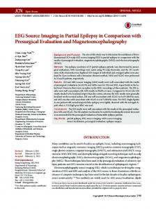
brain. A pre-surgical T1-weighted MRI was recorded with a resolution of 256 slices in axial, 256 slices in sagittal and 120 slices in coronal direction and a voxel-size of 0.86×1.6×0.86 mm. A post-surgical CT was taken with a resolution of 68 slices in axial, 512 in sagittal and 635 in coronal and a voxel-size of 0.49×0.49×2.65 mm. The CT and MR images are registered using methods in the ITK Toolbox (www.itk.org). The registered dataset was segmented into scalp, skull, cerebrospinal fluid (CSF), brain, plastic sheet, and intracranial electrodes. Figure 1 shows the BEM meshes for the skull, plastic sheet, and the scalp. Here, the CSF is not modeled for simplicity. The resulting model is used in forward/inverse problem (FP/IP) calculations to localize independent sources of simultaneously recorded iEEG and sEEG data.
(a)
(b)
(c) Fig. 1. BEM model of the scalp, skull and the plastic sheet, represented by 10000, 30000, and 7000 faces, respectively. (a) Skull and electrode sheet faces, (b) scalp, skull and sheet faces, (c) plastic sheet model of the plastic grid and strip electrode matrices.
A series of simulations were produced to study the effects of head modeling on the forward problem calculations [16]. It was shown that it is crucial to use an accurate head model for correct source localization. III. ICA AND S OURCE L OCALIZATION This section describes the Independent Component Analysis of the iEEG and sEEG recordings. Source localization is done for selected components using the realistic BEM head model. The ICA decomposition and source localization are described in the following sections. A. Independent Component Analysis Infomax Independent Component Analysis (ICA) has proven to be an effective method for removing eye and muscle activity artifacts from scalp EEG data, thus increasing the potential signal-to-noise ratio of subsequent analyses [6]. ICA can also identify and separate functionally independent components, which for normal scalp EEG prove to be most often associated with scalp maps matching the projection of a single equivalent current dipole.
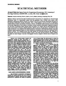
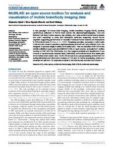
In this work, fifteen minutes of 78 iEEG data from subdural electrodes (Figure 2) from an epilepsy patient during two bursts of seizure were decomposed by extended infomax ICA ([6]) into 78 maximally independent component (IC) processes. ICA decomposition returned a vector of weights giving the relative strength and polarity of the projection of each IC source process to each of the electrodes, and an activation time series giving the time course of activity of each IC process during the data time period. Figure 3 shows maps of the projection patterns of two IC processes to the patient’s cortical surface in (a) and (b), and to the subdural electrode grids in (c) and (d). Patient’s cortex was derived from the patient MR head image by using Freesurfer (http://surfer.nmr.mgh.harvard.edu/). These two components were chosen because they represent orthogonal activity patterns. The IC shown in Figure 3 (a) and (c) projects to a small number of adjacent electrodes all with the same polarity, representing a gyral source. The IC on Figure 3 (b) and (d), on the other hand, is a sulcal source, projecting to two separate pools of adjacent electrodes with opposite polarities. The results of ICA decomposition applied to
is nearly universally assumed that proximal signal sources must dominate signals received by electrodes placed on or in the brain surface, despite the potential influences of volumeconducted potentials from all parts of the brain on each electrode. That is, it is currently assumed that each iEEG channel signal may be considered to be a locally generated signal independent of other more distal source activities. ICA decomposition, by removing or minimizing the presence of volume conducted signal summation at the electrodes, demonstrates that each recorded iEEG signal is in fact the sum of a number of more proximal and more distal source processes. For cortical grid electrodes, the percent variance accounted for by any single (and, typically, proximal) IC source ranges in our experience between about 20% and 80%. The two IC sources above account for at most 47% (left) and 22% (right) of channel variance at their maximally projecting channel. B. Patch-based source modeling We generated a realistic cortical source space including a large number of dipole elements oriented perpendicular to the local cortical surface from subject MR head images using tessellated FreeSurfer gray and white matter surfaces (surfer.nmr.mgh.harvard.edu). To create a multi-scale cortical patch basis on this surface, we selected seed points (single voxel dipoles), then extend each patch conformally to a set of gaussian-tapered patches in three scales with areas in the range 50-200 mm2. Figure 4 shows gaussian patches in three different scales. For our inverse problem analysis we calculated the forward problem for 80130 dipoles and generated a lead field matrix for 240390 patches.
Fig. 2. CT image of the implanted grid electrodes. The two grids (6 × 8, 4×6) and one medial strip (1×8) implanted in the patient for monitoring.
(a)
(a)
(b)
(c)
(d)
(b)
(c) Fig. 3. Potential maps of two IC processes projected on the brain (a) and (b), and on the intracranial electrodes (c) and (d). Red color of potentials represents positive values and the blue color represents negative values. Multiplying these maps by the (two-sided) IC time courses gives the activity at each channel associated with the IC source.
these and similar data force a reinterpretation of the nature of iEEG signals. In both clinical and research practice, it
Fig. 4. Three Gaussian patches in different scales with radius (a) 10 mm, (b) 6 mm, and (c) 3 mm.
C. Sparse patch-based inverse problem solution We solved the EEG inverse problem for this source by identifying sparse cortical regions responsible for generating independent EEG components. For this purpose, we applied
a sparse Bayesian learning (SBL) based method [14]. Figure 5 shows the cortical activity of IC maps shown in Figure 3 (red and blue (not seen here) indicate activity with opposite signs; green indicates no activity). Figure 3 (a) is a sulcal source, whereas (b) is a gyral source.
(a)
(b)
Fig. 5. Inverse problem results of the two components shown in Figure 3.
This may be the first time that volume-conducted and near-field portions of data recorded from the human cortex have been separated and used to localize EEG sources projecting to the cortical electrode grid. We believe these results constitute good preliminary evidence to support our hypothesis that we can use intracranial EEG recordings to image cortical sources, including those located within sulcal folds. IV. CONCLUSIONS AND FUTURE WORK Here, we analyzed intracranial EEG recordings using ICA and numerical forward and inverse solution methods and presented preliminary results of patch-based source localization of seizure data in an epilepsy patient. This research is expected to provide valuable insights into the dynamics of epilepsy and the electrophysiology of the human brain. As a next step, the generation and propagation of seizure will be investigated using this model. We will also investigate state transitions of ictal and interictal EEG data by applying multiple mixture ICA algorithms [17]. This will provide information about the ability of ICA to isolate seizure components. Simultaneously acquired scalp and intracranial EEG data during drowsy resting is also available for this patient. Source localization will be performed using scalp and intracranial EEG data and the relationship between noninvasive and invasive recordings of the electrical brain activity will be explored. R EFERENCES [1] B.A. Assaf, J.S. Ebersole, Continuous Source Imaging of Scalp Ictal Rhythms in Temporal lobe epilepsy, Epilepsia, vol. 38(10), 1997, pp 1114-1123. [2] Y. Zhang, L. Ding, W. van Drongelen, K. Hecox, D. M. Frim, B. He, A cortical potential imaging study from simultaneous extraand intracranial electrical recordings by means of the finite element method, NeuroImage, vol. 31, 2006, pp 1513-1524. [3] Z. Akalin Acar, N.G. Gencer, An advanced BEM implementation for the forward problem of Electro-magnetic source imaging, Physics in Med. and Biol., vol. 49(5), 2004, pp 5011-28. [4] N.G. Gencer, Z. Akalin Acar, Use of the isolated problem approach for multi-compartment BEM models of electro-magnetic source imaging, Physics in Med. and Biol., vol. 50, 2005, pp 3007-22.
[5] J.S. Ebersole, S. Hawes-Ebersole, Clinical application of dipole models in the localization of epileptiform activity. J. Clinical Neurophysiology, vol. 24(2), 2007, pp 120-129. [6] S. Makeig, A. J. Bell, T-P. Jung, and T. J. Sejnowski, Independent component analysis of electroencephalographic data, In: D. Touretzky, M. Mozer and M. Hasselmo (Eds). Advances in Neural Information Processing Systems 8:145-151 MIT Press, Cambridge, MA; 1996. [7] M. Jing, S. Sanei, A novel constrained topographic independent component analysis for seperation of epileptic seizures, Comp. Intelligence and Neuroscience, vol. 2007, 2007. [8] K. Kobayashi, C.J. James, T. Nakahori, T. Akiyama, J. Gotman, Isolation of epileptiform discharges from unaveraged EEG by independent component analysis, Clinical Neurophysiology, vol. 110, 1999, pp 1755-1763. [9] H. Nam, T.G. Yim, S.K. Han, J.B. Oh, S.K. Lee, Independent Component Analysis of ictal EEG in medial temporal lobe epilepsy, Epilepsia, vol. 43, 2002, pp 160-164. [10] C.M. Michel, M.M. Murray, G.L. Lantz, S. Gonzalez, L. Spinelli, R. G. de Peralta, EEG source imaging, Clinical Neurophysiology, vol. 115, 2004, pp 2195-2222. [11] S. Baillet, G. Garnero, A Bayesian approach to introducing anatomofunctional priors in the EEG/MEG inverse problem, IEEE Trans. on Biomed. Eng., vol. 44(7), 1997, pp. 374-385. [12] C-H. Im, C. Lee, H-K. Jung, and S.Y. Lee, A New Neuronal Electrical Source Model Considering Electrophysiology to Simulate Realistic Electroencephalography (EEG) Forward Signals, IEEE Trans. on Magnetics, vol. 44(6), 2008. [13] D. Wipf, S. Nagarajan, A unified Bayesian framework for MEG/EEG source imaging, NueroImage, vol. 44, 2009, pp. 947966 [14] D. Wipf, R.R. Ramirez, J.A. Palmer, S. Makeig, B.D. Rao, Analysis of empirical Bayesian methods for neuroelectromagnetic source localization, NIPS, pp. 1505-1512, 2007. [15] Z. Akalin Acar, S. Makeig, Neuroelectromagnetic forward modeling toolbox, Proc. of IEEE EMBC 2008, Vancouver, Canada. [16] Z. Akalin Acar, S. Makeig, G. Worrell, Head modeling and cortical localization in epilepsy, Proc. of IEEE EMBC 2008, Vancouver, Canada. [17] J.A. Palmer, K. Kreutz-Delgado, B. D. Rao, S. Makeig, Modeling and Estimation of Dependent Subspaces, Proceedings of the 7th International Conference on Independent Component Analysis and Signal Separation, 2007. [18] RR Ramrez and S. Makeig, Neuroelectromagnetic source imaging using multiscale geodesic neural bases and sparse Bayesian learning, 12th Annual Meeting of the Organization for Human Brain Mapping, Florence, Italy, 2006.