Pathogenesis, prevention, and management of ... - Wiley Online Library
Recommend Documents
in mucoid ground substance.29 Collagen fibril diameter is .... degenerated tendon in an effort to restart the healing response, similar to .... and acl reconstruction.
of candidemia is in the range of 5 per 1000 Paediatric Intensive Care. Unit (PICU) ..... mentation of central- line insertion and maintenance bundles that.
Aug 15, 2013 - stream schools, only a fifth consumed at least two servings of fruits and vegetables every day, and a tenth ..... and affordable. With half of Singapore ... agency under the Ministry of Trade and Industry respon- sible for helping ...
was based on an earlier guideline on the management of postpartum haemorrhage (PPH) ... of haemorrhage as it reduces the
Jul 9, 2014 - analyzed using ImageJ software (http://rsbweb.nih.gov/ij/download.html) (bottom). C Confocal images .... ZIP13 interaction is important for both the normal steady-state turn- ... ER-stress-responsive molecules was comparable between the
Traumatic neuroma formation results in persistent post-operative pain after amputation, which reduces quality of life in patients. In article number 1700876, Yixia ...
Curr Opin Immunol 2008;20:3â9. 11. Lich J, Williams K, Moore C, Arthur J, Davis B, Taxman D, et al. Monarch-1 suppresses non-canonical NF-κB activation and ...
Fred Stephen Sarfo,1 Richard Phillips,1. Mark Wansbrough-Jones2 ...... Guenin-Mace, L., Baron, L., Chany, A.C., Tresse, C., Saint-. Auret, S., Jonsson, F., et al.
virus to sialoadhesin activates a clathrin-mediated inter- nalization. The viruses end up in early endosomes where a pH drop is necessary for the disassembly ...
Nov 3, 2011 - ment of a target joint, from 20 091 Canadian dollars ($CDN) .... Diagram of a normal synovial joint: The articular cartilage provides a cushion to resist the load of weight ...... Muir, H. (1995) The chondrocyte, architect of car-.
long-term effectiveness of integrating mental health education with other academic curriculum such as language arts or science. Keywords: mental health ...
weakest inflammatory responses In the popliteal lymph node after challenge with live organisms in the footpad. Nevertheless, this sirain cleared Candida from ...
Jan 23, 2012 - Social determinants of health .... employing media campaigns, screening, counseling, ... and beauty salons as BP screening and control sites.
Mar 25, 2015 - On the other hand, there are obvious advantages of high parenteral doses (1â2 mg) of HOCbl in treating inborn errors of Cbl metabolism.
obesity in adolescents. Keywords: concept mapping, low SES, overweight, youth. Introduction. The high prevalence of childhood and adolescent obesity poses a ...
cost.6. There are also inconsistent data comparing hydrofiber and alginate dressings.7,8. One study ...... operatively using tranexamic acid, cell salvage, and induced ..... Enhancing patient recovery following lower limb arthroplasty with a.
on inventory management, e.g. blood substitutes, pathogen inactivation ...... [email protected] ... software. Our inventory level is being placed on a level of 5 days inventory and for O+, O) and A) it's a 7 days inventory. ..... business continuity.
For copies in excess of 25 or for commercial purposes, please contact Sarah Howell at [email protected] ..... 12 Cooper RS, Liao Y, Rotimi C. Is hypertension.
New insights into the pathogenesis of perinatal hypoxic-ischemic brain injury. Brankica ... natology, 26 Višegradska Street, 11000 Belgrade, Serbia. Email:.
Dec 4, 2013 - Yamagiwa's contribution are attached by way of introduction. It was a milestone for cancer research when Rudolf Virchow, in Berlin, established ...
Department of Microbiology, Kumamoto University School of Medicine, 2-2-1 Honjo, Kumamoto, Kumamoto 860, Japan. Received June 26, 1996. Key words: ...
blue; Griffonia simplicifolia lectin; nucleoside diphosphatase ... isolated retinae were incubated with the Griffonia simplicifolia (GS) lectin or reacted for.
Jan 29, 2014 - ataxia-telangiectasia (A-T) that is characterized by early-onset dystonia and ... Ataxia-Telangiectasia (A-T) is a rare autosomal recessive.
Pathogenesis, prevention, and management of ... - Wiley Online Library
Jun 21, 2017 - Pathogenesis, prevention, and management of bleeding and thrombosis in patients with liver diseases. Ton Lisman PhD | Robert J. Porte MD, ...
|
Received: 24 May 2017 Accepted: 21 June 2017 DOI: 10.1002/rth2.12028
REVIEW ARTICLE
Pathogenesis, prevention, and management of bleeding and thrombosis in patients with liver diseases Ton Lisman PhD
| Robert J. Porte MD, PhD
Surgical Research Laboratory and Section of Hepatobiliary Surgery and Liver Transplantation, Department of Surgery, University of Groningen, University Medical Center Groningen, Groningen, the Netherlands Correspondence Ton Lisman, Department of Surgery, University Medical Center Groningen, Groningen, the Netherlands. Email: [email protected]
Abstract Patients with liver diseases may develop alterations in all components of the hemostatic system. Thrombocytopenia, low levels of coagulation factors and inhibitors, low levels of fibrinolytic proteins, and increased levels of endothelial-derived proteins such as von Willebrand factor are all part of the coagulopathy of liver disease. Due to concomitant changes in pro- and antihemostatic drivers, the net effects of these complex hemostatic changes have long been unclear. According to current concepts, the hemostatic system of patients with liver disease is in an unstable balance, which explains the occurrence of both bleeding and thrombotic complications. This review will discuss etiology and management of bleeding and thrombosis in liver disease and will outline unsolved clinical questions. In addition, we will discuss the role of intrahepatic activation of coagulation for progression of liver disease, a novel paradigm with potential consequences for the general management of patients with liver disease. KEYWORDS
Essentials • Patients with liver diseases may acquire substantial changes in all components of hemostasis. • Hemostasis is in unstable balance due to simultaneous changes in pro- and antihemostatic systems. • Intrahepatic activation of hemostasis may contribute to disease progression. • Optimal strategies for prevention and treatment of bleeding and thrombosis are currently unknown.
1 | INTRODUCTION
The net effects of the complex hemostatic changes in liver diseases have long been unclear. In the next sections, we will provide arguments
The liver is a central organ in the homeostasis of the hemostatic sys-
for a “reset” balance in the hemostatic system in most patients with cir-
tem. The liver synthesizes the majority of plasma proteins involved in
rhosis and acute liver failure.4,5 Interestingly, any of the components of
hemostasis including pro- and anticoagulant factors, pro- and antifi-
the hemostatic system may be simultaneously altered in patients with
brinolytic factors, and thrombopoietin. In patients with advanced liver
liver diseases. The fact that the hemostatic system can deal relatively
diseases complex alterations in the hemostatic system arise which are
well with such extensive changes teaches us about the resilience of the
summarized in Table 1.1 Although it is commonly assumed many of these
hemostatic system in general. In this review we will summarize labora-
changes are related to decreased hepatic synthesis, a consumptive coag-
tory and clinical features of the altered hemostatic system in patients
2,3
ulopathy of systemic or intrahepatic origin may also contribute.
2 | WHY LIVER DISEASES WERE PREVIOUSLY CONSIDERED AS BLEEDING DISORDERS Routine tests of hemostasis such as the prothrombin time (PT), activated partial thromboplastin time (APTT), and platelet count are frequently abnormal in these patients and these test results all indicate a hypocoagulable status. In patients with acute liver failure, abnormal routine hemostasis tests are present per definition since an international normalized ratio (INR) >1.5 is part of the definition of the syndrome. Although the use of the INR in this context seems peculiar since the INR was developed and validated only for monitoring of vitamin K antagonists, the hepatology community misuses this test extensively. It is not only part of the definition of acute liver failure, but also part of the prognostic score that are used to prioritize patients with cirrhosis on the waiting list for a liver transplant. Ironically, there is immense laboratory-to-laboratory variation in the INR in plasma samples from patients with liver disease.6,7 This way, transplant candidates in centers with reagents yielding lower INR results may be at increased risk for dying on the waiting list. Two clinical observations appear to agree with liver diseases as being a bleeding disorder. First, spontaneous bleeding complications in patients with cirrhosis are common. However, it has now been well established that the most common bleeding complication, ruptured
was introduced as a standard clinical procedure in the 1980s, bleeding complicated most, if not all, procedures.11,12 Massive amounts of blood products (red cell concentrates, plasma, and platelet concentrates) were required in many liver transplant procedures. The explanation for the massive blood loss during surgery was believed to be the preoperative coagulopathy which further aggravated during the (lengthy) surgical procedure.13 During the last two decades, however, there has been a tremendous decline in transfusion requirements during liver transplantation. In fact, more and more centers report that in a proportion of patients liver transplant procedures can now be performed without the requirement for any blood products.14–16 Part of the decline in transfusion requirements may be related to improvements in surgical and anesthesiological techniques and improvements in donor organ quality and preservation. However, the key factor in the decrease in transfusion requirements has been the understanding that preoperative correction of the abnormal routine hemostasis test results is not required, and may even do more harm than good. Preoperative correction of thrombocytopenia and elevated PT and APTT test results inevitably requires administration of substantial amounts of volume. In the liver disease patient with portal hypertension, increased plasma volume, and disturbed cardiac function, administration of fluids results in a further increase in portal and central venous pressure. Thus, when platelet concentrates and plasma are administered with the aim to improve the hemostatic status, the increased portal and central venous pres-
esophageal varices, is unrelated to a defective hemostatic system.8
sure may in fact promote bleeding when surgical damage is inflicted.17
Rather, this bleeding event relates to local vascular abnormalities in combination with portal hypertension. Also in acute liver failure, bleeding was common in studies presented in the 1970s.9 At that time, around one-third of patients with acute liver failure died with bleeding as the proximate cause of death. In a recent series, however,
3 | REBALANCED HEMOSTASIS IN LIVER DISEASES
spontaneous and clinically significant bleeding is rare at around 5%, and bleeding very rarely results in death.10 The reasons for this sub-
The PT and APTT are only sensitive for procoagulant proteins, and
stantial reduction in bleeding are unclear, but it has to be noted that
are therefore unlikely to predict the hemostatic status of patients
the intensive care management of patients with acute liver failure has
with complex hemostatic alterations. In patients with liver diseases,
been revolutionized since the 1970s. Second, bleeding during inva-
both pro- and anticoagulant factors may be present in decreased
sive procedures was a substantial problem. When liver transplantation
levels, and the hemostatic status of such patients can only be
Changes that impair hemostasis
Changes that promote hemostasis
Thrombocytopenia
Elevated levels of von Willebrand Factor (VWF)
Platelet function defects
Decreased levels of ADAMTS-13
Enhanced production of nitric oxide and prostacyclin
Elevated levels of factor VIII
Low levels of factors II, V, VII, IX, X, and XI
Decreased levels of protein C, protein S, antithrombin, α2-macroglobulin, and heparin cofactor II
Vitamin K deficiency
Low levels of plasminogen
Dysfibrinogenemia Low levels of α2-antiplasmin, factor XIII, and TAFI Elevated t-PA levels
Source: Modified from the European Association for the Study of the Liver from Lisman et al.1 with permission.
T A B L E 1 Alterations in the hemostatic system in patients with liver disease that impair (left) or promote (right) hemostasis
|
3
LISMAN and PORTE
appreciated by using tests that take the balance between pro- and
of thrombosis, which may be attributable to the high VWF levels,45,46
anticoagulant factors into account. Similarly, the status of the pri-
although alternative explanations for the thrombotic risk of throm-
mary hemostatic system and the fibrinolytic system can only be as-
bopoietin receptor agonists in these patients cannot be excluded. On
sessed using global tests. Using modern thrombin generation tests,
the other hand, it has been suggested that thrombocytopenia (and not
in which thrombomodulin was added to allow for activation of the
prolonged routine coagulation tests) increase the risk for procedure-
protein C system, it was shown that thrombin generation in patient
associated bleeding,47 although not all studies agree.48
plasma was comparable to that of controls, despite substantially pro-
Thrombin generation tests have shown normal-to-increased hemo-
longed PT and APTT values in the patients.18 Thus, a concomitant
static potential in plasma from patients with cirrhosis when thrombin
decline in pro- and anticoagulant factors results in a reset balance
generation tests were performed in the presence of thrombomod-
in the coagulation system, and a similar argument applies for plate-
ulin.18,23–27 Initially, it was concluded that the endogenous thrombin
Some studies have even shown
potential (ETP) in cirrhosis is normal18, but multiple subsequent studies
evidence for enhanced hemostatic capacity in patients with liver dis-
have actually shown increased thrombin generation.23–27 Increasingly,
eases, which is in sharp contrast to the old dogma of liver diseases
the coagulation status of a patient with cirrhosis is not reported as the
let function and fibrinolysis.
as a bleeding disorder.
23–29
19–22
Below we will separately address labora-
thrombin generating capacity, but rather as a ratio between thrombin
tory and clinical evidence for rebalanced hemostasis in cirrhosis and
generation tests performed in absence and presence of thrombomod-
acute liver failure.
ulin, or as ratios between FVIII and protein C.49–51 Unfavorable ratios are referred to as “procoagulant imbalance,” which we feel is confusing
3.1 | The hemostatic status in patients with cirrhosis
terminology. We have argued against the use of ETP or FVIII/protein C ratios as estimates of hemostatic capacity as these ratios may be
Mild to moderate thrombocytopenia is common in cirrhosis, and
misleading, are difficult to interpret, and have no clear clinical correla-
older studies have also indicated functional platelet defects.30–32
tion.52 Rather, we feel that ETP results obtained in the presence of
Thrombocytopenia in cirrhosis is multifactorial as reviewed previ-
thrombomodulin are the most adequate representation of coagulation
ously.1,33 An unexplored potential contributor to the thrombocytope-
status in patients with complex alterations in their hemostatic system.
nia of liver disease are circulating histones. Histones have been shown
Nevertheless, these studies have demonstrated that the PT and APTT
to induce thrombocytopenia in mice,34 and have been implicated in
are inadequate to estimate the hemostatic status of patients with
thrombocytopenia in patients admitted to intensive care.
35
As levels
cirrhosis.
of circulating histones are slightly elevated in patients with cirrhosis,
Besides preserved thrombin generating capacity, we have recently
and substantially elevated in patients with acute liver failure,36 the
demonstrated procoagulant properties of the fibrinogen molecule.
role of histones in the thrombocytopenia of liver disease should be
Despite reduced fibrinogen plasma levels, fibrin clot permeability, a
further explored. Next to their thrombocytopenia, patients with cir-
measure of clot structure and quality, was decreased in patients with
rhosis may have a prolonged skin bleeding time indicative of platelet
cirrhosis, which was attributable to oxidative modifications in the fi-
defects.37 However, more recent data suggest that intrinsic platelet
brinogen molecule.29
28
Cirrhosis has long been thought to be associated with a hyperfi-
although net platelet function may be decreased due to thrombocy-
brinolytic status related to increased levels of tissue-type plasminogen
topenia and/or anemia.39 Technical issues complicate the interpre-
activator which are insufficiently balanced by fibrinolytic inhibitors.53
40
tation of many published studies on platelet function in cirrhosis.
However, using a global plasma-based assay we demonstrated that the
Laboratory studies using flow-based models have shown that the
fibrinolytic system in cirrhosis was rebalanced due to a commensurate
thrombocytopenia of cirrhosis may be balanced by highly elevated
decline in pro- and antifibrinolytic factors.19 Importantly, a typical fi-
function in patients with cirrhosis is normal
20,38
levels of von Willebrand factor (VWF).
38
or even hyperactive,
In addition, plasma levels
brinolytic bleeding rarely occurs in non-surgical patients with cirrho-
of a disintegrin and metalloproteinase with a thrombospondin type 1
sis, which supports the laboratory evidence of rebalanced fibrinolysis.
motif, member 13 (ADAMTS13) are decreased in patients with liver
Nevertheless, some studies using plasma-based or whole blood assays
disease.41 Decreased ADAMTS13 does not result in an increase in the
indicating accelerated fibrinolysis in cirrhosis have questioned our
multimeric size of VWF. In fact, a decrease of higher molecular weight
findings.54,55 Conversely, in large series of patients that were studied
multimers is present in plasma from patients with cirrhosis, which is
with thromboelastography, none had a fibrinolysis rate outside of the
presumably related to proteolysis by other VWF-cleaving proteases
reference range.25,56
such as plasmin.42,43 Nevertheless, decreased ADAMTS13 in cirrhosis
Whole blood clot formation as tested by viscoelastic tests have
may promote primary hemostasis, as ADAMTS13 is also responsible
been demonstrated to be normal in patients with cirrhosis,56 but
for regulation of thrombus growth by proteolysis of VWF within a
also number of studies have demonstrated profound hypocoagula-
growing thrombus.44 Clinical evidence for an important compensa-
bility.57–60 Viscoelastic tests have (whole blood) clot formation as the
tory role of VWF in the thrombocytopenia of cirrhosis may be de-
endpoint, which is an obvious advantage to thrombin generation tests
duced from studies in which thrombopoietin receptor agonists have
or routine diagnostic tests such as the PT, which have thrombin gen-
been used to increase the platelet count in patients with cirrhosis.
eration or the time point of fibrinogen to fibrin conversion in plasma
Elevation of the platelet count was associated with an increased risk
as the endpoint. In addition, viscoelastic tests are sensitive for the
|
LISMAN and PORTE
4
activity of coagulation factor XIII.61 However, an obvious limitation of
weight multimers was detected, which is likely related to VWF pro-
viscoelastic tests in patients with complex disorders of hemostasis is
teolysis by proteases other than ADAMTS13.20 Interestingly, levels
that it is not a true representation of hemostatic balance as it lacks
of ADAMTS13 measured on admission to the hospital have been re-
activation of the anticoagulant protein C system, and is insensitive for
lated to outcome, which has been proposed to be a consequence of
von Willebrand factor. Given the profound changes in VWF and the
increased formation of intrahepatic platelet thrombi in those patients
protein C pathway in cirrhosis, it appears unlikely that thromboelas-
with low levels of ADAMTS13.
tography forms a valid representation of overall hemostatic balance in these patients.
The alterations in coagulation proteins are more extensive in patients with acute liver failure compared to patients with cirrho-
It has to be noted that most studies on the hemostatic balance
sis. Levels of the liver-derived factors are substantially decreased,
in patients with cirrhosis have been performed in mixed cohorts of
and can become as low as 1–10% of normal.21,68 Nevertheless,
relatively well-compensated patients. Although hemostatic changes in
thrombomodulin-modified thrombin generation in patients with acute
patients with cirrhosis from different etiologies are similar, they are
liver failure was shown to be normal or increased relative to healthy
certainly not identical. For example, patients with cholestatic cirrho-
controls.21,68 In addition, thromboelastography test results are consis-
sis appear in a more hypercoagulable state as compared to patients
tent with rebalanced hemostasis in acute liver failure.69
with noncholestatic cirrhosis,
62
and patients with nonalcoholic fatty
In contrast to cirrhosis, acute liver failure is characterized by a pro-
liver disease associated cirrhosis are somewhat more prothrombotic
found hypofibrinolytic status, which is likely related to substantially
as compared to patients with alcohol-induced cirrhosis.63 Also, it
elevated plasma levels of PAI-1 and low plasminogen levels.21
has been hypothesized that the hemostatic balance may be no longer maintained in patients with decompensated disease. However, in recent studies we demonstrated normo- to hypercoagulability in patients with acutely decompensated cirrhosis and acute-on-chronic
3.3 | Clinical evidence of intact hemostatic capacity in cirrhosis and acute liver failure What clinical evidence exists to support the theory of rebalanced
liver failure using thrombin generation tests (unpublished data). Although the hemostatic status of patients with cirrhosis appears
hemostasis in liver disease and to refute the value of routine diag-
in balance, there are clear hypo- and hypercoagulable features which
nostic tests such as the PT and platelet count in assessing hemo-
may contribute to bleeding or thrombosis. Hypocoagulable features
static competence in liver diseases? First, more and more centers
include hypofibrinogenemia,29 decreased clot formation and sta-
report transfusion-free liver transplantation in a substantial pro-
bility in some studies using viscoelastic testing,60 delayed fibrin po-
portion of patients.14–16 Although transfusion requirements still
64
lymerization,
65
and hyperfibrinolysis.
include platelet hyperreactivity,
28
Hypercoagulable features
vary widely between centers,70 there is no doubt that this lengthy
23–27
procedure with substantial surgical damage does not per se result
enhanced thrombin generation, 66
increased production of intravascular tissue factor,
and prothrom-
botic properties of the fibrin clot.29
in major blood loss. A recent study from a single center showed that almost 80% of a series of 700 consecutive patients were transplanted without any transfusion requirements.16 These data strongly argue against liver disease being associated with a bleed-
3.2 | The hemostatic status of patients with acute liver failure
ing diathesis. Secondly, although bleeding is common is cirrhosis,
Thrombocytopenia is less common in patients with acute liver failure
(eg, bleeding from ruptured esophageal varices). Bleeding problems
as compared to cirrhosis. The pathophysiology of thrombocytopenia
such as bruising, purpura, epistaxis, gingival bleeding, menorrhagia,
in acute liver failure has not been extensively addressed, but recent
and bleeding associated with invasive procedures may be related
most bleeding episodes are unrelated to hemostatic dysfunction
studies indicate that platelet activation may contribute.67 Substantially
to defective hemostasis. However, in some cases, elevated venous
elevated levels of highly procoagulant microparticles, mainly from
pressure may contribute to bleeding problems that at first sight ap-
platelet origin, have been demonstrated in plasma from patients with
pear as a consequence of deranged hemostasis. Interestingly, the
acute liver failure. These microparticles have been suggested to be
extent of coagulopathy as measured by the PT or platelet count
the results of platelet fragmentation in a process resembling dissemi-
does not appear predictive of bleeding complications.71 In acute
nated intravascular coagulation of sepsis. Indeed, intravascular (or in-
liver failure, clinically significant bleeding is rare, and in contrast
trahepatic) activation of hemostasis has been demonstrated in animal
to patients with cirrhosis bleeding from esophageal varices is vir-
models of acute liver failure as will be outlined in the section on intra-
tually absent, which is explained largely by the usual absence of
hepatic thrombosis as a contributor of disease progression. Whether
portal hypertension. Third, patients with liver disease are not pro-
the circulating procoagulant microparticles have a role in hemostasis
tected from thrombotic events, and increasing clinical data refute
has not been directly investigated.
the twentieth-century concept of patients with liver diseases being
Patients with acute liver failure, similar to patients with cirrhosis,
“auto-anticoagulated.” Cirrhosis has been identified as a risk fac-
have highly elevated levels of VWF and substantially decreased lev-
tor for venous thrombosis,72 and in patients with acute liver failure,
els of ADAMTS13.22 Although ADAMTS13 levels are undetectable
thrombotic complications are even more common than bleeding in
in a proportion of patients, a reduced proportion of high molecular
patients with acute liver failure in recent series.69
|
5
LISMAN and PORTE
F I G U R E 1 The hemostatic balance in patients with liver disease as compared to that of healthy individuals. This cartoon depicts the stable hemostatic balance in healthy individuals and shows that although the hemostatic system in patients with liver disease is (re)balanced, the balance is fragile and may easily tip to either a hypo- or a hypercoagulable status. Modified from the European Association for the Study of the Liver from Lisman et al.116 with permission
Prohemostatic factors
Antihemostatic factors
Prohemostatic factors
Normal liver
3.4 | The limited stability of the hemostatic balance in liver diseases Although laboratory and clinical evidence supports the concept of rebalanced hemostasis in the “average” patient with liver disease,
Antihemostatic factors
Cirrhotic liver
4 | PREVENTION AND MANAGEMENT OF BLEEDING IN LIVER DISEASES 4.1 | Bleeding in cirrhosis
the new hemostatic balance appears much more fragile as com-
We will limit our discussion to the management of “hemostatic” bleed-
pared to the hemostatic balance in patients with intact liver function
ing. Bleeding due to mechanical causes such as ruptured esophageal
(Figure 1). The limited balance of hemostasis in liver disease is in
varices and true surgical bleeds are beyond the scope of this review.
part explained by the low levels of pro- and antihemostatics, but
Prevention of bleeding during invasive procedures starts with a very
may very well also be related to the distinct hypo- and hyperco-
restrictive fluid infusion policy the rationale for which is extensively
agulable features of the patient with liver disease as discussed in
outlined by us elsewhere.75,76 As is evident from the liver transplant
the paragraph on “The hemostatic status in patients with cirrhosis.”
data, routine correction of abnormal routine test of hemostasis (plate-
As depicted in Figure 1, the patient with liver disease is constantly
let count, PT, APTT) by administration of platelet concentrates or
struggling to remain in hemostatic balance as a result on the low lev-
plasma is not indicated and might even do harm.77 Undesired side-
els of hemostatic factors on each end of the hemostatic scale and by
effects of blood product administration include fluid overload and ex-
dynamic changes in pro- and antihemostatic processes. Therefore,
acerbation of portal hypertension (which may paradoxically increase
it is likely difficult to predict using laboratory tests or clinical scores
bleeding risk) in addition to general transfusion-related side effects. In
which patient is more likely to tip towards a bleeding diathesis and
addition, complete correction of prolonged coagulation tests is almost
which one is more likely to develop thrombosis. The clinical reality is
never achieved.78 Although some investigators argue that a correc-
that patients may present with bleeding and thrombosis simultane-
tion of the platelet count may be helpful in preventing bleeding in
ously, and obviously management of such patients is a particularly
high risk procedures,71 clinical data substantiating this are lacking. It
difficult clinical challenge.
is even debated whether a low preprocedural platelet count increases
Furthermore, a number of disease-related factors can actively tip
the balance towards bleeding or thrombosis. Factors that promote
tion of platelet concentrates are still common, and thrombopoietin
bleeding include renal failure and infection, both of which are com-
receptor agonists have been tested in clinical trials and are consid-
mon in liver disease.73,74 In addition, alterations in flow, endothelial
ered as potential alternatives for platelet transfusions,79 although they
activation, disruption of the endothelial glycocalix, and generation of
may be associated with an risk for thrombotic events.46 Based on our
procoagulant microparticles all potentially predispose to a thrombotic
experience in liver transplantation, we believe it is best to wait with
phenotype.
blood product administration until a hemostatic bleed actually occurs.
|
LISMAN and PORTE
6
Prophylactic administration of antifibrinolytics have been shown to
drugs may be desired or deemed essential in selected cases. The un-
substantially reduce blood product use during liver transplantation,
predictable pharmacokinetics of anticoagulant drugs necessitates
and given the relative safety of such agents, they may be helpful in
careful monitoring, which is a particular challenge in patients with liver
a prophylactic setting.80 Prophylactic correction of coagulation by
diseases. Monitoring of vitamin K antagonists is difficult since the INR
low volume products such as prothrombin complex concentrates has
is already prolonged at baseline in patients with advanced disease. In
theoretical advantages over administration of plasma and the effect
such patients, the appropriate target INR has not been established. In
of such agents on blood loss during liver transplantation is currently
addition, there is tremendous laboratory-to-laboratory variation in the
being investigated in a randomized controlled clinical trial.81 Other
INR in liver disease patients, which complicates studies on optimal tar-
measures that are likely helpful in preventing bleeding is adequate
get ranges.6 Recent studies have indicated that monitoring of heparins
control of infections and renal failure. Finally, a structured bleeding
by standard anti-Xa assays give a substantial underestimation of the
history should be part of the work-up of patients with liver diseases
true circulating heparin mass.91,92 In clinical practice the use of anti-Xa
for (elective) invasive procedures, although admittedly the value of
monitoring of heparins may therefore lead to incorrect and potentially
this screening method for predicting bleeding in this population has
dangerous dose escalations. Only anti-Xa assays in which excess exog-
not been thoroughly investigated.
enous antithrombin is present in the reagent give accurate anti-Xa lev-
When bleeding does occur, it is a challenge to decide on the most
els in plasma from patients with cirrhosis.91 The second caveat of using
effective pro-hemostatic therapy as bleeding may be caused by failure
antihemostatic drugs in patients with liver disease is altered drug po-
of any of the hemostatic systems (platelets, coagulation, fibrinolysis).
tency. The complex hemostatic changes of liver disease were recently
Thromboelastography may be helpful in deciding which factor(s) re-
shown to impact drug potency (at least in vitro).92–94 Using in vitro
quire correction. Although thromboelastography-based transfusion
thrombin generation tests it has been demonstrated that the extent by
algorithms are rapidly gaining popularity,82,83 convincing clinical ev-
which thrombin generation is inhibited by various anticoagulant drugs
idence that such algorithms lead to an optimal restoration of hemo-
may differ tremendously between patients and controls. The differ-
static capacity is lacking. Pros and cons of various prohemostatic
ential effects were proportional to the severity of disease indicating
strategies in patients with liver diseases have been discussed by us
that the extent of alteration in the hemostatic system determines the
elsewhere.84
deviation in potency. Confusingly, some drugs appear to have an increased anticoagulant potency, whereas others are less effective in
4.2 | Bleeding in acute liver failure
plasma from patients with cirrhosis. Although it has not yet been established whether these in vitro alterations in potency are relevant in
Prophylactic administration of blood products in patients with acute
vivo, it may be that for optimal management of anticoagulant drugs in
liver failure is not recommended.10 Not only is the risk of spontane-
patients with liver diseases a monitoring test (or combination of tests)
ous or procedure-related bleeding low, administration of blood prod-
will be required that assesses both drug levels and drug potency.
ucts may cause worsening of intracranial hypertension. Moreover, the
As patients with cirrhosis are at risk for venous thrombosis, stan-
INR is an important prognostic indicator in acute liver failure, which
dard thromboprophylaxis should be applied upon immobilization,
is obscured by administration of plasma. Some authorities have rec-
hospitalization, and post-surgery, even in patients with a prolonged
ommended cautious prophylaxis with recombinant factor VIIa prior
INR. Anticoagulant drugs (LMWH, vitamin K antagonists) have been
to high-risk procedures, notably intracranial pressure monitors.85
used to treat established portal vein thrombosis, with resolution of the
However, the uncertain benefit and risk for thrombosis requires fur-
thrombus in a proportion of patients.95 Although the safety profile of
ther evaluation of this strategy before broad use in this setting can be
LMWH in this context has been excellent and much better than that of
86
recommended.
vitamin K antagonists, prolonged treatment may be required which is a burden for the patient. For this reason, the use of direct oral antico-
5 | PREVENTION AND MANAGEMENT OF THROMBOSIS IN LIVER DISEASES
agulants is considered by an increasing number of centers,96,97 despite the fact that there is virtually no clinical experience with these agents in patients with advanced liver disease, which were all excluded from the large randomized trials, and the theoretical issues with dosing and
Thrombotic events, including venous thrombosis and portal vein
monitoring. One study has suggested that LMWH is also effective in
thrombosis are frequent in patients with cirrhosis.72,87–90 In addition,
prevention of portal vein thrombosis (PVT),98 but the results of that
anticoagulation for arterial events and atrial fibrillation may be required.
study await confirmation.
There are two major issues with antihemostatic treatment of patients with liver diseases. First, the liver and kidneys are involved in metabolic activation or clearance of a number of the drugs we use on a day-to-day basis in general thrombosis management. This results
6 | INTRAHEPATIC THROMBOSIS AS A CONTRIBUTOR TO DISEASE PROGRESSION
in unpredictable pharmacokinetics and for that reason only, some of these drugs are contraindicated for patients with liver diseases.
The most exciting advance in the field of thrombosis in liver diseases
However, despite these contraindications, the use of contraindicated
is the accumulating evidence of intrahepatic activation of coagulation
|
7
LISMAN and PORTE
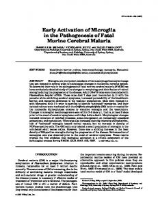
(A)
(B)
IIa
F I G U R E 2 Potential mechanisms involved in progression of liver disease by intrahepatic activation of hemostasis. (A) Intrahepatic activation of endothelial cells results in the formation of platelet microthrombi in the sinusoid. Such microthrombi result in disease progression via the results of microischemia of the downstream tissue. (B) Hepatocellular injury results in decryption of hepatocyte tissue factor, the generation of thrombin (IIa), and eventually fibrin deposition in the sinusoid. (C) Concomitant activation of hepatocytes and hepatic endothelium results in the formation of platelet and fibrin-containing microthrombi in the sinusoid. (D) Thrombin generated via decryption of hepatocyte tissue factor activates hepatic stellate cells to synthesize collagen
(C)
(D)
IIa
IIa
as a contributor to disease progression. In the 1980s it was first rec-
disease in acute liver failure. Activation of coagulation in this model
ognized that microthrombi can be found in rodent models of liver dis-
was demonstrated to rely on tissue factor expressed on hepato-
ease,99,100 and in the 1990s the first reports of microthrombi within
cytes.108 Whereas most tissue factor in a healthy liver is in an en-
human livers appeared.
101
A functional role of these microthrombi
crypted state, insults such as hepatocyte necrosis induced by acute
has been suggested based on experimental animal models and clinical
liver failure appear to result in tissue factor decryption, resulting in
studies. In models of chronic liver disease, anticoagulant or antiplate-
coagulation activation.
let drugs slow down the progression of disease.101,102 Conversely,
Observational studies in humans have suggested a faster disease
animals homozygous for factor V Leiden showed accelerated disease
progression of patients with fibrosis who were also carriers of factor V
progression.102 Confusingly, in some models of cholestasis-induced
Leiden, although not all studies agree.109,110 In addition, patients with
fibrosis, complete absence of platelets or fibrinogen paradoxically in-
hemophilia and hepatitis-related fibrosis appeared to have a slower
creases disease progression, suggesting that microthrombi may not
disease progression compared to patients with hepatitis without he-
always be harmful.103,104 Fibrin and platelets have also been impli-
mophilia.111 The combined results of experimental animal studies
cated in tissue repair mechanisms, which may partly explain the con-
and observational human studies have led to the proposal that an-
tradictory findings in some models.105,106 Nevertheless, the results of
ticoagulant drugs may be used as adjunct therapy in patients with
human studies outlined in the subsequent paragraphs of this section
early cirrhosis to prevent disease progression, decompensation, and
suggest that in humans the harmful actions of intrahepatic coagula-
delay or prevent liver transplantation or death.112 Indeed, a recent
tion activation are dominant.
randomized clinical study showed that prolonged daily administra-
In rodent models of acute liver failure, intrahepatic fibrin forma-
tion of low-molecular-weight heparin substantially delayed decom-
tion has also been demonstrated.107 Similar to result in rodent models
pensation and death, without significant side effects.98 Although
of fibrosis, administration of anticoagulants reduced progression of
the results of this study require confirmation, these findings could
• Which patients are at risk for procedural or spontaneous bleeding? • Can we identify such patients by laboratory tests or an algorithm including patient characteristics and laboratory data?
• Which patients are at risk for venous thrombosis or portal vein thrombosis? • Can we identify such patients a clinical score, laboratory tests or an algorithm including patients’ characteristics and laboratory data? • Does a (relative) hypercoagulable state accelerate disease progression in humans?
Management of bleeding
Management of thrombosis
• In vitro hemostatic responses to prohemostatic drugs may be very different in patients with liver–are dose-adjustments required because of altered hemostatic responses? • If so, are dose adjustments dependent on etiology and severity, and how should we decide on the extent of dose adjustment?
• In vitro hemostatic responses to antihemostatic drugs may be very different in patients with liver disease-related hemostatic changes–are dose-adjustments required because of altered hemostatic responses? • If so, are dose adjustments dependent on etiology and severity, and how should we decide on the extent of dose adjustment?
• How should treatment in the bleeding patient be guided (routine diagnostic tests, viscoelastic tests, others)?
• How to monitor VKAs in patients with elevated baseline INR? • anti-Xa tests underestimate and APTT overestimates heparin levels–alternative tests required? • Monitoring of DOACs required in these complex patients? • Accumulation of DOACs in patients with combined liver and renal failure?
• Which agents (FFP + platelets vs factor concentrates, role of antifibrinolytics and recombinant factor VIIa)? • What are optimal transfusion triggers and target ranges for the different agents?
• Which agents for which indication (VKAs vs. heparins vs. DOACs)? • Duration of anticoagulation? • Is anticoagulation for portal vein thrombosis (always) required?
• Is pre-procedural correction of hemostasis required to avoid procedural bleeding? • If so, which agents, and what about the risk/benefit ratio? • Alternatively, is a “wait-and-see” approach safe and effective? • Differences in approach between well compensated and acutely ill patients? • Differences in disease etiology (including chronic and acute liver diseases)? • Which patients require thromboprophylaxis to prevent venous thrombosis? • Does prophylactic anticoagulation prevent portal vein thrombosis? • If so, does this improve outcome? • Does prophylactic anticoagulation decrease rate of decompensation and death? • Which agents for which indication (VKAs vs. heparins vs. DOACs)? • Duration of anticoagulation? • Differences in approach between well compensated and acutely ill patients? • Differences in disease etiology (including chronic and acute liver diseases)?
Dosing
Monitoring
Treatment
Prevention
|
VKA, vitamin K antagonist; DOAC, direct oral anticoagulant.
Prediction
T A B L E 2 Unsolved clinical questions on hemostatic management in patients with liver diseases
8
LISMAN and PORTE
|
9
LISMAN and PORTE
potentially revolutionize the management of cirrhosis, possibly as part of a “polypill” program as proposed previously.112 Currently, two randomized controlled trials are assessing efficacy and safety of LMWH and rivaroxaban in patients with established cirrhosis with decompensation and survival as primary endpoints (clinicaltrials.gov identifiers NCT02643212 and NCT02271295). Whether antihemostatic treatment will also become part of the management of acute liver failure
ADDENDUM T. Lisman and R.J. Porte contributed equally to the development of the concepts outlined in the paper. T. Lisman wrote the initial version of the manuscript and R.J. Porte revised the paper. No funding for writing this review was obtained.
is less certain and clinical application may be limited by the perceived bleeding risk. Furthermore, a small study conducted in the 1970s found no evidence for a beneficial effect of heparin in patients with acute liver failure.113 Two hypotheses for the mechanism by which intrahepatic ac-
RELATIONSHIP DISCLOSURE We have no conflicts of interest to report
tivation of coagulation leads to disease progression have been proposed. Initially, the theory of “parenchymal extinction” was proposed, in which physical blockade of the microcirculation by platelets, fibrin, or a platelet and fibrin-containing thrombus is held responsible for the progression of disease.
114
In this theory, tissue ischemia caused
by these microthrombi is held responsible for the effects on disease progression. In recent years, a second hypothesis in which cellular activation by coagulation proteases is key in driving disease progression is gaining popularity. Thrombin (and factor Xa)-mediated activation of protease activated receptors on stellate cells results in activation of these cells resulting in collagen production by the stellate cells.115 Alternatively, the mechanism may involve thrombin activation of protease activated receptors on platelets resulting in the release of profibrogenic molecules such as platelet-derived growth factor.3 In models of acute liver failure activation of protease-activated receptors also contribute to disease progression, although it has not been established activation of which cell types are responsible for the effect.107 The potential mechanisms explaining the pathogenic role of coagulation activation for progression of liver disease are summarized in Figure 2. Clearly, the different pathways are not mutually exclusive.
7 | CONCLUSION The complex hemostatic changes that frequently occur in patients with liver diseases results in a “rebalanced” hemostatic system. This reset hemostatic balance, however, has a limited stability which explains the occurrence of both bleeding and thrombotic complications in these patients. Prediction, prevention management, and monitoring of bleeding or thrombosis in patients with liver diseases are complicated as a result of the extensive baseline changes in the hemostatic system. As such, it is very difficult to provide guidelines for management. Some management advice has been published previously,71,84,116–120 but we would like to stress that most advice is based on expert opinion only. Table 2 lists unsolved clinical questions that require additional study. Importantly, carefully designed clinical studies on the efficacy and safety of pro- and antihemostatic strategies need to be performed. An exciting area of research regards the use of antihemostatic agents to prevent progression of disease. Whether such strategies are truly effective and sufficiently safe awaits confirmation in larger clinical trials.
REFERENCES 1. Lisman T, Leebeek FW, de Groot PG. Haemostatic abnormalities in patients with liver disease. J Hepatol. 2002;37:280–7. 2. Bakker CM, Knot EA, Stibbe J, Wilson JH. Disseminated intravascular coagulation in liver cirrhosis. J Hepatol. 1992;15:330–5. 3. Anstee QM, Wright M, Goldin R, Thursz MR. Parenchymal extinction: coagulation and hepatic fibrogenesis. Clin Liver Dis. 2009;13:117–26. 4. Lisman T, Porte RJ. Rebalanced hemostasis in patients with liver disease: evidence and clinical consequences. Blood. 2010;116:878–85. 5. Lisman T, Stravitz RT. Rebalanced hemostasis in patients with acute liver failure. Semin Thromb Hemost. 2015;41:468–73. 6. Trotter JF, Olson J, Lefkowitz J, Smith AD, Arjal R, Kenison J. Changes in international normalized ratio (INR) and model for endstage liver disease (MELD) based on selection of clinical laboratory. Am J Transplant. 2007;7:1624–8. 7. Tripodi A, Baglin T, Robert A, Kitchen S, Lisman T, Trotter JF. Subcommittee on Control of Anticoagulation of the Scientific and Standardisation Committee of the International Society on Thrombosis and Haemostasis. Reporting prothrombin time results as international normalized ratios for patients with chronic liver disease. J Thromb Haemost. 2010;8:1410–2. 8. Garcia-Tsao G, Bosch J. Management of varices and variceal hemorrhage in cirrhosis. N Engl J Med. 2010;362:823–32. 9. Gazzard BG, Portmann B, Murray-Lyon IM, Williams R. Causes of death in fulminant hepatic failure and relationship to quantitative histological assessment of parenchymal damage. Q J Med. 1975;44:615–26. 10. Munoz SJ, Stravitz RT, Gabriel DA. Coagulopathy of acute liver failure. Clin Liver Dis. 2009;13:95–107. 11. Lewis JH, Bontempo FA, Cornell F, et al. Blood use in liver transplantation. Transfusion. 1987;27:222–5. 12. Farrar RP, Hanto DW, Flye MW, Chaplin H. Blood component use in orthotopic liver transplantation. Transfusion. 1988;28:474–8. 13. Porte RJ, Knot EA, Bontempo FA. Hemostasis in liver transplantation. Gastroenterology. 1989;97:488–501. 14. Ramos HC, Todo S, Kang Y, Felekouras E, Doyle HR, Starzl TE. Liver transplantation without the use of blood products. Arch Surg. 1994;129:528–32. 15. de Boer MT, Molenaar IQ, Hendriks HG, Slooff MJ, Porte RJ. Minimizing blood loss in liver transplantation: Progress through research and evolution of techniques. Dig Surg. 2005;22:265–75. 16. Massicotte L, Thibeault L, Roy A. Classical notions of coagulation revisited in relation with blood losses, transfusion rate for 700 consecutive liver transplantations. Semin Thromb Hemost. 2015;41:538–46. 17. Massicotte L, Lenis S, Thibeault L, Sassine MP, Seal RF, Roy A. Effect of low central venous pressure and phlebotomy on blood product
|
10
18.
19.
20.
21.
22.
23.
24.
25.
26.
27.
28. 29.
30.
31.
32.
33.
34. 35.
36.
transfusion requirements during liver transplantations. Liver Transpl. 2006;12:117–23. Tripodi A, Salerno F, Chantarangkul V, et al. Evidence of normal thrombin generation in cirrhosis despite abnormal conventional coagulation tests. Hepatology. 2005;41:553–8. Lisman T, Leebeek FW, Mosnier LO, et al. Thrombin-activatable fibrinolysis inhibitor deficiency in cirrhosis is not associated with increased plasma fibrinolysis. Gastroenterology. 2001;121: 131–9. Lisman T, Bongers TN, Adelmeijer J, et al. Elevated levels of von Willebrand factor in cirrhosis support platelet adhesion despite reduced functional capacity. Hepatology. 2006;44:53–61. Lisman T, Bakhtiari K, Adelmeijer J, Meijers JC, Porte RJ, Stravitz RT. Intact thrombin generation and decreased fibrinolytic capacity in patients with acute liver injury or acute liver failure. J Thromb Haemost. 2012;10:1312–9. Hugenholtz GC, Adelmeijer J, Meijers JC, Porte RJ, Stravitz RT, Lisman T. An unbalance between von willebrand factor and ADAMTS13 in acute liver failure: implications for hemostasis and clinical outcome. Hepatology. 2013;58:752–61. Gatt A, Riddell A, Calvaruso V, Tuddenham EG, Makris M, Burroughs AK. Enhanced thrombin generation in patients with cirrhosis- induced coagulopathy. J Thromb Haemost. 2010;8:1994–2000. Youngwon N, Kim JE, Lim HS, Han KS, Kim HK. Coagulation proteins influencing global coagulation assays in cirrhosis: hypercoagulability in cirrhosis assessed by thrombomodulin-induced thrombin generation assay. Biomed Res Int. 2013;2013:856754. Kleinegris MC, Habets CA, van de Sande AJ, et al. Liver cirrhosis is associated with hypercoagulability, decreased clot strength and normal fibrinolysis. J Thromb Haemost. 2013;11:49–50. Groeneveld D, Porte RJ, Lisman T. Thrombomodulin-modified thrombin generation testing detects a hypercoagulable state in patients with cirrhosis regardless of the exact experimental conditions. Thromb Res. 2014;134:753–6. Lebreton A, Sinegre T, Pereira B, Lamblin G, Duron C, Abergel A. Plasma hypercoagulability in the presence of thrombomodulin but not of activated protein C in patients with cirrhosis. J Gastroenterol Hepatol. 2017;32:916–24. Raparelli V, Basili S, Carnevale R, et al. Low-grade endotoxemia and platelet activation in cirrhosis. Hepatology. 2017;65:571–81. Hugenholtz GC, Mccrae FL, Adelmeijer J, et al. Procoagulant changes in fibrin clot structure in patients with cirrhosis are associated with oxidative modifications of fibrinogen. J Thromb Haemost. 2016;15:1054–66. Laffi G, Cominelli F, Ruggiero M, et al. Altered platelet function in cirrhosis of the liver: Impairment of inositol lipid and arachidonic acid metabolism in response to agonists. Hepatology. 1988;8:1620–6. Laffi G, Marra F, Gresele P, et al. Evidence for a storage pool defect in platelets from cirrhotic patients with defective aggregation. Gastroenterology. 1992;103:641–6. Laffi G, Marra F, Failli P, et al. Defective signal transduction in platelets from cirrhotics is associated with increased cyclic nucleotides. Gastroenterology. 1993;105:148–56. Violi F, Basili S, Raparelli V, Chowdary P, Gatt A, Burroughs AK. Patients with liver cirrhosis suffer from primary haemostatic defects? Fact or fiction? J Hepatol. 2011;55:1415–27. Fuchs TA, Bhandari AA, Wagner DD. Histones induce rapid and profound thrombocytopenia in mice. Blood. 2011;118:3708–14. Alhamdi Y, Abrams ST, Lane S, Wang G, Toh CH. Histone-associated thrombocytopenia in patients who are critically ill. JAMA. 2016;315:817–9. Wen Z, Lei Z, Yao L, et al. Circulating histones are major mediators of systemic inflammation and cellular injury in patients with acute liver failure. Cell Death Dis. 2016;7:e2391.
LISMAN and PORTE
37. Mannucci PM, Vicente V, Vianello L, et al. Controlled trial of desmopressin in liver cirrhosis and other conditions associated with a prolonged bleeding time. Blood. 1986;67:1148–53. 38. Lisman T, Adelmeijer J, de Groot PG, Janssen HL, Leebeek FW. No evidence for an intrinsic platelet defect in patients with liver cirrhosis– studies under flow conditions. J Thromb Haemost. 2006;4:2070–2. 39. Escolar G, Cases A, Vinas M, et al. Evaluation of acquired platelet dysfunctions in uremic and cirrhotic patients using the platelet function analyzer (PFA-100): influence of hematocrit elevation. Haematologica. 1999;84:614–9. 40. Caldwell S, Lisman T. The cirrhotic platelet: Shedding light on an enigma. Hepatology. 2017;65:407–10. 41. Uemura M, Fujimura Y, Matsumoto M, et al. Comprehensive analysis of ADAMTS13 in patients with liver cirrhosis. Thromb Haemost. 2008;99:1019–29. 42. Federici AB, Berkowitz SD, Lattuada A, Mannucci PM. Degradation of von Willebrand factor in patients with acquired clinical conditions in which there is heightened proteolysis. Blood. 1993;81:720–5. 43. Tersteeg C, de Maat S, De Meyer SF, et al. Plasmin cleavage of von Willebrand factor as an emergency bypass for ADAMTS13 deficiency in thrombotic microangiopathy. Circulation. 2014;129:1320–31. 44. Shida Y, Nishio K, Sugimoto M, et al. Functional imaging of shear- dependent activity of ADAMTS13 in regulating mural thrombus growth under whole blood flow conditions. Blood. 2008;111:1295–8. 45. Afdhal NH, Giannini EG, Tayyab G, et al. ELEVATE Study Group. Eltrombopag before procedures in patients with cirrhosis and thrombocytopenia. N Engl J Med. 2012;367:716–24. 46. Lisman T, Porte RJ. Eltrombopag before procedures in patients with cirrhosis and thrombocytopenia. N Engl J Med. 2012;367:2055–6. 47. Giannini EG, Greco A, Marenco S, Andorno E, Valente U, Savarino V. Incidence of bleeding following invasive procedures in patients with thrombocytopenia and advanced liver disease. Clin Gastroenterol Hepatol. 2010;8:899–902. 48. Napolitano G, Iacobellis A, Merla A, et al. Bleeding after invasive procedures is rare and unpredicted by platelet counts in cirrhotic patients with thrombocytopenia. Eur J Intern Med. 2017;38:79–82. 49. Tripodi A, Primignani M, Chantarangkul V, et al. An imbalance of pro- vs anti-coagulation factors in plasma from patients with cirrhosis. Gastroenterology. 2009;137:2105–11. 50. Tripodi A, Chantarangkul V, Primignani M, et al. Thrombin generation in plasma from patients with cirrhosis supplemented with normal plasma: considerations on the efficacy of treatment with fresh- frozen plasma. Intern Emerg Med. 2012;7:139–44. 51. Kalambokis GN, Oikonomou A, Christou L, et al. Von Willebrand factor and procoagulant imbalance predict outcome in patients with cirrhosis and thrombocytopenia. J Hepatol. 2016;65:921–8. 52. Potze W, Sanyal AJ, Lisman T. Reply to: “Procoagulant imbalance in patients with non-alcoholic fatty liver disease”. J Hepatol. 2017;66:250–1. 53. Leebeek FW, Kluft C, Knot EA, de Maat MP, Wilson JH. A shift in balance between profibrinolytic and antifibrinolytic factors causes enhanced fibrinolysis in cirrhosis. Gastroenterology. 1991;101:1382–90. 54. Colucci M, Binetti BM, Branca MG, et al. Deficiency of thrombin activatable fibrinolysis inhibitor in cirrhosis is associated with increased plasma fibrinolysis. Hepatology. 2003;38:230–7. 55. Rijken DC, Kock EL, Guimaraes AH, et al. Evidence for an enhanced fibrinolytic capacity in cirrhosis as measured with two different global fibrinolysis tests. J Thromb Haemost. 2012;10:2116–22. 56. Stravitz RT. Potential applications of thromboelastography in patients with acute and chronic liver disease. Gastroenterol Hepatol (N Y). 2012;8:513–20. 57. Tripodi A, Primignani M, Chantarangkul V, et al. The coagulopathy of cirrhosis assessed by thromboelastometry and its correlation with conventional coagulation parameters. Thromb Res. 2009;124:132–6.
LISMAN and PORTE
58. Lentschener C, Flaujac C, Ibrahim F, et al. Assessment of haemostasis in patients with cirrhosis: Relevance of the ROTEM tests?: A prospective, cross-sectional study. Eur J Anaesthesiol. 2016;33:126–33. 59. De Pietri L, Bianchini M, Rompianesi G, Bertellini E, Begliomini B. Thromboelastographic reference ranges for a cirrhotic patient population undergoing liver transplantation. World J Transplant. 2016;6:583–93. 60. Kleinegris MC, Bos MH, Roest M, et al. Cirrhosis patients have a coagulopathy that is associated with decreased clot formation capacity. J Thromb Haemost. 2014;12:1647–57. 61. Bedreli S, Sowa JP, Malek S, et al. Rotational thromboelastometry can detect factor XIII deficiency and bleeding diathesis in patients with cirrhosis. Liver Int. 2017;37:562–8. 62. Ben-Ari Z, Panagou M, Patch D, et al. Hypercoagulability in patients with primary biliary cirrhosis and primary sclerosing cholangitis evaluated by thrombelastography. J Hepatol. 1997;26:554–9. 63. Potze W, Siddiqui MS, Boyett SL, et al. Preserved hemostatic status in patients with non-alcoholic fatty liver disease. J Hepatol. 2016;65:980–7. 64. Martinez J, Palascak JE, Kwasniak D. Abnormal sialic acid content of the dysfibrinogenemia associated with liver disease. J Clin Invest. 1978;61:535–8. 65. Violi F, Ferro D, Basili S, et al. Hyperfibrinolysis resulting from clotting activation in patients with different degrees of cirrhosis. the CALC group. Coagulation abnormalities in liver cirrhosis. Hepatology. 1993;17:78–83. 66. Saliola M, Lorenzet R, Ferro D, et al. Enhanced expression of monocyte tissue factor in patients with liver cirrhosis. Gut. 1998;43:428–32. 67. Stravitz RT, Bowling R, Bradford RL, et al. Role of procoagulant microparticles in mediating complications and outcome of acute liver injury/acute liver failure. Hepatology. 2013;58:304–13. 68. Habib M, Roberts LN, Patel RK, Wendon J, Bernal W, Arya R. Evidence of rebalanced coagulation in acute liver injury and acute liver failure as measured by thrombin generation. Liver Int. 2014;34:672–8. 69. Stravitz RT, Lisman T, Luketic VA, et al. Minimal effects of acute liver injury/acute liver failure on hemostasis as assessed by thromboelastography. J Hepatol. 2012;56:129–36. 70. Ozier Y, Pessione F, Samain E, Courtois F; French Study Group on Blood Transfusion in Liver Transplantation. Institutional variability in transfusion practice for liver transplantation. Anesth Analg. 2003;97:671–9. 71. Northup PG, Caldwell SH. Coagulation in liver disease: A guide for the clinician. Clin Gastroenterol Hepatol. 2013;11:1064–74. 72. Ambrosino P, Tarantino L, Di Minno G, et al. The risk of venous thromboembolism in patients with cirrhosis. A systematic review and meta-analysis. Thromb Haemost. 2017;117:139–48. 73. Noris M, Remuzzi G. Uremic bleeding: Closing the circle after 30 years of controversies? Blood. 1999;94:2569–74. 74. Goulis J, Armonis A, Patch D, Sabin C, Greenslade L, Burroughs AK. Bacterial infection is independently associated with failure to control bleeding in cirrhotic patients with gastrointestinal hemorrhage. Hepatology. 1998;27:1207–12. 75. Westerkamp AC, Lisman T, Porte RJ. How to minimize blood loss during liver surgery in patients with cirrhosis. HPB (Oxford). 2009;11:453–8. 76. Alkozai EM, Lisman T, Porte RJ. Bleeding in liver surgery: prevention and treatment. Clin Liver Dis. 2009;13:145–54. 77. de Boer MT, Christensen MC, Asmussen M, et al. The impact of intraoperative transfusion of platelets and red blood cells on survival after liver transplantation. Anesth Analg. 2008;106:32–44. 78. Youssef WI, Salazar F, Dasarathy S, Beddow T, Mullen KD. Role of fresh frozen plasma infusion in correction of coagulopathy of chronic liver disease: a dual phase study. Am J Gastroenterol. 2003;98:1391–4.
|
11
79. Qureshi K, Patel S, Meillier A. The use of thrombopoietin receptor agonists for correction of thrombocytopenia prior to elective procedures in chronic liver diseases: review of current evidence. Int J Hepatol. 2016;2016:1802932. 80. Porte RJ, Molenaar IQ, Begliomini B, et al. Aprotinin and transfusion requirements in orthotopic liver transplantation: a multicentre randomised double-blind study. EMSALT study group. Lancet. 2000;355:1303–9. 81. Arshad F, Ickx B, van Beem RT, et al. Prothrombin complex concentrate in the reduction of blood loss during orthotopic liver transplantation: PROTON-trial. BMC Surg 2013;13:22. 82. Wang SC, Shieh JF, Chang KY, et al. Thromboelastography-guided transfusion decreases intraoperative blood transfusion during orthotopic liver transplantation: Randomized clinical trial. Transplant Proc. 2010;42:2590–3. 83. Saner FH, Gieseler RK, Akiz H, Canbay A, Gorlinger K. Delicate balance of bleeding and thrombosis in end-stage liver disease and liver transplantation. Digestion. 2013;88:135–44. 84. Lisman T, Caldwell SH, Burroughs AK, et al. Coagulation in Liver Disease Study Group. Hemostasis and thrombosis in patients with liver disease: the ups and downs. J Hepatol. 2010;53:362–71. 85. Shami VM, Caldwell SH, Hespenheide EE, Arseneau KO, Bickston SJ, Macik BG. Recombinant activated factor VII for coagulopathy in fulminant hepatic failure compared with conventional therapy. Liver Transpl. 2003;9:138–43. 86. Pavese P, Bonadona A, Beaubien J, et al. FVIIa corrects the coagulopathy of fulminant hepatic failure but may be associated with thrombosis: a report of four cases. Can J Anaesth. 2005;52:26–9. 87. Northup PG, McMahon MM, Ruhl AP, et al. Coagulopathy does not fully protect hospitalized cirrhosis patients from peripheral venous thromboembolism. Am J Gastroenterol 2006;101:1524-8; quiz 1680. 88. Sogaard KK, Horvath-Puho E, Gronbaek H, Jepsen P, Vilstrup H, Sorensen HT. Risk of venous thromboembolism in patients with liver disease: a nationwide population-based case-control study. Am J Gastroenterol. 2009;104:96–101. 89. Gulley D, Teal E, Suvannasankha A, Chalasani N, Liangpunsakul S. Deep vein thrombosis and pulmonary embolism in cirrhosis patients. Dig Dis Sci. 2008;53:3012–7. 90. Tsochatzis EA, Senzolo M, Germani G, Gatt A, Burroughs AK. Systematic review: portal vein thrombosis in cirrhosis. Aliment Pharmacol Ther. 2010;31:366–74. 91. Potze W, Arshad F, Adelmeijer J, et al. Routine coagulation assays underestimate levels of antithrombin-dependent drugs but not of direct anticoagulant drugs in plasma from patients with cirrhosis. Br J Haematol. 2013;163:666–73. 92. Senzolo M, Rodriguez-Castro KI, Rossetto V, et al. Increased anticoagulant response to low-molecular-weight heparin in plasma from patients with advanced cirrhosis. J Thromb Haemost. 2012;10:1823–9. 93. Potze W, Arshad F, Adelmeijer J, et al. Differential in vitro inhibition of thrombin generation by anticoagulant drugs in plasma from patients with cirrhosis. PLoS ONE. 2014;9:e88390. 94. Potze W, Adelmeijer J, Lisman T. Decreased in vitro anticoagulant potency of rivaroxaban and apixaban in plasma from patients with cirrhosis. Hepatology. 2015;61:1435–6. 95. Rodriguez-Castro KI, Simioni P, Burra P, Senzolo M. Anticoagulation for the treatment of thrombotic complications in patients with cirrhosis. Liver Int. 2012;32:1465–76. 96. Intagliata NM, Henry ZH, Maitland H, et al. Direct oral anticoagulants in cirrhosis patients pose similar risks of bleeding when compared to traditional anticoagulation. Dig Dis Sci. 2016;61:1721–7. 97. De Gottardi A, Trebicka J, Klinger C, et al. VALDIG Investigators. Antithrombotic treatment with direct-acting oral anticoagulants in
|
12
98.
99.
100.
101.
102.
103.
104.
105. 106. 107.
108. 109.
patients with splanchnic vein thrombosis and cirrhosis. Liver Int. 2017;37:694–9. Villa E, Camma C, Marietta M, et al. Enoxaparin prevents portal vein thrombosis and liver decompensation in patients with advanced cirrhosis. Gastroenterology. 2012;143:1253–60. MacPhee PJ, Dindzans VJ, Fung LS, Levy GA. Acute and chronic changes in the microcirculation of the liver in inbred strains of mice following infection with mouse hepatitis virus type 3. Hepatology. 1985;5:649–60. Wanless IR, Belgiorno J, Huet PM. Hepatic sinusoidal fibrosis induced by cholesterol and stilbestrol in the rabbit: 1. morphology and inhibition of fibrogenesis by dipyridamole. Hepatology. 1996;24:855–64. Wanless IR, Wong F, Blendis LM, Greig P, Heathcote EJ, Levy G. Hepatic and portal vein thrombosis in cirrhosis: Possible role in development of parenchymal extinction and portal hypertension. Hepatology. 1995;21:1238–47. Anstee QM, Goldin RD, Wright M, Martinelli A, Cox R, Thursz MR. Coagulation status modulates murine hepatic fibrogenesis: Implications for the development of novel therapies. J Thromb Haemost. 2008;6:1336–43. Kodama T, Takehara T, Hikita H, et al. Thrombocytopenia exacerbates cholestasis-induced liver fibrosis in mice. Gastroenterology. 2010;138:2487–98. Luyendyk JP, Kassel KM, Allen K, et al. Fibrinogen deficiency increases liver injury and early growth response-1 (egr-1) expression in a model of chronic xenobiotic-induced cholestasis. Am J Pathol. 2011;178:1117–25. Drew AF, Liu H, Davidson JM, Daugherty CC, Degen JL. Wound- healing defects in mice lacking fibrinogen. Blood. 2001;97:3691–8. Lesurtel M, Graf R, Aleil B, et al. Platelet-derived serotonin mediates liver regeneration. Science. 2006;312:104–7. Ganey PE, Luyendyk JP, Newport SW, et al. Role of the coagulation system in acetaminophen-induced hepatotoxicity in mice. Hepatology. 2007;46:1177–86. Sullivan BP, Kopec AK, Joshi N, et al. Hepatocyte tissue factor activates the coagulation cascade in mice. Blood. 2013;121:1868–74. Wright M, Goldin R, Hellier S, et al. Factor V leiden polymorphism and the rate of fibrosis development in chronic hepatitis C virus infection. Gut. 2003;52:1206–10.
LISMAN and PORTE
110. Goulding C, O’Brien C, Egan H, et al. The impact of inherited prothrombotic risk factors on individuals chronically infected with hepatitis C virus from a single source. J Viral Hepat. 2007;14:255–9. 111. Assy N, Pettigrew N, Lee SS, Chaudhary RK, Johnston J, Minuk GY. Are chronic hepatitis C viral infections more benign in patients with hemophilia? Am J Gastroenterol. 2007;102:1672–6. 112. Tsochatzis EA, Bosch J, Burroughs AK. New therapeutic paradigm for patients with cirrhosis. Hepatology. 2012;56:1983–92. 113. Gazzard BG, Clark R, Borirakchanyavat V, Williams R. A controlled trial of heparin therapy in the coagulation defect of paracetamol- induced hepatic necrosis. Gut. 1974;15:89–93. 114. Wanless IR, Wong F, Blendis LM, Greig P, Heathcote EJ, Levy G. Hepatic and portal vein thrombosis in cirrhosis: possible role in development of parenchymal extinction and portal hypertension. Hepatology. 1995;21:1238–47. 115. Fiorucci S, Antonelli E, Distrutti E, et al. PAR1 antagonism protects against experimental liver fibrosis. role of proteinase receptors in stellate cell activation. Hepatology. 2004;39:365–75. 116. Shah NL, Intagliata NM, Northup PG, Argo CK, Caldwell SH. Procoagulant therapeutics in liver disease: a critique and clinical rationale. Nat Rev Gastroenterol Hepatol. 2014;11:675–82. 117. Lisman T, Kamphuisen PW, Northup PG, Porte RJ. Established and new-generation antithrombotic drugs in patients with cirrhosis - possibilities and caveats. J Hepatol. 2013;59:358–66. 118. Weeder PD, Porte RJ, Lisman T. Hemostasis in liver disease: Implications of new concepts for perioperative management. Transfus Med Rev. 2014;28:107–13. 119. Donohue CI, Mallett SV. Reducing transfusion requirements in liver transplantation. World J Transplant. 2015;5:165–82. 120. Valla D. Splanchnic vein thrombosis. Semin Thromb Hemost. 2015;41:494–502.
How to cite this article: Lisman T, Porte RJ. Pathogenesis, prevention, and management of bleeding and thrombosis in patients with liver diseases. Res Pract Thromb Haemost. 2017;00:1–12. https://doi.org/10.1002/rth2.12028