Perspective For reprint orders, please contact
[email protected]
Pathological glial reactions in neurodegenerative disorders: prospects for future therapeutics Peter Schubert† and Stefano Ferroni
CONTENTS Current therapeutic targets Glial reactions as current therapeutic targets? Glial reaction: a way between skylla & charybdis Evolving pathomechanisms: a base for therapy
Pathological activation of the immune-competent glial cells is an obligatory event in neurodegenerative diseases. The secondary recruitment of astrocytes, resulting from an upgraded microglial activation, represents a critical point. Reactive astrocytes have to give up physiologically important functions (control of extracellular homeostasis and of synaptic transmission) and build a synergistic alliance with microglia in promoting oxidative, excitotoxic and β-amyloid-induced neuronal damage. Growing understanding of the pathogenically relevant molecular signaling pathways opens new possibilities of pharmacological corrections at the second messenger level. Here, the respective know-how of endogenous modulators, such as adenosine, might be used. The aim should be a titration of the glia reaction in order to maintain supposed beneficial functions of reactive microglia and to prevent the dangerous involvement of astrocytes. Expert Rev. Neurotherapeutics 3(3), 279–287 (2003)
The problem with the current therapy of neurodegenerative diseases is that pharmacological treatments, if available at all, remain only symptomatic. A causal therapy is still missing, this would, however, be required to halt the progression of these diseases, which determines their inevitable outcome. Therefore, the prevalent task is the identification of the relevant pathomechanisms, which are responsible for maintained nerve cell damage and continued loss of synaptic function. On the basis of this knowledge, new lines of therapy could be developed.
KEYWORDS: calcium signaling, IL-1βinhibition, protective adenosine, reactive astrocytes, reactive microglia, receptor cross-talk, reinforced cAMP signaling, reinforced cGMP signaling, TNF-α -inhibition, upregulated glia reaction
One of the favored therapeutic targets today, is cell death by apoptosis, which has been reported to occur in all types of neurodegenerative diseases [1,2]. However, it should be emphasized that the observed non-necrotic form of nerve cell death does not fulfill the strict criteria of apoptosis as classically defined [3]. Apoptosis has been postulated to be a major pathomechanism in Morbus Parkinson, a partly hereditary disease leading to an almost total loss of dopaminergic neurons in the substantia nigra over decades [4]. These neurons
are particularly sensitive to oxidative stress, which together with mitochondrial dysfunction, belong to relevant pathogenetic factors for the generation of apoptosis [5]. The currently predominant therapy is symptomatic, such as application of leuo-3,4 dihydroxyphenylalanine (L-DOPA), a blood–brain barrier permeable precursor of the lacking dopamine. Minocycline is under clinical study for the treatment of Huntington’s chorea, resulting from a preferential loss of γ-aminobutyric acid (GABA)-ergic neurons in the striatum. This antibiotic drug inhibits the formation of free radicals and interferes with the apoptotic cascade [6]. The degeneration of motorneurons in amyotrophic lateral sclerosis also appears to be related to so-called apoptosis and oxidative stress. The latter results from a malfunction of the superoxide-dismutase, which regulates the formation of oxygen radicals [7]. In our aging societies, one of the most serious challenges is the development of a causal therapy for Alzheimer’s disease (AD), which hits 10% of people over 65 years and more than 50% of those over 85 years [8]. AD patients suffer from progressive dementia due to an ongoing loss of preferentially cholinergic
www.future-drugs.com
© Future Drugs Ltd. All rights reserved. ISSN 1473-7175
279
Where & how to interfere with glial reactions Expert opinion Five-year view Key issues References Affiliations
Current therapeutic targets †Author for correspondence
Max Planck Institute of Neurobiology, 82152 Martinsried, Germany Tel.: +49 172 866 2393
[email protected]
Schubert & Ferroni
neurons and synaptic connections in the hippocampus, basal forebrain and cortex. The present therapy is almost exclusively focused on the resulting cholinergic deficit, the cardinal symptom of AD. In the meantime, various better tolerated acetylcholine (Ach) esterase inhibitors have been developed in the last decade with the aim of regaining a sufficient extracellular Ach concentration to maintain correct cholinergic signaling. This is required, among others, for the physiological processing of the β-amyloid precursor protein (APP). An impaired APP processing, which could be genetically determined and reinforced in cases with a mutation of the preseniline proteins, favors the generation of neurotoxic β-amyloid plaques. This is considered as a major pathomechanism of AD. The present targets for therapeutic intervention are outlined by Irizarry and Hyman [9]. Of them, is the diminished expression of the neuroprotective nerve growth factor (NGF) and the brain-derived growth factor (BDGF). Their reduced levels make neurons more sensitive to oxidative stress. Although a previous trial with intracerebral NGF infusion revealed severe side effects, a therapy by implanting NGF producing cells is now being studied. Recent approaches to prevent excitotoxic neuronal damage by antagonizing the excessive stimulation of glutamate receptors with the rediscovered drug memantine, a noncompetitive inhibitor of the N-methyl-D-aspartate (NMDA)-type glutamate receptor are promising [10]. When trying to delineate basic pathomechanisms for the generation of progressive nerve cell death, it turns out that oxidative stress, overstimulation of glutamate receptors and the consecutive excessive loading of neurons with calcium are common to all types of neurodegenerative diseases [11]. In AD, β-amyloid toxicity plays an additional role. All these pathomechanisms could be reinforced by reactive glial cells. This perspective focuses on experimentally delineated possible modes of interference with glia related neurotoxicity, mainly in respect to its significance for AD. However, since a strong glia reaction is another common event, most of the conclusions drawn should be also be relevant as therapeutic prospects for other neurodegenerative diseases. Glial reactions as current therapeutic targets?
A more or less intensive reaction of glial cells is an almost obligatory event under pathological conditions. It is seen after trauma, ischemia, infection, intoxication and also during the course of neurodegenerative diseases. The glia reaction reflects a response of the immune system, in which microglia and the astrocytes serve as brain endogenous representatives of immune-competent cells. Although basically a defensive response, reactive glial cells adopt potentially autotoxic functions. They may add to secondary nerve cell damage, which is responsible for the progressive neuronal death occurring in addition to or even beyond the pathogenic action of the primary trigger mechanisms. When reviewing the literature for indications from clinical studies for an involvement of reactive glia as therapeutic target, the outcome is sparse. The finding that chronic treatment with
280
nonsteroidal anti-inflammatory drugs (NSAIDs) reduces the risk of dementia in AD patients may be linked to an effect on reactive microglia, since they are known to mediate brain inflammation by the production of proinflammatory cytokines [12]. This is supported by some studies demonstrating a reduction of reactive microglia in the vicinity of amyloid plaques after NSAID treatment [13]. A relation betweeen amyloid neurotoxicity and microglial activation is suggested by the observation that those amyloid plaques, which have been found in the brains of nondemented elderly people, were not associated with reactive microglia [14]. Conclusive evidence for glia-linked therapeutic effects, to our knowledge, has not been reported. Attempts have been made by Hoechst/Aventis to launch the supposed glial cell modulator, propentofylline, for a causal treatment of AD. This neuroprotective compound had been shown in preceding extensive in vitro studies to antagonize potentially neurotoxic functions of activated microglia and to favor the maintenance of astrocytes in a differentiated state [15,16]. Propentofylline had previously been developed to limit postischemic and traumatic brain damage and was successfully used in Japan for the prevention and therapy of stroke [101]. After several promising clinical studies on AD patients in Europe, propentofylline finally failed international admission for the therapy of AD because of negative results in clinical US studies [17]. The question remains: are pathological glial reactions a promising target for future lines of therapy in neurodegenerative diseases? Our guess is: yes! The reason is that reactive glial cell functions could contribute to all the basic pathomechanisms, which are operative in neurodegenerative diseases. Thus, reactive microglia and particularly secondarily activated astrocytes could add to oxidative and excitatory neuronal damage. In addition, they may increase β-amyloid toxicity, which is specifically relevant for AD. Reactive microglia, however, may exert ambiguous functions, called Janus-faced in Greek mythology. There is a long ongoing debate whether their pathological activation is detrimental or, in contrast, beneficial. The recent provocative report that a pharmacologically enhanced proliferation of microglia causes the removal of neurotoxic amyloid plaques in a transgenic mouse model has thrown oil on the fire of this debate [18]. In this context, a salomonic statement has been made by one of the coauthors, Morgan: “a little is good but too much is bad. The question is: How to titrate the microglial reaction?” We think, before considering how to titrate, it should be determined where is the point on the calibration scale for titration optimal for interfering with an escalating glial reaction. As will be discussed, a critical turning point where reactive glial cell functions become dangerous, is reached – according to our opinion – when astrocytes are secondarily recruited as immune-effective cells. Glial reaction: a way between skylla & charybdis
Microglial cells are the first-line representatives of the brain endogenous immune system. They are highly sensitive
Expert Rev. Neurotherapeutics 3(3), (2003)
Glia reactions as a therapeutic target
sensors for pathological conditions and in a stand-by position, are ready to bring into play immunological defense mechanisms. Upon pathological stimulation, microglia react with a graded response [19]. In relation to the activation state reached, different surface receptors are successively expressed among them the major histocompatibility complex (MHC) class-II, a sign of advanced activation. Reactive microglia acquire a number of potentially neurotoxic properties, such as the formation of releasable oxygen radicals, nitric oxide and proinflammatory cytokines. The astrocytes, on the other hand, stay in the second line for immunological recruitment. However, under physiological conditions, they also fulfill important functions. There is steadily growing evidence that neuronal signaling relies largely upon the functioning of an interactive neuron–glia network, in which the astrocytes play an essential role [20]. By buffering neuronally released potassium and glutamate, they maintain the extracellular ion homeostasis, which is a prerequisite for correct nerve cell firing. In addition, they appear to play a more active role in the regulation of synaptic transmission. Thus, upon activation of chemokine receptors, astrocytes are forced to release the cytokine tumor necrosis factor (TNF)-α which, in turn, triggers (via calcium-dependent exocytosis) the release of glutamate from astrocytes [21]. This could provide a powerful physiological mechanism to strengthen the transmission at excitatory synapses. Such a neuron–glia co-operation may fulfill extreme physiological demands, for example, by providing sufficient glutamate concentrations at learning synapses, which have to activate their NMDA- and (α-amino-3-hydroxy-5methylisoxazole-4-proprionic acid (AMPA)-type glutamate receptors for memory formation. If, however, reactive microglia come into play, this primarily physiological mechanism may convert into a pathogenic mechanism. Here, the additional formation of TNF-α by reactive microglia is the critical event forcing astrocytes to produce excessive amounts of glutamate, which may lead to excitotoxic neuronal damage. Thus, TNF-α is a highly dangerous cytokine. It serves as a positive feed-forward signal, that stimulates the cellular release of other cytokines, such as interleukin (IL)-1β, IL-6, colony stimulating factors (CSF) and TNF itself from other cells, including astrocytes [22]. Since TNF-α is not only a paracrine but also an autocrine signal, it can cause a further upgrading of the microglial activation state up to the level that the cytokine IL-1β is produced [19,23]. This is the key trigger which initiates the secondary recruitment of astrocytes into the phalanx of immune-reactive cells. Astrocytes, in turn, may then add to TNF-α production and further increase, in a vicious circle, the process of an escalating glia activation. If so, the second safety limit is passed because reactive microglia and astrocytes build a synergistic alliance. They start to co-operate in the production of nitric oxide (NO) which in conjunction with the massive release of oxygen radicals from reactive microglia, allows the formation of the particularly aggressive peroxynitrates, thus favoring oxidative nerve cell damage.
www.future-drugs.com
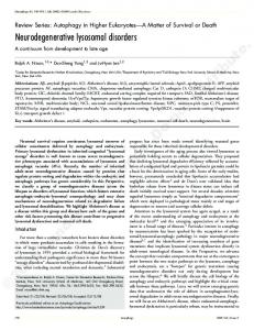
Before this critical point of a secondary astrocyte activation is reached, the potentially dangerous NO production by reactive microglia is not supported but inhibited by nonreactive and differentiated astrocytes via a transforming growth factor (TGF)-β–mediated physiological feed-back control [24]. Another negative feed-back control, which inhibits the TNF-α signaling, seems to be provided by the (otherwise proinflammatory) cytokine IL-6 (FIGURE 1) [22,25–27]. The growing understanding of complex glia–glia and neuron–glia communication, as well as the further elucidation of the diverse and interactive pathogenic signaling pathways involved, can be expected to provide a basis for the development of future lines of therapy. They should address the presumed essential role of glial cell reactions in maintaining the process of continued neuronal death during the course of neurodegenerative diseases. Possible sites of interference are indicated in FIGURE 1. It appears, however, that we have to face the same problem as Odysseus on his ancient cruises through unknown territory: avoiding sailing too close to Skylla implied the risk of getting too close to Charybdis. Accordingly, suppression of pathological glial reactions implies the risk of lacking defense, whereas allowance of an escalating glial response, including the astrocytes, may add to progressive nerve cell death. The optimal route inbetween needs to be found. Evolving pathomechanisms : a base for therapy Glial contribution to oxidative neuronal damage
Reactive microglia release massive amounts of reactive oxygen radicals. They form in conjunction with concomitantly released NO the particularly aggressive peroxynitrates, which cause cell membrane perturbation and nitrosation reactions changing the function of membrane transporters and ion channels. Autotoxic effects are probably prevented as long as the TGF-β-mediated inhibitory control of microglial NO production by astrocytes is functioning. This control seems to be very effective. As observed in our own experiments on cultured rat glial cells [UNPUBLISHED DATA], the large NO release from microglia upon stimulation with lipopolysaccharide and interferon(IFN)-γ was heavily suppressed by conditioned medium obtained from differentiated type-2 astrocyte cultures. However, a significant inhibitory effect was no longer seen in astrocyte conditioned medium which had been harvested from proliferative cultures. This suggests that the negative feedback control of microglial NO production gets lost upon the secondary activation of astrocytes, when they are forced to give up their mature resting state [27]. Accordingly, an aggravation of peroxynitrate induced neuronal damage by astrocytes has already been reported some years ago to induce nerve cell death if the astrocytes were pathologically activated by stimulation with the microglial cytokine IL-1β and INF-γ [28]. Glial contribution to excitotoxic neuronal damage
The stimulation of excitatory neuronal receptors by their agonist glutamate is physiologically a highly controlled event. A
281
Schubert & Ferroni
Reactive astrocytes
NO
Chaperones
Oxidative damage
ONOO TNF Microglial triggers •
Il-1
Reactive microglia
Toxic Aβ
•
Ischemia
•
Brain Damage
•
Complement cascade
O2
Defence protection
Excitotoxic damage
NO
[ cGMP]
TGFb Il-1
Excessive Ca 2+ loading
Phagocytosis Resting astrocytes
NMDA-R AMPA-R
Extracellular homeostasis
Astrocyte activation
[K +] o
Reactive astrocytes Loss of homeostasis
Membrane depol .
[GLU] o
Figure 1. Endogenous regulation of glial cell reactions, their potential contribution to pathological nerve cell death and possible sites of interference. Cytokine-mediated positive and negative feed-back control regulates the graded activation of microglia and may escalate in a secondary recruitment of astrocytes. As long as the pathologically triggered glia activation remains restricted to the microglia (i.e., within the dark black frame), reactive microglia functions, such as the release of oxygen radicals and phagocytosis, may provide powerful instruments of a defensive immune response. With the secondary activation of astrocytes, the reaction gets out of this frame and becomes dangerous. Reactive astrocytes are forced to give up important physiological functions (e.g., the homeostatic control of extracellular potassium and glutamate, and also their negative feed-back control on microglial functions). They form an alliance with reactive microglia and contribute to potentially neurotoxic functions, which favor oxidative and excitotoxic nerve cell damage. Possible points of (pharmacological) interference are indicated by asterisks. AMPA: a-amino-3-hydroxy-5-methylisoxazole-4-proprionic acid; Ca: Calcuim; GLU: Glutamate; GMP: Glucose Monophosphate; IL: Interleukin; K: Potassium; NMDA: N-methyl-D-aspartate; NO: Nitrous oxide; ONOO: Peroxynitrate; TNF: Tumor necrosis factor.
number of vicious circles are responsible that this control can be over-run under pathological conditions. They lead to an excessive accumulation of glutamate and of membrane depolarizing potassium in the extracellular space. As a consequence, the dangerous glutamate receptors are over stimulated, which leads to an exaggerated neuronal calcium influx through voltage-operated, NMDA- and AMPA-receptor controlled ion channels. This causes a consecutive disturbance of the intracellular calcium homeostasis – the key event causing neuronal death in degenerative diseases [29]. Reactive astrocyte functions are apparently involved in all these vicious circles. Upon pathological activation, the astrocytes are forced to give up their mature differentiated state. This can be expected to impair the maintenance of the extracellular homeostasis by astrocytes, since their physiological buffering function depends on the presence of a mature ion channel pattern [26]. As a consequence, elevated extracellular concentrations of glutamate and potassium will enhance excitatory receptor stimulation as well as membrane depolarization. This, in turn, will further reduce the voltage-sensitive reuptake of glutamate from the extracellular space. In addition,
282
reactive astrocytes can actively contribute to the pathological load of excitatory transmitters by the above mentioned TNF-α stimulated release of glutamate [21]. This may be exaggerated under oxidative stress, as the release of excitatory transmitters from astrocytes is also stimulated by peroxynitrates [30]. A TNF-α-mediated increased expression of neuronal AMPA receptors and a sensitization of NMDA-receptor activation by lipophilic amines released from pathologically stimulated microglia would further favor excitotoxic neuronal damage [23]. Glial contribution to β-amyloid toxicity
A pathogenic role of a synergistic action of reactive microglia and astrocytes is already suggested by their obligatory presence in the vicinity of neurotoxic β-amyloid plaques, as has already been observed by Alzheimer himself in his first case of AD. An advanced state of glial cell activation around the plaques is indicated by the expression of respective microglial markers, such as MHC class II, macrophage-colony-stimulating factor (MCSF) and other surface receptors [19]. The stimulatory role of β-amyloid and its associated proteins in triggering an escalating glial reaction is evaluated in detail by
Expert Rev. Neurotherapeutics 3(3), (2003)
Glia reactions as a therapeutic target
Meda and collaborators [25]. The interaction between β-amyloid and pathologically activated glia works in both directions, for example, β-amyloid is one of the molecular signals which initiates a microglial reaction, increasing their capacity to generate oxidative damage [23,25,31,32]. In turn, reactive glial cells favor β-amyloid toxicity. Here, the secondarily activated astrocytes probably play a predominant role. First, they show an upregulated APP expression. If the processing of APP is pathological, as favored by oxidative stress and cholinergic deficiency, the production of amyloidogenic fragments by the big mass of astrocytes can be expected to add significantly to βamyloid toxicity [26,27]. Second, reactive astrocytes are the brain endogenous producers of the chaperones antichymotrypsin and apolipoprotein-E, which are regularly found in neurotoxic dense core plaques and have been shown by Potters group to promote the transformation of aggregating β-amyloid peptides into the neurotoxic filamentous form [26,27,33]. Third, if the active physiological role of astrocytes in determining synaptic efficacy is altered upon their pathological activation, this would affect the synaptic function. For the development of dementia in AD, the β-amyloid induced impairment of (astrocyte related) synaptic function is thought to be more important than nerve cell death [34]. Taken together, an escalated glia reaction involving the astrocytes should be considered as a major pathogenic mechanism promoting β-amyloid toxicity. On the other hand, the recent report that a pharmacologically induced microglial proliferation caused the phagocytosis of β-amyloid deposits in a transgenic mouse model indicates that the glial immune response has presumably also important beneficial effects [18]. A prerequisite may be that the glial reaction is limited to a relatively low state of activation. Where & how to interfere with glial reactions Suggested principal strategies
In view of the broad repertoire of potentially pathogenic mechanisms, which can be adopted during an escalating glial activation by reactive astrocytes, it should be a prevalent task for therapeutic intervention to keep the reaction limited to the microglia. As the pathological glial activation is a graded response, this should be possible by a selective interference with the identified signaling paths, which mediate the upgrading of the microglial activation state and the secondary involvement of astrocytes. As a consequence, if the reactive microglia cannot rely on the co-operative support by activated astrocytes, the risk that a pathological glia reaction becomes autotoxic will probably be reduced and neuronal protection by beneficial functions of the reactive microglia may be dominating. An obvious way to prevent an escalation of the glial reaction would be to rebalance the interplay of the regulatory cytokines, to inhibit production, release or action of the feed-forward signals TNF-α and IL-1β, and to support negative feed-back signaling as well as mechanisms which favor a differentiated state of astrocytes. Generally, a correcting guidance of pathologically
www.future-drugs.com
altered signaling pathways and using the respective know-how of endogenous regulatory mechanisms are probably fruitful targets for future lines of therapy. Thus, nature has invented a sophisticated receptor cross-talk, which allows a fine-tuning of multiple G-protein coupled signaling pathways [35]. As a consequence, the intracellular signaling of one G-protein coupled pathway may be amplified by the concomitant activation of another path, which is linked to a different receptor. This could be a fruitful therapeutic target for pharmacological correction in order to reinforce a pathologically impaired signaling pathway. Again, a prerequisite for successful pharmacological manipulation of the complex signaling cross-talk at the second messenger level is a detailed knowledge of its physiological and pathophysiological functioning and hence, the support of (primarily nonprofitable) basic research in system physiology and pathophysiology besides the favored focus on genetic engineering of today. Specifically, one should bear in mind a possible guidance by the brain-endogenous small molecules acting as regulatory modulators, which have learned, how to do this job during long years of evolution. For example, adenosine and chemokines belong to this group of intelligent small molecular signals. Adenosine exerts a number of neuroprotective actions [26,36,37] and has been proposed for a potential treatment of several disorders including brain ischemia, Morbus Parkinson and other neurodegenerative diseases [38]. Chemokines, first recognized as inflammatory mediators, have later been demonstrated to also exert physiological regulatory functions, which could be relevant as therapeutic target [21]. Possible modes of interference Strengthening of the cyclic nucleotide signaling
A strengthening of the cyclic adenosine monophosphate (cAMP)-signaling has been found in vitro to influence activated microglial properties in a differential manner. TNF-α and IL-1β-release from pathologically stimulated rat microglia were strongly inhibited, whereas the production of the negative feed-back signal IL-6 was not affected [39]. This differentiated action enables cAMP to function as an emergency break, which can be used to stop an unwanted escalating glia reaction. Furthermore, there was a cAMP-mediated inhibition of the heavy microglial release of free oxygen radicals but not of NO release. Sparing NO could add to cyclic guanosine monophosphate (cGMP)-mediated protection, since NO is an ambiguous compound, which may add to the formation of peroxynitrates as well as to cGMP synthesis. If the microglial release of free oxygen radicals is selectively inhibited by a strengthened cAMP signaling, only a small percentage of the released NO can be taken for the generation of the cell damaging peroxynitrates. The remaining NO would be free to activate the guanylcyclase and to increase the cGMP-signaling. To illustrate this ambiguous NO action: we found complete nerve cell death in (microglia containing) neuronal cultures after addition of NO-releasing nitroprusside. This damage was almost completely prevented either upon scavenging oxygen radicals or upon maintaining the NO-generated cGMP by inhibiting its degradation [40]. The
283
Schubert & Ferroni
protective effects of cGMP include a reinforcement of neurotrophic pathways and an antiapoptotic action, as has recently been demonstrated in cultured astrocytes [41]. Strengthened cAMP signaling reduces the risk of a secondary astrocyte activation not only by inhibiting the upgrading cytokine release from microglia but also by a direct effect on astrocytes stabilizing their differentiated state. This is supported by our finding that a prolonged treatment of cultured rat astrocytes with cAMP-analogs induced cell differentiation and the expression of a mature ion channel pattern, which is required for the physiological maintenance of the extracellular homeostasis [26]. The cyclic nucleotide signaling can be pharmacologically strengthened by blocking the enzymatic degradation of cAMP by phosphodiesterase (PDE). The naturally available large family of identified PDE-subtypes reflects their apparently important function in the brain–endogenous regulation of cyclic nucleotide signaling. The interactive crosstalk between the individually regulated PDE subtypes and their concerted function still needs to be clarified. On the basis of this knowledge, selective PDE blockers could be developed, which will probably provide powerful instruments for subtle pharmacological corrections of cyclic nucleotide signaling according to specific pathophysiological demands. A strengthening of cellular cAMP signaling can be further achieved by introducing the physiological G-protein-linked regulators of the adenylcyclase activity. This includes the favored glial cell modulator adenosine, which increases intracellular cAMP levels via (low-threshold) A2a receptors and (high-threshold) A2b receptors. However, adenosine also has a Janus face: it may exert both protective and adverse effects [36]. This endogenous regulator may serve as one of the triggers initiating a pathological glia reaction. An A2a receptor mediated cAMP elevation has been reported to activate the cyclooxygenase (COX), known to stimulate microglial proliferation. On the other hand, we observed an inhibition of microglial proliferation in cultures, which were pathologically stimulated with phorbol esters. We also found that adenosine induced apoptosis in proliferative microglial cultures. The latter, however, was no longer observed, if the microglia had been brought into a less activated state by pretreating the cultures with cAMP analogs [26,27,36]. From these experiments, it can be concluded that the mode of regulatory adenosine actions depends on the microglial activation state – stimulatory on resting microglia but inhibitory on pathologically activated microglia. Using the adenosine mediated signaling crosstalk
Most interesting is the ability of adenosine to mediate a crosstalk to other pathways from different receptors. For example, stimulation of the Gi-protein coupled adenosine A2a receptor has been reported to transactivate a Trk receptor-linked neurotrophic pathway, even in the absence of neurotrophins [42]. The physiological agonists of this pathway are NGF and BDNF, which can apparently be replaced by the cross-talk
284
through adenosine. This may provide an elegant and less risky approach to support the currently considered beneficial supply of these neurotrophic factors in the treatment of AD. By an adenosine mediated stimulation of neurotrophic signaling, it may also be possible to maintain the microglial reaction in the range of a lower activation state, since NGF and BDNF have been reported to inhibit the MHC class II inducibility in isolated microglia [43]. Another adenosine-mediated receptor cross-talk between intracellular calcium raising pathways and the (calcium dependent) muscarinic signaling may be used as a therapeutic target in order to recover deficient cholinergic signaling in AD. Previous experiments on cultured rat astrocytes had already demonstrated that slight elevations of the extracellular adenosine concentration up to 1 µM collaborated via the Gi-protein-coupled adenosine A1 receptors with metabotropic glutamate receptor pathways in generating a potentiated intracellular calcium mobilization [44]. The latter is deficient in the muscarinic signaling pathway of AD patients due to the lack of the physiological agonist acetylcholine. Therefore, we tested whether an analog potentiation of the intracellular calcium signal can be achieved by concomitant adenosine A1 receptor stimulation in conjunction with a deficient stimulation of muscarinic receptors by low acetylcholine [45]. The results of these experiments were positive. The combined acetylcholine/adenosine action led to a robust calcium signal, which was equivalent to that generated by a 30-fold higher concentration of acetylcholine alone. In our opinion, this calcium potentiating adenosine effect can be used in combination with the conventional treatment of AD patients by acetylcholine-esterase inhibitors to regain an effective muscarinic signaling [102]. The latter is essentially required for correct APP processing, which produces the nonamyloidogenic soluble APPs and avoids β-amyloid toxicity. At present, however, it is still a problem to achieve the required (slight) rise in the extracellular adenosine concentration under nonischemic conditions. The development of uptake blockers, which inhibit the cellular reuptake of the brain–endogenous extracellular adenosine and are more effective than propentofylline in this respect, would be helpful since systemically applied artificial adenosine agonists are usually not able to pass the blood–brain barrier [16]. Dependent on the stage of the neurodegenerative process, the specific pathomechanisms, which should be antagonized, are probably different. At some stages, it might be indicated to block potentially adverse adenosine effects, such as the A2a receptor mediated COX activation. However, despite the fact that adenosine belongs to the group of possible triggers initiating a microglial reaction, the beneficial actions appear to be prevalent and justify the testing of the capabilities of this cell modulator as a target for future therapeutics. Other therapeutic targets
There are certainly a number of other targets which allow an interference with potentially toxic properties of reactive glial
Expert Rev. Neurotherapeutics 3(3), (2003)
Glia reactions as a therapeutic target
Key issues • Presumed common pathomechanisms for the progressive nerve cell death in neurodegenerative diseases are: excitotoxic and oxidative damage. β-amyloid toxicity plays an additional role in Alzheimer's disease. All these pathomechanisms may be reinforced by the generally observed reactions of microglia and astrocytes. • Reactive microglia can turn the strengthening of synaptic efficacy by a tumour necrosis factor (TNF)-α evoked astrocytic glutamate release from a physiological into a pathogenic mechanism, which favors excitotoxic neuronal damage. • The secondary pathological activation of astrocytes by the microglial cytokine interleukin (IL)-1β represents a critical point, at which a synergistic alliance between reactive microglia and astrocytes is formed promoting oxidative, excitotoxic and β-amyloid-induced neuronal damage. • Reactive microglia are reported to exert also beneficial actions, such as phagocytosis of β-amyloid plaques. • A prevalent task of pharmacological interference should be a titration of the microglial reaction to a point that allows beneficial actions but prevents TNF-α overload and IL-1β release. • Prerequisite for such a selective pharmacological interference is an identification of the different microglial activation states, their labeling with specific markers of representative surface receptors and their correlation with respective microglial functions. • The recently discovered signaling cross-talk at the second messenger level may provide a new target for the pharmacological treatment of neurodegenerative disorders, which are characterized by a progressive loss of functionally important signaling pathways. • Specifically, a strengthening of the pathologically often impaired cyclic nucleotide signaling and a rebalancing of the calcium/cAMP interplay could provide neuroprotection and may allow a recovery of deficient signaling pathways. • Here, one could use the know-how of endogenous cell modulators, such as adenosine, which have learned during evolution how to do this difficult job. cells, such as the above mentioned chemokine receptors and TNF-α receptors [21,46]. A suppression of the COX activation may help to maintain a glia-linked inflammatory response within safe limits. Interestingly, COX inhibitors from the NSAIDs group have recently been found to exert an additional effect, apparently unrelated to an influence on COX activity. Thus, the NSAID ibrufen decreased the highly amyloidogenic β-amyloid peptide in cell cultures, and an NO-coupled NSAID stimulated the phagocytotic activity of reactive microglia and reduced β-amyloid deposits in transgenic mice [18,47]. It has been hypothesized that treatment with such NSAIDs may also limit escalating microglial activation. Another way to keep microglial reaction in the range of a (presumably beneficial) lower activation state may be to inhibit the MHC class II inducibility by a therapeutic stimulation of the neurotrophic signaling pathways [43]. Expert opinion
The common aim of pharmacological treatment should be to prevent an escalation of the glia reaction up to the point where it becomes autotoxic [48,49]. We think that this point is indicated by the secondary involvement of astrocytes, which may provide a useful marker on the calibration scale for an optimal titration of the pathological glia reaction by pharmacological interference. Five-year view
The evolving important role of astrocytes as active regulators of synaptic function and as a source for neurotrophic factors will presumably focus protective therapies not only on neurons but also on the maintenance of physiological astrocyte functions.
www.future-drugs.com
The endogenous regulation of potentially neurotoxic pathological glia reactions by the cytokine-mediated complex interaction of positive and negative feed-back loops, which regulate the graded immune response of microglia and astrocytes, will be better understood and (hopefully) allow a fine-tuning by pharmacological interference. The further elucidation of the sophisticated cross-talk between the second messengers calcium, cAMP and cGMP will provide a chance to influence pathologically altered signaling pathways by rebalancing the second messenger interplay. A useful therapeutic approach to strengthen an impaired cyclic nucleotide signaling could be a blockade of enzymatic cAMP and cGMP breakdown by newly developed phosphodiesterase-inhibitors. They should be subtype-specific and capable of modifing, in a better understood manner, the concerted interaction of the large family of individually regulated phosphodiesterase-subtypes. The recent discovery that the cross-talk between different signaling pathways can be mediated by adenosine (and probably other small molecule signals which have to be identified) is promising. A further improved understanding of such endogenous regulatory actions may provide new therapeutic strategies based on the pharmacological strengthening of beneficial homeostatic cell modulators. Generally, upstream interference with pathogenic mechanisms will presumably become the prevalent therapeutic strategy, as it minimizes the risk of adverse side effects.
285
Schubert & Ferroni
References Papers of special note have been highlighted as: • of interest •• of considerable interest Reed JC. Apoptosis-based therapies. Nat. 1 Rev. Drug Disc. 11, 111–121 (2002). 2
Mattson MP. Apoptosis in neurodegenerative disorders. Nat. Rev. Mol. Cell Biol. 1, 120–129 (2000).
3
Graeber MB, Moran LB. Mechanisms of cell death in neurodegenerative diseases: fiction and facts. Brain Pathol. 12, 385–390 (2002).
4
Hirsch EC, Hunot S, Faucheux B et al. Dopaminergic neurons degenerate by apoptosis in Parkinson’s disease. Mov. Disord. 14, 383–385 (1999).
5
Giasson BE, Lee VM. Parkin and the molecular pathways of Parkinson’s disease. Neuron 31, 885–888 (2001).
6
Lin S, Zhang Y, Dodel R, Farlow MR, Paul SM, Du Y. Minocycline blocks nitric oxideinduced neurotoxicity by inhibition p38 MAPK in rat cerebellar granule neurons. Neurosci. Lett. 315, 61–64 (2001).
7
Sathasivam S, Ince PG, Shaw PJ. Apoptosis in amyotrophic lateral sclerosis: a review of evidence. Neuropathol. Appl. Neurobiol. 27, 257–274 (2001).
8
Antuono P, Beyer J. The bourdon of dementia. A medical and research perspective. Theor. Med. Bioeth. 20, 3–13 (1999).
9
Irizarry MC, Hyman BT. Alzheimer’s disease therapeutics. J. Neuropathol. Exp. Neurol. 60, 923–928 (2001).
10
Winblad B, Poritis N. Memantine in severe dementia: results of the 9M-Best Study. Int. J. Geriatr. Psychiatry 14, 135–146 (1999).
11
Mattson MP, Duan W, Pedersen WA, Culmsee C. Neurodegenerative disorders and ischemic brain diseases. Apoptosis 6, 69–81 (2001).
12
Breitner JC. Epidemiologic clues to the causes and routes to prevention of Alzheimer’s disease. J. Neural Transm. Suppl. 59, 251–254 (2000).
13
Mackenzie IR, Munoz DG. Nonsteroidal anti-inflammatory drug use and Alzheimertype pathology in aging. Neurology 50, 986–990 (1998).
14
Sasaki A, Yamaguchi H, Ogawa A, Sugihara S, Nakazato Y. Microglial activation in early stages of amyloid-β protein deposition. Acta Neuropathol. 94, 316–322 (1997).
286
15
Schubert P, Rudolphi K. Interfering with the pathologic activation of microglial cells and astrocytes in dementia. Alz. Dis. Ass. Disorders 12, 21–28 (1998).
16
17
24
Schubert P, Ogata T, Rudolphi K, Marchini C, McRae A, Ferroni S. Support of homeostatic glial cell signalling: a novel therapeutic approach by propentofylline. Ann. NY Acad. Sci. 826, 337–347 (1997).
Vincent VAM, Tilders FJH, van Dam AM. Inhibition of endotoxin-induced oxide synthase production in microglial cells by the presence of astroglial cells: a role for transforming growth factor β. Glia 19, 190–198 (1997).
25
Kittner B, Roessner M, Rother M. Clinical trials in dementia. Ann. NY Acad. Sci. 826, 307–316 (1997).
•
Meda L, Baron P, Scarlato G. Glial activation in Alzheimer’s disease: the role of Aβ and its associated proteins. Neurobiol. Aging 22, 885–893 (2001). Evaluation of the escalating activation of microglia and astrocytes by toxic β-amyloid. Schubert P, Morino T, Miyazaki H et al. Cascading glia reactions: a common pathomechanism and its differentiated control by cyclic nucleotide signaling. Ann. NY Acad. Sci. 903, 24–34 (2000).
Jantzen PT, Konnor KE, DiCarlo G et al. Microglial activation and β-amyloid deposit reduction caused by a nitric oxidereleasing nonsteroidal anti-inflammatory drug in amyloid precursor protein plus preseniline-1 transgenic mice. J. Neurosci. 22, 2246–2254 (2002). • Good introduction to the problem of reactive glial cell function (good or bad, or both?). 19 Raivich G, Bohatschek M, Kloss et al. Neuroglial activation repertoire in the injured brain: graded response, molecular mechanisms and cues to physiological function. Brain Res. Rev. 30, 77–105 (1999). • The activation state of reactive microglia, which determines their potentially neurotoxic power, is reflected by the graded expression of different cell-surface markers. 20 Haydon PG. Glia: listening and talking to the synapse. Nat. Rev. Neurosci. 2, 185–193 (2001). • Reviews the evolving essential role of astrocytes for physiological brain function and their active participation in synaptic transmission. 21 Bezzi P, Domercq M, Brambilla L et al. CXCR4-activated astrocyte glutamate release via TNF-α: amplification by microglia triggers neurotoxicity. Nat. Neurosci. 4, 702–710 (2001). •• Illustration of the dangerous TNF-α mediated (and astrocyte-linked) mechanisms, which allow strengthening of synaptic efficacy but may also turn into a damaging pathomechanism. 22 Aschner M. Astrocytes as mediators of immune and inflammatory responses in the CNS. Neurotoxicology 19, 269–289 (1998). 18
23
Giullian D. A strategy for identifying immunosuppressive therapies for Alzheimer’s disease. Alzheimer’s disease and Associated Disorders 12(Suppl. 2), 7–14 (1998).
26
27
•
28
•
29
Schubert P, Ogata T, Marchini C, Ferroni S. Glia-related pathomechanisms in Alzheimer’s disease: a therapeutic target? Mech. Aging Develop. 123, 47–57 (2001). Evaluation of possible modes of protection by influencing (potentially neurotoxic) reactive glial cell functions at the second messenger level. Here, the action of the cell modulator, adenosine, which influences the generation and interaction of the second messengers calcium and cyclic AMP, may be a promising target for pharmacological intervention. Hewett SJ, Csernansky CA, Choi DW. Selective potentiation of NMDA induced neuronal injury following induction of astrocytic iNOS. Neuron 13, 487–494 (1994). Pioneering paper that suggests a possible pathogenic role of reactive astrocytes to oxidative neuronal damage. LaFerla FM. Calcium dyshomeostasis and intracellular signalling in Alzheimer’s disease. Nature Rev. Neurosci. 3, 862–872 (2002).
30
Haskew RE, Mongin AA, Kimelberg HK. Peroxynitrite enhances astrocytic volume-sensitive excitatory amino acid release via a src tyrosine kinasedependent mechanism. J. Neurochem. 82, 903–912 (2002).
31
Akama KT, van Eldik LJ. β-amyloid stimulation of inducible nitric-oxide synthase in astrocytes is interleukin-1β and tumor necrosis factor-α dependent and involves a TNF-α receptor-associated factor and NFkB inducing kinase-dependent signaling mechanism. J. Biol. Chem. 275, 7918–7924 (2000).
Expert Rev. Neurotherapeutics 3(3), (2003)
Glia reactions as a therapeutic target
32
33
•
34
•
35
•
36
37
•
38
39
Ishii K, Muelhauser F, Liebl U, Beyreuther K. Subacute NO generation induced by Alzheimer’s β-amyloid in the living brain: reversed by inhibition of the inducible NO synthase. FASEB J. 14, 1485–1489 (2000). Ma J, Yee A, Brewer HB, Das S, Potter H. Amyloid associated proteins α-1antichymotrypsin and apolipoprotein-E assembly of β-protein into filaments. Nature 372, 92–94 (1994). Pioneering paper showing that specific proteins, which can be formed by reactive astrocytes, aggravate β-amyloid toxicity. Small DH, Mok SS, Bornstein JC. Alzheimer’s disease and Aβ toxicity: from top to bottom. Nat. Rev. Neurosci. 2, 595– 598 (2001). It is interesting that an impairment of synaptic function is proposed as the major pathomechanism responsible for the generation of dementia. Selbie LA, Hill S. G-protein coupled receptor crosstalk: the fine-tuning of multiple receptor-signalling pathways. TIPS 19, 87–93 (1998). The evolving sophisticated signaling crosstalk may be a promising target for future therapy. Ongini E, Schubert P. Neuroprotection induced by stimulating A1 or blocking A2a adenosine receptors: an apparent paradox. Drug Devel. Res. 45, 387–393 (1998). Sebastiao A, Ribieiro J. Fine tuning neuromodulation by adenosine. Trends Pharmacol. Sci. 21, 341–361 (2000). Recently reviewed the fine-tuning modulation of various cell functions by adenosine. Moreau JL, Huber G. Central adenosine A (A2A) receptors: an overview. Brain Res. Rev. 31, 65–82 (1999). Si Q, Nakamura Y, Ogata T, Kataoka K, Schubert P. Differential regulation of microglial activation by propentofylline via cAMP signaling. Brain Res. 812, 97–104 (1998).
www.future-drugs.com
40
41
Ogata T, Kohgami S, Okumura H et al. Nitric oxide-induced neurotoxicity is inhibited by propentofylline via cyclic GMP elevation. Neurobiol. Aging 19, 255 (1998). Takuma K, Phuagphong P, Lee E, Mori K, Baba A, Matsuda T. Anti-apoptotic effect of cGMP in cultured astrocytes: inhibition by cGMP-dependent protein kinase of mitochondrial permeability transition pore. J. Biol. Chem. 276, 48093–48099 (2001).
Lee SF, Chao MV. Activation of Trk neurotrophin receptors in the absence of neurotrophins. Proc. Natl Acad. Sci. 98, 3555–3560 (2001). •• Very interesting idea of the newly evolving capability of adenosine to mediate a crosstalk between different signaling pathways at the second messenger level. Reinforcement of this adenosine action may provide a chance to recover pathologically impaired pathways, such as NGF producing neurotrophic pathways. 43 Neumann H, Misgeld T, Matsauro K, Wekerle H. Neurotrophins inhibit major histocompatibility class II inducibility of microglia: involvement of the p75 neurotrophin receptor. Proc. Natl Acad. Sci. (USA) 95, 5779–5784 (1998). 42
Ogata T, Nakamura,Y, Schuber P. Potentiated cAMP rise in metabotropically stimulated rat cultured astrocytes by a Ca2+-related A1/A2 adenosine receptor cooperation. Eur. J. Neurosci. 8, 1124–1131 (1996). • Experimental evaluation of the adenosine mediated interaction between the second messengers calcium and cAMP in cultured astrocytes. 45 Ferroni S, Marchini C, Ogata T, Schubert P. Recovery of deficient cholinergic calcium signaling by adenosine in cultured rat cortical astrocytes. J. Neurosci. Res. 68, 615– 621 (2002). •• A further example of the newly evolving capability of adenosine to mediate a crosstalk between different signaling pathways at the second messenger level. Reinforcement of this adenosine action 44
46
may provide a chance to recover pathologically impaired pathways, such as the deficient muscarinic signaling in Alzheimer’s disease. Rossi D, Zlotnik A. The biology of chemokines and their receptors. Ann. Rev. Immunol. 18, 217–242 (2000).
Weggen S, Eriksen JL, Das P et al. A subset of NSAIDs lower amyloidogenic Abeta42 independent of cyclooxygenase activity. Nature 414, 159–160 (2001). • An evolving possibility of pharmacological interference with β-amyloid toxicity. 48 McGeer P, McGeerEG. Inflammation, autotoxicity and Alzheimer’s disease. Neurobiol. Aging 22, 799–809 (2001). •• Highly authentic view of the presumed significance of reactive microglia for neuronal damage. 49 Bin L, Jau-Shyong H. Role of microglia in inflammation-mediated neurodegenerative diseases: mechanisms and strategies for therapeutic intervention. JPET 304, 1–7 (2003). • Recently reviewed the presumed significance of reactive microglia for neuronal damage. 47
Patents 101
Schubert P (Max Planck Institute od Neurobiology, Martinsried, Germany) and Hoechst/Aventis: ‘Use of Xanthine Derivatives for the Treatment of Secondary Nerve Cell Damage after Cranio-Cerebral Traumas’ USA. 5,409,935; EU A61K31/52
102
Schubert P, Aventis: ‘Combination Drug for Treatment of Dementia’, EU 98102620.6-2107
Affiliations •
•
Peter Schubert, Max-Planck-Institute of Neurobiology, 82152 Martinsried, Germany, Tel.: +49 172 866 2393,
[email protected] Stefano Ferroni, Department of Human and General Physiology, University of Bologna, Via San Donato 19/2, 40127 Bologna, Italy, Tel.: +39 051 253 568, Fax: +39 051 251 731,
[email protected]
287