APPLIED AND ENVIRONMENTAL MICROBIOLOGY, Feb. 2000, p. 850–854 0099-2240/00/$04.00⫹0 Copyright © 2000, American Society for Microbiology. All Rights Reserved.
Vol. 66, No. 2
Pathovars of Pseudomonas syringae Causing Bacterial Brown Spot and Halo Blight in Phaseolus vulgaris L. Are Distinguishable by Ribotyping ´ LEZ,1* ELENA LANDERAS,2 ANA J. GONZA
AND
M. CARMEN MENDOZA3
Servicio Regional de Investigacio ´n y Desarrollo Agrario, Consejerı´a de Agricultura, Ganaderı´a y Pesca, 33300Villaviciosa,1 Laboratorio de Sanidad Vegetal, Consejerı´a de Agricultura, Ganaderı´a y Pesca, 33071-Oviedo,2 and Departamento de Biologı´a Funcional, Area Microbiologı´a, Facultad de Medicina, Universidad de Oviedo, 33006-Oviedo,3 Principado de Asturias, Spain Received 26 July 1999/Accepted 19 November 1999
Ribotyping was evaluated as a method to differentiate between Pseudomonas syringae pv. phaseolicola and pv. syringae strains causing bacterial brown spot and halo blight diseases in Phaseolus vulgaris L. Ribotyping, with restriction enzymes BglI and SalI and using the Escherichia coli rrnB operon as the probe, differentiated 11 and 14 ribotypes, respectively, and a combination of data from both procedures yielded 19 combined ribotypes. Cluster analysis of the combined ribotypes differentiated the pathovars phaseolicola and syringae, as well as different clonal lineages within these pathovars. The potential of ribotyping to screen for correlations between lineages and factors such as geographical region and/or bean varieties is also reported. Phytopathogenic bacteria of the Pseudomonas syringae group cause diseases in all major groups of higher plants and can be divided into more than 50 pathovars. The term pathovar is used to refer to strains with similar features that are differentiated at the subspecies level on the basis of differences in plant host range and types of symptoms, and additionally by biochemical profiles (7, 12, 16). Such a phenotypically based classification is of practical interest, but it does not reveal the genetic relatedness within or between pathovars (1, 3). In this work, a two-way ribotyping procedure was evaluated as a method to distinguish between P. syringae pv. syringae and pv. phaseolicola organisms isolated from Phaseolus vulgaris L., which are the causal agents of bacterial brown spot and halo blight, respectively. Isolation procedure and phenotypic characterization of P. syringae strains. The P. syringae isolation procedure used has been described elsewhere (6, 10, 14). The samples of bean plants analyzed were collected at different times from different bean fields in a geographical area of about 100,000 km2 in the north of Spain, over the period 1991 to 1997. This area includes two different climatic regions: the Cantabrian coast area, wet and temperate (WT), and the northern inland meseta, dry and continental (DC). P. syringae organisms were isolated from 19.0 and 40.9% of sets of infected plant material (leaves and/or pods showing either typical or suspected symptoms of bacterial brown spot and halo blight diseases) collected in the WT and DC regions, respectively, and from 26.1% of damaged seed sets, each supplied by a different farmer in the WT region. Of the isolated organisms, only the 61 strains compiled in Table 1 were characterized in this work. The initial strain identification was based on the Hugh-Leifsson reaction and the LOPAT scheme (production of levane, oxidase, pectinases, and arginine dihydrolase, as well as tobacco hypersensitivity) (7). Pathovar confirmation included tests for esculin hydrolysis and acid production from mannitol, erythritol, and myoinositol (6, 10) as well as an enzyme-linked
immunosorbent assay–double-antibody sandwich indirect procedure using an antiserum specific for flagellar antigens of pathovar phaseolicola strain CFBP 1390. These tests led us to assign the P. syringae strains to two well-defined pathovars; phaseolicola (46 strains) and syringae (15 strains). It is noticeable that in our series a total correlation between negative esculin hydrolysis and pathovar phaseolicola and between positive esculin hydrolysis and pathovar syringae was revealed; besides, this test is rapid, cheap, and easily performed. The assignment of the pathovar phaseolicola strains to defined races according to the scheme published in reference 15 was carried out in the Servicio de Investigacio ´n, Desarrollo y Tecnologı´a Agraria, Valladolid, Spain. By this procedure, 32 strains were differentiated into races 2, 5, 6, 7, and 9, whereas 10 other strains could not be assigned to a defined race and were labeled as nonidentified (NI). Findings of epidemiological interest were that in leaves and/or pods from the two climatic regions both pathovars were found but that pathovar phaseolicola was much more frequent in the DC region. Conversely, from seeds (all collected from a single autochthonous bean variety named Granja Asturiana within the WT region) only pathovar syringae organisms were identified. Two-way ribotyping of P. syringae group strains. Isolation of genomic DNA and ribotyping were carried out as reported in reference 9. Ribotyping was performed with the enzymes BglI and SalI (Stratagene, La Jolla, Calif.), and hybridization was performed with an rrnB operon from Escherichia coli and use of the nonradioactive DNA labeling and detection kit of Boehringer GmbH (Mannheim, Germany). The polymorphic restriction sites along the ribosomal DNA regions were inferred from the presence or absence of bands in the overall ribotypes from each enzyme. In addition to the 61 strains cited above, three other strains of P. syringae were analyzed and used as outgroup strains: pathovar phaseolicola strain ATCC 19304 (Coleccio ´n Espan ˜ola de Cultivos Tipo; CECT 321); pathovar tomato strain ATCC 10862 (CECT 126); and one unspecifiedpathovar strain, Boelema P2 (CECT 94), collected from Pisum sativum. With BglI, 13 different B ribotypes were found in the series (Fig. 1A), two of which (B1 and B2) were represented by pathovar phaseolicola and nine of which (B21 to B29) were
* Corresponding author. Mailing address: SERIDA, Apartado 13, 33300 Villaviciosa, Asturias, Spain. Phone: 34-985890066. Fax: 34985891854. E-mail:
[email protected]. 850
RIBOTYPING OF TWO PSEUDOMONAS SYRINGAE PATHOVARS
VOL. 66, 2000
851
TABLE 1. Origin and features of P. syringae strains causing diseases in the common beana Pathovar (race)b
Strain/yr
Phaseolicola (5) Phaseolicola (7) Phaseolicola (7) Phaseolicola (7) Phaseolicola (7) Phaseolicola (7) Phaseolicola (7) Phaseolicola (7) Phaseolicola (7) Phaseolicola (7) Phaseolicola (7) Phaseolicola (7) Phaseolicola Phaseolicola (NI) Phaseolicola Phaseolicola Phaseolicola (NI) Phaseolicola (NI) Phaseolicola (NI) Phaseolicola (5) Phaseolicola (7) Phaseolicola (9) Phaseolicola (NI) Phaseolicola (9) Phaseolicola (6) Phaseolicola (6) Phaseolicola (6) Phaseolicola (6) Phaseolicola (7) Phaseolicola (6) Phaseolicola (6) Phaseolicola (5) Phaseolicola (5) Phaseolicola (6) Phaseolicola (6) Phaseolicola (7) Phaseolicola (NI) Phaseolicola (7) Phaseolicola (7) Phaseolicola (NI) Phaseolicola Phaseolicola (NI) Phaseolicola (NI) Phaseolicola (NI) Phaseolicola (2) Phaseolicola (2) Syringae Syringae Syringae Syringae Syringae Syringae Syringae Syringae Syringae Syringae Syringae Syringae Syringae Syringae Syringae
21/95 24/95 25/95 26/95 28/95 29/95 33/95 34/95 35/95 36/95 41/95 43/95 44/95 45/95 46/95 65/96 55/95 14/95 15/95 69/96 10/95 17/95 19/95 20/95 11/95 39/95 40/95 42/95 37/95 13/95 16/95 61/96 64/96 66/94 22/95 27/95 23/95 50/95 51/95 67/96 31/95 59/96 62/96 63/96 48/95 49/95 30/95 57/96 73/97 53/95 54/95 38/95 56/95 7/91 58/96 60/96 6/91 5/91 1/91 2/91 4/91
Host features Variety
Sample
MV MV MV MV Taylorc Taylorc Canela Canela Canela Canela Canela pinta Canela pinta Pinta Pinta Pinta Blanca Blanca Pinta Rin ˜o ´n
Leaves Leaves Leaves Leaves Leaves Leaves Leaves Leaves Leaves Leaves Leaves Leaves Leaves Leaves Leaves Pods Leaves Leaves Leaves Leaves/pods Leaves Leaves Leaves Leaves Leaves Leaves Leaves Leaves Leaves Leaves Leaves Leaves Leaves Leaves/pods Leaves Leaves Leaves Leaves Leaves Leaves/pods Leaves Leaves Leaves Leaves Leaves Leaves/pods Leaves Leaves Leaves/pods Leaves/pods Leaves/pods Leaves Leaves Seed Leaves Leaves/pods Seed Seed Seed Seed Seed
Canela Canela Canela Canela Taylor Planchada Planchada Canela Canela Pinta Pinta Granja A Granja A MV Rin ˜o ´n MV Canela Canela Granja A. Granja A. Granja A. Canela Canela 6J-64-94 Granja A. Granja A. Canela Canela Canela Blanca Larga Granja A. Granja A. Granja A. Granja A. Granja A. Granja A. Granja A. Granja A.
CA/Province
ELISA-DASI
DC/Leo ´n DC/Leo ´n DC/Leo ´n DC/Leo ´n DC/Leo ´n DC/Leo ´n DC/Leo ´n DC/Palencia DC/Palencia DC/Palencia DC/Palencia DC/Palencia DC/Palencia DC/Palencia DC/Palencia DC/Palencia DC/Soria DC/Zamora DC/Zamora WT/Paı´s Vasco DC/Avila DC/Valladolid DC/Valladolid DC/Valladolid DC/Avila DC/Palencia DC/Palencia DC/Palencia DC/Salamanca DC/Zamora DC/Zamora WT/P. Asturias WT/P. Asturias WT/Paı´s Vasco DC/Leo ´n DC/Leo ´n DC/Leo ´n DC/Zamora DC/Zamora WT/Paı´s Vasco DC/Leo ´n WT/P. Asturias WT/P. Asturias WT/P. Asturias DC/Zamora DC/Zamora DC/Leo ´n WT/P. Asturias WT/Lugo DC/Salamanca DC/Salamanca DC/Salamanca DC/Soria WT/P. Asturias WT/P. Asturias WT/P. Asturias WT/P. Asturias WT/P. Asturias WT/P. Asturias WT/P. Asturias WT/P. Asturias
⫹ ⫹ ⫹ ⫹ ⫹ ⫹ ⫹ ⫹ ⫹ ⫹ ⫹ ⫹ ⫹ ⫹ ⫹ ⫹ ⫹ ⫹ ⫹ ⫹ ⫺ ⫹ ⫹ ⫺ ⫹ ⫹ ⫹ ⫹ ⫹ ⫹ ⫹ ⫹ ⫹ ⫹ ⫹ ⫹ ⫹ ⫹ ⫹ ⫹ ⫹ ⫹ ⫹ ⫹ ⫹ ⫹ ⫺ ⫹ ⫺ ⫺ ⫺ ⫺ ⫺ ⫺ ⫹ ⫺ ⫺ ⫺ ⫺ ⫺ ⫺
Fermentation
Ribotype
Man.
Ery.
BglI
SalI
⫺ ⫺ ⫺ ⫺ ⫺ ⫺ ⫺ ⫺ ⫺ ⫺ ⫺ ⫺ ⫺ ⫺ ⫺ ⫺ ⫹ ⫺ ⫺ ⫺ ⫺ ⫺ ⫺ ⫺ ⫺ ⫺ ⫺ ⫺ ⫺ ⫺ ⫺ ⫺ ⫺ ⫺ ⫺ ⫺ ⫹ ⫺ ⫺ ⫺ ⫺ ⫺ ⫺ ⫺ ⫺ ⫺ ⫹ ⫹ ⫹ ⫹ ⫹ ⫹ ⫹ ⫹ ⫹ ⫹ ⫹ ⫹ ⫹ ⫺ ⫺
⫺ ⫺ ⫺ ⫺ ⫺ ⫺ ⫺ ⫺ ⫺ ⫺ ⫺ ⫺ ⫺ ⫺ ⫺ ⫺ ⫺ ⫺ ⫺ ⫺ ⫺ ⫺ ⫺ ⫺ ⫺ ⫺ ⫺ ⫺ ⫺ ⫺ ⫺ ⫺ ⫺ ⫺ ⫺ ⫺ ⫺ ⫺ ⫺ ⫺ ⫺ ⫺ ⫺ ⫺ ⫺ ⫺ ⫹ ⫹ ⫹ ⫺ ⫹ ⫹ ⫹ ⫺ ⫹ ⫹ ⫹ ⫹ ⫹ ⫹ ⫹
B1 B1 B1 B1 B1 B1 B1 B1 B1 B1 B1 B1 B1 B1 B1 B1 B1 B1 B1 B1 B1 B1 B1 B1 B1 B1 B1 B1 B1 B1 B1 B1 B1 B1 B1 B1 B1 B1 B1 B1 B1 B1 B1 B1 B2 B2 B21 B21 B21 B21 B21 B22 B22 B23 B24 B25 B26 B27 B28 B29 B29
S1 S1 S1 S1 S1 S1 S1 S1 S1 S1 S1 S1 S1 S1 S1 S1 S1 S1 S1 S1 S2 S2 S2 S2 S3 S3 S3 S3 S3 S3 S3 S3 S3 S3 S4 S4 S5 S5 S5 S5 S6 S6 S6 S6 S3 S3 S26 S26 S26 S27 S27 S26 S28 S25 S23 S21 S24 S21 S22 S21 S23
a Abbreviations: MV, Manto de la Virgen; Granja A., Granja Asturiana; CA, climatic area; P. Asturias, Principado de Asturias; Man, mannitol; Ery, erythritol; DASI, double-antibody sandwich indirect; ELISA, enzyme-linked immunosorbent assay. b The assignment of the pathovar phaseolicola strains to defined races was carried out by the method described in reference 15. c Plant breeding.
852
´ LEZ ET AL. GONZA
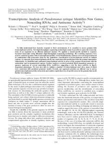
FIG. 1. Ribotypes from P. syringae strains causing disease in the common bean. Ribotypes were generated by BglI (A) and SalI (B). Lane T, ribotype from pathovar tomato strain ATCC 10862; lane UP, ribotype from the unspecifiedpathovar Boelema P2 strain; Phas. and Phaseolicola, ribotypes from pathovar phaseolicola strains; Syringae, ribotypes from pathovar syringae strains. Values to the left of the gels indicate sizes, in kilobases. Data for phytopathogenic strains representing each ribotype are compiled in Table 1.
represented by pathovar syringae organisms. Pathovar phaseolicola strain ATCC 19304 generated the B1 ribotype, while the unspecified-pathovar strain Boelema 2 and pathovar tomato strain ATCC 10862 generated ribotypes not represented among the Spanish strains. The B ribotypes included between four and eight fragments that appeared in the region below 5.2 kb. Only one fragment of about 1.4 kb was common to all of them, and 20 polymorphic restriction sites were revealed. The number and size of the fragments indicate the presence of one or more BglI sites within the five rrn operons of P. syringae strains. With SalI, 15 different S ribotypes were found in the series (Fig. 1B), six of these (S1 to S6) being represented by pathovar phaseolicola and eight (S21 to S28) being represented by pathovar syringae. Pathovar phaseolicola strain ATCC 19304 fell into the S6 ribotype, the unspecified-pathovar strain Boelema P2 generated an S ribotype not represented among the Spanish strains, and pathovar tomato strain ATCC 10862 could not be identified by this procedure (successive SalI digestions failed). The S ribotypes included between three and five fragments that appeared in the region between 14.2 and 6.6 kb. None of the fragments were common to all S ribotypes, but a fragment of about 7 kb appeared in the S ribotypes represented by pathovar phaseolicola strains and another fragment of about 12 kb appeared in the S ribotypes from pathovar syringae strains. These data suggest that the strains have no SalI cutting points within rrn operons and that each band usually carries only one rnn operon (2). However, in S ribotypes with fewer than five bands some fragments could carry
APPL. ENVIRON. MICROBIOL.
two neighbor rrn operons or, alternatively, could correspond to two different DNA fragments of very similar sizes. The performance of two-way ribotyping was evaluated according to several criteria (13). With the enzymes used, all the P. syringae group strains, except pathovar tomato strain ATCC 10862, which failed with SalI, could be assigned to well-differentiated ribotypes. Reproducibility and ease of interpretation of the band profiles were very good. The profiles from each enzyme showed a certain degree of similarity with one another and were pathovar specific, thus revealing the relatedness of P. syringae organisms. The discriminatory power (not including the three reference strains) was tested by considering the number of ribotypes generated using each enzyme and by the calculation of the discrimination index (DI) (3, 5). The number of ribotypes (and DIs) obtained was lower with BglI than with SalI: 11 and 14 ribotypes (DI ⫽ 0.48 and 0.84), respectively. By combining profiles from both enzymes, 19 combined ribotypes (DI ⫽ 0.86) were generated, and the two pathovars were subdivided: pathovar phaseolicola into 7 (DI ⫽ 0.73) and pathovar syringae into 12 (DI ⫽ 0.96) combined ribotypes. Within pathovar phaseolicola, when ribotypes were combined with races, a further differentiation was revealed, 15 subtypes (DI ⫽ 0.93). For these calculations, only the 42 strains tested for races could be used, and the NI strains were considered a single and different type. Ribotyping presents other advantages such as accessibility (requiring basic DNA analysis equipment, as well as commercial materials and reagents) and flexibility (it can be used for different bacterial species). On the other hand, it cannot be overlooked that ribotyping is a laborious procedure, requiring multiple steps. It must also be noted that BglI ribotyping can be a useful tool to identify pathovar phaseolicola organisms, on the basis of the specific band profiles generated. However, it is not a good tool to differentiate pathovar phaseolicola organisms, because in the series studied all except two strains generated a single band profile. A correlation between ribotypes and races was not revealed, with the following exceptions. The two race 2 strains generated the B2 ribotype, which was not represented by any other strain of the series. The five strains from the Granja Asturiana bean variety were assigned to race 5, B1-S3 combined ribotype and race NI, B1-S6 combined ribotype, which are feature combinations not registered among the strains from the remaining bean varieties. However, strains of different races fell into a single combined ribotype (races 5, 7, and NI into B1-S1 and races 6 and 7 into B1-S3), while strains assigned to NI generated a single B ribotype (B1) but four different S ribotypes (S1, S3, S5, and S6). For the phylogenetic analysis, data from ribotypes were used. A combined numerical analysis of the different banding profiles revealed by each enzyme was performed with a software package as previously described (9). A high heterogeneity of ribosomal DNA regions of the strains under study (similarity between 22 and 93%) was registered, and the similarity dendrogram showed that different groupings could be observed by varying levels of similarity (Fig. 2). At a low level (S ⫽ 0.46), the strains were distributed into two clusters labeled A (including only pathovar phaseolicola strains) and B (including all pathovar syringae strains as well as the Boelema P2 strain). These groupings support previous studies assigning strains of the pathovars phaseolicola and syringae to different genospecies (1, 3, 4, 8, 11). At a higher level (S ⫽ 0.66), both clusters were differentiated into subclusters and branches. Organisms falling into each one of these groupings could be considered members of the same clonal lineage. The above-mentioned data have also been applied to further our knowledge in the contemporary molecular epidemiology of
VOL. 66, 2000
RIBOTYPING OF TWO PSEUDOMONAS SYRINGAE PATHOVARS
853
FIG. 2. Dendrogram obtained from cluster analysis of BglI and SalI ribotypes of P. syringae group strains. Each branch represents a combined ribotype. A and B and A1, B1, and B2 are the clusters and subclusters revealed at similarity coefficients of 0.43 and 0.68, respectively. The pathovar, race, and climatic area where the strains falling into each branch were collected are indicated. Other features of the strains and clusters are shown in Table 1 and/or described in the text.
P. syringae causing bacteriosis in the common bean in two Spanish climatic regions which are very important bean production areas. Some findings to emphasize are the following. (i) Ribotyping procedures are useful tools for distinguishing individual phytopathogenic strains of P. syringae, allowing the assignment of organisms with defined ribotypes to specific pathovars and the grouping of strains with identical ribotypes into clones. (ii) Different pathogen clones have been selected, but only some pathovar phaseolicola clones appeared with a wide geographic spread. (iii) The cluster analysis grouped clones into subclusters which were considered clonal lineages, enabling us to screen relationships between lineages with some bean varieties and/or climatic regions. Two lineages (pathovar phaseolicola subcluster A1 and pathovar syringae subcluster B1) presented a wide range of hosts and were widespread, while a third lineage (pathovar syringae subcluster B2) showed a specific host range and could be considered endemic in the WT region. Organisms of this last lineage were collected only from the Granja Asturiana bean variety (which is large, with a highly appreciated sensory quality). It is also noteworthy that the only two pathovar phaseolicola race 2 strains appeared as members of a fourth lineage. These findings support ribotyping as a useful tool for specific epidemiological studies such as establishing the endemic types versus those introduced for the first time in a country or geographical area and ascertaining the pathogenic strains carried by bean seeds for sowing or in seeds from other countries with different endemic P. syringae lineages. This last point could be particularly useful to protect the production of specific autochthonous dry bean varieties of high market value, such as the Spanish Granja Asturiana. We thank M. Altwegg for the pKK3535 plasmid, the source of the rrn probe; F. Uruburu of CECT for the P. syringae reference strains; C. Jorda´ of the Universidad Polite´cnica de Valencia for the specific antiserum of pathovar phaseolicola strain CFBP 1390; C. Asensio of the
Servicio de Investigacio ´n, Desarrollo y Tecnologı´a Agraria, Valladolid, for the identification of pathovar phaseolicola races and samples of infected plants from the DC region; and R. Marquinez of the Instituto de Semillas y Plantas de Vivero, Vitoria, for the Paı´s Vasco strains. We are also indebted to M. R. Rodicio for her critical revision of the manuscript. This work has been supported by a grant from the Instituto Nacional de Investigacio ´n Agraria y Alimentaria, Ministerio de Agricultura, Pesca y Alimentacio ´n, Spain (reference SC-94-051). REFERENCES 1. Clerc, A., C. Manceau, and X. Nesme. 1998. Comparison of randomly amplified polymorphic DNA with amplified fragment length polymorphism to assess genetic diversity and genetic relatedness within genospecies III of Pseudomonas syringae. Appl. Environ. Microbiol. 64:1180–1187. 2. De Ita, M. E., R. Marsch-Moreno, P. Guzma ´n, and A. Alvarez-Morales. 1998. Physical map of the chromosome of the phytopathogenic bacterium Pseudomonas syringae pv. phaseolicola. Microbiology 144:493–501. 3. Denny, T. P., M. N. Gilmour, and R. K. Selander. 1988. Genetic diversity and relationships of two pathovars of Pseudomonas syringae. J. Gen. Microbiol. 134:1949–1960. 4. Gardan, L., H. L. Shafik, S. Belouin, R. Broch, F. Grimont, and P. A. D. Grimont. 1999. DNA relatedness among the pathovars of Pseudomonas syringae and description of Pseudomonas cannabina, sp. nov. (ex Sutic and Dowson 1959). Int. J. Syst. Bacteriol. 49:469–478. 5. Hunter, P. R., and M. A. Gaston. 1988. Numerical index of the discriminatory ability of typing systems: an application of Simpson’s index of diversity. J. Clin. Microbiol. 26:2465–2466. 6. Jansing, H., and H. Rudolph. 1990. A sensitive and quick test for determination of bean seed infestation by Pseudomonas syringae pv. phaseolicola. Z. Pflanzenkr. Pflanzenschutz 97:42–55. 7. Lelliot, R. A., E. Billing, and A. C. Hayward. 1966. A determinative scheme for fluorescent plant pathogenic bacteria. J. Appl. Bacteriol. 29:470–478. 8. Manceau, C., and A. Horvais. 1997. Assessment of genetic diversity among strains of Pseudomonas syringae by PCR-restriction fragment length polymorphism analysis of rRNA operons with special emphasis on P. syringae pv. tomato. Appl. Environ. Microbiol. 63:498–505. 9. Mendoza, M. C., R. Alzugaray, E. Landeras, and M. A. Gonza ´lez-Hevia. 1996. Discriminatory power and application of ribotyping of Yersinia enterocolitica O:3 in an epidemiological study. Eur. J. Clin. Microbiol. Infect. Dis. 15:220–226. 10. Ministerio de Agricultura, Pesca y Alimentacio ´n. 1991. Manual de labora-
854
´ LEZ ET AL. GONZA
torio. Diagno ´stico de hongos, bacterias y nematodos fitopato ´genos. Ministerio de Agricultura, Pesca y Alimentacio ´n, Madrid, Spain. 11. Pecknold, P. C., and R. G. Grogan. 1973. Deoxyribonucleic acid homology groups among phytopathogenic Pseudomonas species. Int. J. Syst. Bacteriol. 23:111–121. 12. Sands, D. C., M. N. Schroth, and D. C. Hildebrand. 1988. Pseudomonas, p. 60–78. In N. W. Schaad (ed.), Laboratory guide for identification of plant pathogenic bacteria, 2nd ed. American Phytopathological Society, St. Paul, Minn. 13. Struelens, M. J., et al. 1996. Consensus guidelines for appropriate use and
APPL. ENVIRON. MICROBIOL. evaluation of microbial epidemiologic typing systems. Clin. Microbiol. Infect. 2:2–11. 14. Taylor, J. D. 1970. The quantitative estimation of the infection of bean seed with Pseudomonas phaseolicola (Burkh) Dowson. Annu. Appl. Biol. 66:29–36. 15. Taylor, J. D., D. M. Tevenson, D. J. Allen, and M. A. Pastor Corrales. 1996. Identification and origin of races of Pseudomonas syringae pv. phaseolicola from Africa and other bean growing areas. Plant Pathol. 45:469–478. 16. Young, J. M., Y. Takikawa, L. Gardan, and D. E. Stead. 1992. Changing concepts in the taxonomy of plant pathogenic bacteria. Annu. Rev. Phytopathol. 30:67–105.