Patient Specific Simulation and Navigation of Ventriculoscopic Interventions 1 S.P. DIMAIO a , J. WADA b , N. HATA a , G. SZÉKELY c , R. KIKINIS a , F. JOLESZ a a Brigham and Women’s Hospital, Harvard Medical School, USA b Department of Neurosurgery, Tokyo Medical University c Computer Vision Laboratory, ETH Zurich, Switzerland
R. SIERRA
a,2 ,
Abstract. In this paper a comprehensive framework for pre-operative planning, procedural skill training, and intraoperative navigation is presented. The goal of this system is to integrate surgical simulation with surgical planning in order to improve the individual treatment of patients. Various surgical approaches and new, more complex procedures can be assessed using a safe and objective platform that will allow the physicians to explore and discuss possible risks and benefits prior to the intervention. A simulation environment extends the pre-operative planning in a natural way, as it allows for direct evaluation of the surgical approach envisioned for each case. In addition, by providing intraoperative navigation based on this simulation, surgeons can carry out the previously optimized plan with higher precision and greater confidence. Keywords. patient-specific simulation, ventricles, fluid simulation, neurofiberscopic surgery, endoscope navigation
1. Introduction To date, research has focused on either surgical training simulation or surgical planning, which have in some cases been combined with intraoperative guidance [1]. Surgical interventions are becoming increasingly complex in order to treat a broader range of diseases with minimal invasiveness while trying to reduce iatrogenic injuries. In particular, technological advances, e.g., in the miniaturization of devices, have provided the necessary means to reach remote areas in the human body through small incisions and to perform complex operations with appropriately adapted manipulators. For the success of these interventions, it is crucial that the surgery is not only thoroughly planned, but also that an appropriate environment is provided for the surgeons to practice and test the necessary skills. Ventriculoscopy – the minimally invasive endoscopic inspection and treatment of the ventricles of the brain – serves as a driving application for this research. From the large range of ventriculoscopic procedures performed nowadays, ventriculostomy (the surgical 1 This work was partially funded by NIH (U41-RR019703) and the Swiss National Science Foundation Fellowship (PBEZ2-110771) 2 Correspondence to: Raimundo Sierra,
[email protected]
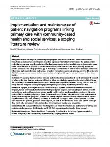
establishment of an opening in a ventricle, e.g., for the treatment of hydrocephalus) and tumor biopsy are particularly challenging tasks, as evidenced by the high rates of failure and complication [2]. The long term goal of this research is to enable endoscopic, trans-ventricular intraparenchymal brain tumor resection. Our vision is to provide a system for the safe treatment of pathologies that are unreachable using current techniques; where their removal would cause unacceptable morbidity when targeted directly from the skull. For such an intervention, accurate planning – including patient-specific training of skills and complication management – as well as intraoperative navigation, will be indispensable. Bleeding, for example, can impede the direct visualization of the surgical site through the endoscopic camera, thus requiring enhanced navigation in a simulated environment. 2. Materials and Methods The position of the camera and the instruments, located at the tip of the flexible endoscope, is tracked using a miniaturized tracking coil introduced through the working channel of the endoscope. The position and heading direction of the coil, i.e., 5 degrees of freedom (DOF), are measured by the NDI Aurora tracking system [3]. In our experiments, the complete ventricular system consisting of both lateral, the third and the fourth ventricle were segmented based on the patient’s pre-operative MR data using an active contour segmentation tool using a level set approach as implemented in the 3D Slicer [4]. The resulting surface meshes were in excellent agreement with a manual segmentation performed by a clinical expert. A corresponding hollow plastic model was built by stereolithography and fixed inside a Styrofoam head model. The resulting anthropomorphic phantom was imaged in a CT scanner. The resulting images were registered with the head phantom – within the tracker’s coordinate frame – and the endoscope position was tracked in order to be able to generate virtual endoscopic views from the image data. The underlying software framework is based on a high fidelity surgical simulator developed for procedural training of hysteroscopic interventions [5]. In the current implementation, the cerebrospinal fluid flow, bleeding, and soft tissue deformations can be simulated in real-time; however, given the complexity of the anatomical structures and the size of the meshes (approx. 175,000 triangles), tissue deformation had to be limited to a region of interest in order to maintain real-time performance. 3. Results The current system is illustrated in Figure 1, where the field generator of the Aurora tracker can be seen on the left side. The true endoscopic camera view is presented on the left screen, while the virtual environment is illustrated on the right screen with the navigation view on the left side and the simulated endoscopic perspective on the right side. A subjective assessment of the real and virtual view shows excellent agreement. The missing information of the 6th degree of freedom is clearly visible as a rotation of the virtual view around the camera axis with respect to the actual camera view. This problem will be solved by using a new generation of coils which will allow for tracking of 6 DOF. The accuracy of endoscope localization was measured at a number of known landmarks visible in the CT images, yielding a mean overall error of 1.25mm (1.36mm RMS), which includes image registration errors [6]. Distortion due to camera optics has been
Figure 1. Ventriculoscopy navigation and simulation system.
measured separately. Lighting parameters and depth of view have been set empirically but will be measured in the future. 4. Discussion Several aspects of navigated endoscopy can be explored using the framework developed here. By leaving out the real camera and navigation view, surgical skills can be trained in a realistic virtual, simulated environment. In addition, different entry points and instrument workspace traversals can be evaluated for planning and procedure prototyping. Intra-procedural brain shift is less pronounced in endoscopic procedures as compared to open surgeries; nevertheless, the system will have to account for this. Several options will be investigated, including image-based registration of the intraoperative view with the simulation or the control of the cerebrospinal fluid volume to maintain a constant fluid pressure and thus a constant ventricle shape. The ability to track the position of the instruments during surgery will be crucial in order to relate the video sequence to the virtual model which is necessary to quantify the predictive capabilities of the simulator. References [1] [2] [3] [4] [5] [6]
D. T. Gering et al. An Integrated Visualization System for Surgical Planning and Guidance Using Image Fusion and an Open MR. J. Mag. Res. Imag., 13:967–975, 2001. D. Hellwig et al. Endoscopic third ventriculostomy for obstructive hydrocephalus. Neurosurg Rev, 28:1– 34, 2005. http://www.ndigital.com/aurora.php K. Krissian et al. Fast Sub-Voxel Re-initialization of the Distance Map for Level Set Methods. Pattern Recognition Letters, 26:10,1532–1542, 2005. http://www.hystsim.ethz.ch J. Wada et al. Development of a Slicer-based Navigation System for Neurofiberscopic Surgery. Symp. of the Int. Brain Mapping and Intraoerative. Surgical Planning Society, 2005.