Stability and tissue penetration characteristics of azithromycin are desirable in the treatment of bacteri- al diseases of reptiles. The ball python (Python regius).
Pharmacokinetics and tissue concentrations of azithromycin in ball pythons (Python regius) Rob L. Coke, DVM; Robert P. Hunter, PhD; Ramiro Isaza, DVM, MS; David E. Koch, MS; Marie A. Goatley, BA; James W. Carpenter, MS, DVM
ObjectiveTo determine pharmacokinetics and tissue concentrations of azithromycin in ball pythons (Python regius) after IV or oral administration of a single dose. Animals2 male and 5 female ball pythons. ProceduresUsing a crossover design, each snake was given a single dose of azithromycin (10 mg/kg) IV. After a 4-week washout period, each snake was given a single dose of azithromycin (10 mg/kg) orally. Blood samples were collected prior to dose administration and 1, 3, 6, 12, 24, 48, 72, and 96 hours after azithromycin administration. Azithromycin was quantitated by use of liquid chromatography-mass spectrometry. ResultsAfter IV administration, azithromycin had an apparent volume of distribution of 5.69 L/kg and a plasma clearance of 0.19 L/h/kg. Harmonic means for the terminal half-life were 17 hours following IV administration and 51 hours following oral administration. Mean residence times were 37 and 94 hours following IV and oral administration, respectively. Following oral administration, azithromycin had a peak plasma concentration (Cmax) of 1.04 µg/mL, a time to Cmax of 8.4 hours, and a prolonged mean absorption time of 57 hours. Mean oral bioavailability was 77%. Tissue concentrations ranged from 4 to 140 times the corresponding plasma concentration at 24 and 72 hours after azithromycin administration. Conclusions and Clinical Relevance—Azithromycin is well absorbed and tolerated by ball pythons. On the basis of plasma pharmacokinetics and tissue concentration data, we suggest an azithromycin dosage in ball pythons of 10 mg/kg, orally, every 2 to 7 days, depending upon the site of infection and susceptibil ity of the infective organism. (Am J Vet Res 2003;64:225–228)
A
zithromycin is a member of a subclass of macrolide antimicrobials classified as azalides. The chemical
Received May 13, 2002. Accepted September 6, 2002. From the Zoological Pharmacology Laboratory, Department of Clinical Sciences (Coke, Isaza, Carpenter), and the Department of Anatomy & Physiology (Hunter, Koch, Goatley), College of Veterinary Medicine, Kansas State University, Manhattan, KS 66506. Dr. Coke’s present address is the San Antonio Zoo, 3903 N St Mary’s St, San Antonio, TX 78212-3199. Supported by the Department of Anatomy & Physiology’s Clinical Research Grant Program, College of Veterinary Medicine, Kansas State University. Presented in part at the Joint Conference of the American Association of Zoo Veterinarians, American Association of Wildlife Veterinarians, Association of Reptilian and Amphibian Veterinarians, and National Association of Zoo and Wildlife Veterinarians, Orlando, September, 2001. Address correspondence to Dr. Hunter. AJVR, Vol 64, No. 2, February 2003
structure of azithromycin is similar to erythromycin but with the addition of a methyl-substituted nitrogen in the lactone ring creating a 15-membered azalide.1 The change in structure improves acid stability and tissue penetration, compared with erythromycin. The mechanism of action is also similar to the inhibition of protein synthesis by binding of the 50S ribosomal subunit.2,3 Azithromycin provides broad-spectrum antibiosis with some activity against anaerobic organisms.4 Results of recent studies indicate that azithromycin has activity against Mycoplasma, Chlamydia, Toxoplasma, Borrelia, Cryptosporidium, Giardia, and Plasmodium spp, and the Mycobacterium avium complex.5-12 Azithromycin administration results in sustained drug concentrations in tissues that are greater than the corresponding plasma concentration. The drug rapidly moves from plasma into the intracellular compartments, especially in the pulmonary, lymphatic, and genital tissues.13,14 At equilibrium in humans, azithromycin concentrations in tissues are up to 200X greater than in plasma. Azithromycin also accumulates within WBCs, which allows azithromycin to be carried directly to the site of infection.15,16 Stability and tissue penetration characteristics of azithromycin are desirable in the treatment of bacterial diseases of reptiles. The ball python (Python regius) is a species that is representative of the Boidae family. Therefore, the purpose of the study presented here was to determine the pharmacokinetics and tissue concentrations of azithromycin in ball pythons after IV or oral administration of a single dose. Data derived from our study will be used in designing therapeutic dosage regimens for treating infectious bacterial diseases of ball pythons. Materials and Methods AnimalsThe Institutional Animal Care and Use Committee of Kansas State University approved our study. Seven ball pythons (2 males, 5 females) weighing 0.67 to 0.96 kg were used. Each snake was housed individually in a 114-L aquaria with newspaper substrates and screen tops. Snakes had access to a hide box and water ad libitum. Cages were housed in a thermostatically controlled room at 30oC with a 12-hour light-dark cycle.17 A physical examination and CBC determination were performed for each snake prior to the start of our study. Study designIn the first phase of our study, a crossover design was used to evaluate the pharmacokinetics of azithromycin. Each snake was given a single dose of azithromycina (10 mg/kg) via cardiocentesis. After a 4-week washout period, each snake was given a single dose of azithromycin (10 mg/kg) orally by use of the same preparation. Blood samples (0.75 mL) were collected prior to dose administration (at least 1 week) and at 1, 3, 6, 12, 24, 48, 72, 225
and 96 hours after azithromycin administration. Samples were immediately transferred to evacuated lithium heparin tubes.b Plasma was separated via centrifugation (10 minutes, approx 2000 X g) and stored at –70oC until analyzed. In the second phase of our study, 6 snakes were used to determine tissue concentrations of azithromycin. Snakes had not received a dose of azithromycin for a period of 4 months. Each snake was given a single dose of azithromycin (10 mg/kg) orally. Three snakes were euthanatized 24 hours after dose administration, and the remaining 3 snakes were euthanatized 72 hours after dose administration. Blood samples and liver, kidney, lung, and skin specimens were collected and stored at –70oC until analyzed. For euthanasia, tiletamine-zolazepamc (25 mg/kg, IM) was administered first to anesthetize the snakes. After a cut down to the vena cava and blood collection from this vein, pentobarbital and phenytoin solutiond (1 mL/4.5 kg) were administered via the vena cava to euthanatize each snake. Plasma and tissue analysis for azithromycinBlood samples and tissue specimens were analyzed via liquid chromatography-mass spectrometry by use of previously described methods for extraction of the samples13 and chromatographic conditions.18 Accuracy and precision were within ± 15% of actual values, and recovery was > 80% across the range of the assay in all fluids and tissues. The plasma standard curve had a linear range of 0.015 to 2.00 µg/mL. The limit of quantitation was 0.013 µg/mL for plasma. The tissue standard curve had a linear range of 0.25 to 15 µg/g with a limit of detection of 0.22 µg/g. Pharmacokinetic calculationsValues of pharmacokinetic parameters were determined for each snake by use of noncompartmental analysis19,20 with a commercial software program.e Values calculated following the IV administration of azithromycin were as follows: plasma area under the concentrations versus time curve (AUC); area under the first moment curve (AUMC); mean residence time (MRT), where MRT = AUMC/AUC; apparent volume of distribution at steady state (Vd), where Vd = (dose X AUMC/AUC2); plasma clearance (Clp), where Clp = dose/AUC; elimination rate constant (kel) calculated as the slope of the terminal phase of the plasma concentration curve that included a minimum of 3 time points; and terminal half-life (t½), where t½ = 0.693/kel. Following oral administration of azithromycin, the following parameters were determined: AUC; AUMC; MRT; mean absorption time (MAT), where MAT = MRTPO – MRTIV; and bioavailability (F), where F = (AUCPO/AUCIV) X 100. The AUC and AUMC were calculated by use of the trapezoidal rule with extrapolation to infinity.
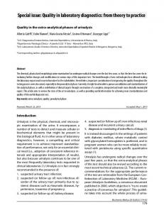
Figure 1Mean (± SD) plasma concentrations (µg/mL) of azithromycin in ball pythons following IV and oral (PO) administration of a single dose (10 mg/kg). 226
Results All snakes were clinically normal upon physical examination prior to the start of our study. The CBC results were within reference range limits for our institution. Two weeks after IV administration of azithromycin, 1 snake died from apparent nonregenerative anemia. All other snakes remained clinically normal during and after both phases of our study. In the first phase of our study, plasma AUCs (Fig 1) and pharmacokinetic parameters (Table 1) following IV and oral administration of azithromycin were determined. In the second phase of our study, concentrations of azithromycin in plasma samples and liver, kidney, lung, and skin tissues were determined following oral administration (Fig 2). Tissue-to-plasma concentration ratios for azithromycin were determined for each snake (Table 2). Because of the limited numTable 1Azithromycin pharmacokinetic parameters in ball pythons following IV and oral administration of a single dose (10 mg/kg) Values Parameters
Mean
SD
IV administration (n = 7) AUC0-∞ (h X µg/mL) AUMC0-∞ (h2 X µg/mL) MRT (h) Vd (L/kg) Clp (L/h/kg) kel (h–1) t½ (h)
Median Minimum Maximum
70 2909 37 5.69 0.190 0.041 17
37 2563 19 3.16 0.114 0.061 HM
72 2441 35 4.70 0.139 0.0183 38
25 130 5.2 2.08 0.0837 0.0081 3.9
119 8211 69 10.1 0.401 0.180 86
Oral administration (n = 6) AUC0-∞ (h X µg/mL) AUMC0-∞ (h2 X µg/mL) MRT (h) MAT (h) Cmax (µg/mL) Tmax (h) kel (h–1) t½ (h) F (%)
45 4908 94 57 1.04 8.4 0.014 51 77
22 5187 50 43 0.382 8.8 0.0063 HM 27
43 2360 76 52 0.948 6.0 0.014 49 69
20 1688 53 17 0.538 1.0 0.0059 32 49
82 14851 181 112 1.50 24 0.022 117 128
AUC0-∞ = Area under the plasma concentration versus time curve from time of administration to infinity. AUMC0-∞ = Area under the first moment curve from time of administration to infinity. MRT = Mean residence time. Vd = Apparent volume of distribution at steady state. Clp = Plasma clearance. kel = Elimination rate constant. t½ = Half-life. HM = Harmonic mean. MAT = Mean absorption time. Cmax = Peak plasma concentration. Tmax = Time of peak plasma concentration. F = Bioavailability.
Figure 2Individual plasma and tissue concentrations (µg/mL or µg/g, respectively) of azithromycin in ball pythons following oral administration of a single dose (10 mg/kg). AJVR, Vol 64, No. 2, February 2003
Table 2Individual azithromycin tissue-to-plasma concentration ratios in ball pythons following oral administration of a single dose (10 mg/kg) Tissue
24 h
72 h
Lung
13, 22, 29
19, 31, 51
Skin
4.1, 5.0, 9.2
4.6, 4.7, 5.0
Liver
22, 28, 34
52, 62, 102
Kidney
43, 48, 72
88, 118, 143
ber of time points for tissue concentrations, t½ values were not determined. Discussion In human medicine, azithromycin is approved for use in treating respiratory tract, skin, and sexually transmitted diseases.14,21 It has a long terminal plasma t½ (70 hours) in humans.22 Azithromycin is excreted approximately 75% unchanged in the bile of mammals, which indicates that the primary xenobiotic component is the unaltered drug compound.1,13 Compared with other species, the t½ for a single dose of orally administered azithromycin in the ball python is 51 hours, which is comparable to humans. The Vd determined in the snakes of our study (5.69 L/kg) is not large, compared with the Vd of rats (84 L/kg), cats (23 L/kg), and dogs (12 L/kg).1,13,23 However, azithromycin in pythons appears to be distributed to a greater extent than amikacin (0.41 L/kg) or piperacillin (2.6 L/kg).24,25 The larger Vd of azithromycin in the ball python supports the high concentrations of azithromycin found in tissue specimens (Fig 2) and the potential for greater efficacy against susceptible organisms, compared with other antimicrobials evaluated in pythons to date. It is unknown why the Vd of azithromycin in ball pythons is so much smaller in this species of reptile, compared with the Vd of azithromycin in the 3 mammalian species mentioned. The smaller Vd in ball pythons could be a result of a high rate of metabolism of azithromycin or the result of species differences in protein binding. Finally, the smaller Vd reported for ball pythons in our study could be the result of reptilian cellular anatomy, which may decrease the penetration of azithromycin into some or all tissues. The mean bioavailability of azithromycin was 77% following oral administration of a single dose. This value is greater than the mean bioavailability of azithromycin in humans (37%) or cats (58%), but less than that in dogs (97%).1,13,22 The variation in bioavailability of azithromycin in the snakes of our study may be the result of the prolonged absorption indicated by the values of MAT and time to reach maximal concentration. Also, feeding may affect intestinal absorption. When a snake consumes a meal, the small intestinal mucosa will increase in thickness by 2 to 3 times the prefeeding thickness, but total length of the small intestine does not change. Also, the villi length increases to 2 times greater than the prefeeding length. An increase in the surface area of the small intestine in response to a recent meal would allow for an increase in absorption of azithromycin. The intestine of snakes will regress in size during prolonged periods of not eating.26-28 This factor may greatly enhance or restrict AJVR, Vol 64, No. 2, February 2003
intestinal absorption of azithromycin or any pharmacologic agent in the gastrointestinal tract, depending upon the period between drug administration and the last feeding. As with intestinal changes, metabolism is also affected by feeding.27,28 Metabolism will peak about 1 week after feeding; however, there is minor metabolic investment in digestion after a period of not eating and energy reserve depletion. With variable states of metabolism, drug metabolism and elimination could be directly affected by feeding intervals. Snakes of our study received a dose of azithromycin 6 days after feeding in an attempt to achieve consistent rates of metabolism and elimination. The affect of feeding status will widely affect the way orally administered pharmacologic agents are absorbed by snakes. Because of the accumulation of azithromycin in tissues, it is not evaluated on the basis of plasma concentrations alone. Data from our study indicate that concentrations of azithromycin are higher in tissues than in plasma (Fig 2). Even at 72 hours following administration, the concentration of azithromycin in the lung, liver, skin, and renal tissues is greater than the corresponding plasma values. Aeromonas hydrophila is 1 of the most common reptile pathogens, especially in infections of the respiratory tract and in cases of stomatitis.29 The minimum inhibitory concentration that will result in death of 90% of the organisms (MIC90) of azithromycin for A hydrophila is 4 µg/mL.30 In our study, the concentration of azithromycin in skin specimens was slightly less than the reported MIC90 for A hydrophila at 24 hours after administration and much lower at 72 hours after administration. However, at 24 and 72 hours after administration, concentrations of azithromycin in lung specimens were 2 to 4 times greater than the reported MIC90 for A hydrophilia. Although azithromycin has been reported to be active against Pseudomonas infections in humans,31 more research is need to determine the efficacy of azithromycin against this pathogen in snakes. Compared with other antimicrobials, azithromycin (10 mg/kg, PO, q 2 to 7 d) may be effective for the treatment of susceptible microbes in reptiles because of its broad spectrum of activity, increased tissue penetration, and prolonged residence in tissues. The dose administration interval should be optimized on the basis of the MIC of azithromycin for the target organism and the location of the infection (eg, skin, q 3 d; respiratory tract, q 5 d; liver and kidney, q 7 d). a
Zithromax, Pfizer Inc, New York, NY. Vacutainer, Becton Dickinson & Co, Franklin Lakes, NJ. c Telazol, Fort Dodge Animal Health, Fort Dodge, Iowa. d Beuthanasia-D Special, Schering-Plough Animal Kenilworth, NJ. e WinNonlin, version 3.1, Pharsight, Mountain View, Calif. b
Health,
References 1. Shepard RM, Falkner FC. Pharmacokinetics of azithromycin in rats and dogs. J Antimicrob Chemother 1990;25(suppl A):49–60. 2. Champney WS, Burdine R. Azithromycin and clarithromycin inhibition of 50S ribosomal subunit formation in Staphylococcus aureus cells. Curr Microbiol 1998;36:119–123. 3. Girard AE, Girard D, English AR, et al. Pharmacokinetics and in vivo studies with azithromycin (CP-62,993), a new macrolide 227
with an extended half-life and excellent tissue distribution. Antimicrob Agents Chemother 1987;31:1948–1954. 4. Retsema J, Girard A, Schelkly W, et al. Spectrum and mode of action of azithromycin (CP-62, 993), a new 15-membered-ring macrolide with improved potency against gram-negative organisms. Antimicrob Agents Chemother 1987;31:1939–1947. 5. Anderson SL, Berman J, Kuschner R, et al. Prophylaxis of Plasmodium falciparum malaria with azithromycin administered to volunteers. Ann Intern Med 1995;123:771–773. 6. Chang HR. The potential role of azithromycin in the treatment of prophylaxis of toxoplasmosis. Int J STD AIDS 1996;7 (suppl1):18–22. 7. Dever LL, Jorgensen JH, Barbour AG. Comparative in vitro activities of clarithromycin, azithromycin, and erythromycin against Borrelia burgdorferi. Antimicrob Agents Chemother 1993;37:1704–1706. 8. Hyde TB, Gilbert M, Schwartz SB, et al. Azithromycin prophylaxis during a hospital outbreak of Mycoplasma pneumoniae pneumonia. J Infect Dis 2001;183:907–912. 9. Ikerd TR, Koletar SL. In-vitro activity of ciprofloxacin, temafloxacin, azithromycin, clarithromycin and metronidazole against Giardia lamblia. J Antimicrob Chemother 1993;31:615–617. 10. Niki Y, Kimura M, Miyashita N, et al. In vitro and in vivo activities of azithromycin, a new azalide antibiotic, against Chlamydia. Antimicrob Agents Chemother 1994;38:2296–2299. 11. Rehg J. A comparison of anticryptosporidial activity of paromomycin with that of other aminoglycosides and azithromycin in immunosuppressed rats. J Infect Dis 1994;170:934–938. 12. Van der Heyden N. New strategies in the treatment of avian mycobacteriosis. Semin Avian Exot Pet Med 1997;6:25–33. 13. Hunter RP, Lynch MJ, Ericson JF, et al. Pharmacokinetics, oral bioavailability, and tissue distribution of azithromycin in cats. J Vet Pharmacol Ther 1995;18:38–46. 14. Schentag JJ, Ballow CH. Tissue-directed pharmacokinetics. Am J Med 1991;91:5–11. 15. Carbon C. Clinical relevance of intracellular and extracellular concentrations of macrolides. Infection 1995;23:S10–S14. 16. Girard AE, Cimochowski CR, Faiella JA. Correlation of increased azithromycin concentrations with phagocyte infiltration into sites of localized infection. J Antimicrob Chemother 1996; 37(suppl C):9–19. 17. de Vosjoli P, Klingenberg R, Barker D, et al. The ball python manual. Santee, Calif: Advanced Vivarium Systems, 1997;76.
228
18. Fouda HG, Shepard RM, Ferraina RA, et al. Atmospheric pressure HPLC/MS/MS identification of azithromycin rat biliary metabolites, in Proceedings. 38th Am Soc Mass Spectrom Conf Allied Topics, 1990. 19. Gibaldi M, Perrier P. Pharmacokinetics. 2nd ed. New York: Marcel Dekker Inc, 1982;409–417. 20. Riviere JE. Comparative pharmacokinetics: principles, techniques, and applications. Ames, Iowa: Iowa State University Press, 1999;327. 21. Physicians’ desk reference. 56th ed. Montvale, NJ: Medical Economics Co, 2002;2739–2751. 22. Foulds G, Shepard RM, Johnson RB. The pharmacokinetics of azithromycin in human serum and tissues. J Antimicrob Chemother 1990;25(suppl A):73–82. 23. Boothe DM. Small animal clinical pharmacology and therapeutics. Philadelphia: WB Saunders Co, 2001;806. 24. Hilf M, Swanson D, Wagner R, et al. Pharmacokinetics of piperacillin in blood pythons (Python curtus) and in vitro evaluation of efficacy against aerobic gram-negative bacteria. J Zoo Wildl Med 1991;22:199–203. 25. Johnson JH, Jensen JM, Brumbaugh GW, et al. Amikacin pharmacokinetics and the effects of ambient temperature on the dosage regimen in ball pythons (Python regius). J Zoo Wildl Med 1997;28:80–88. 26. Starck JM, Beese K. Structural flexibility of the intestine of Burmese python in response to feeding. J Exp Biol 2001;204:325–335. 27. Secor SM, Diamond J. Determinants of the post-feeding metabolic response of Burmese pythons, Python molurus. Physiol Zool 1997;70:202–212. 28. Starck JM, Beese K. Structural flexibility of the small intestine and liver of garter snakes in response to feeding and fasting. J Exp Biol 2002;205:1377–1388. 29. Cooper JE. Bacteria. In: Cooper JE, Jackson OF, eds. Diseases of the reptilia. Vol 1. London: Academic Press Inc, 1981;165–191. 30. Jones K, Felmingham D, Ridgway G. In vitro activity of azithromycin (CP-62,993), a novel macrolide, against enteric pathogens. Drugs Exp Clin Res 1988;14:613–615. 31. Tateda K, Ishii Y, Matsumoto T, et al. Direct evidence for antipseudomonal activity of macrolides: exposure-dependent bactericidal activity and inhibition of protein synthesis by erythromycin, clarithromycin, and azithromycin. Antimicrob Agents Chemother 1996;40:2271–2275.
AJVR, Vol 64, No. 2, February 2003