AbstractâA vertically aligned nanotube (NT) array platform with peptide-modified ends is used for sensing metal ions in aqueous environments. The NT arrays ...
Peptide modified SWNT array-based copper sensor Monessha Nambiar, Joe Shapter School of Chemical and Physical Science The Flinders University of South Australia, Adelaide SA 5042, Australia 50mg of SWNTs were refluxed in 2M HNO3 for 24 hours to remove any associated impurities. This was followed by sonication for 8 hours in a 50ml acid mixture of concentrated nitric acid and concentrated sulphuric acid in a 1:3 ratio during which the temperature in the sonicator was maintained in the 0oC - 5oC range. After the excision process, the SWNT/acid mixture was diluted with over 1000ml of water to reduce their acidity. The nanotubes were filtered, washed with water several times and dried. 2) Self-assembly of nanotubes on gold Gold electrodes that were polished, rinsed in piranha solution (3:1 conc. H2SO4 and 30% H2O2) for 1-2min at 6080oC, followed by electrochemical cleaning were incubated in a 1mM cysteamine solution in 95% aqueous ethanol for 3 hrs. A solution mixture containing 1mg of cut nanotubes, 2.5mg of DCC and 5 ml of DMF was prepared via sonication for four hours to ensure uniform dispersion of nanotubes. The cysteamine modified gold surface was incubated in the nanotube suspension overnight in order to achieve covalent attachment of the nanotubes to the electrode surface. 3) Peptide Attachment and Metal Ion Accumulation The carboxylic acid terminated-carbon nanotube array, self-assembled on gold, was incubated in GlyGlyHis peptide solution of 2mg/mL in HEPES buffer set to neutral pH for 30 minutes. This step is carried out immediately after exposure to the nanotube suspension in order to ensure that the carboxylic acid ends of the nanotubes remain activated towards peptide immobilisation. Subsequently, the electrode was rinsed in a plain HEPES buffer without peptide, followed by rinsing in milli-Q water and then dried under a gentle stream of nitrogen. The peptide-modified electrodes were immersed into a known concentration of the Cu2+ solution in 50mM ammonium acetate buffer (pH 7.0) for a specific length of time. The electrode was removed, rinsed with the metal ion free ammonium acetate buffer, followed by purified water and air-drying.
Abstract—A vertically aligned nanotube (NT) array platform with peptide-modified ends is used for sensing metal ions in aqueous environments. The NT arrays were assembled on gold substrates and characterised using confocal Raman microscopy to confirm the electrode fabrication process. A copper ion selective peptide – GlyGlyHis was used as the sensing recognition element. Cyclic voltammetry was used to measure the sensor’s electrochemical response of the bound copper and to determine the stability of the sensor. The sensor showed minimum interference from other species and was proven to be very sensitive, enabling detection in the nanomolar range. Keywords—carbon nanotubes; copper sensor; GlyGlyHis; cyclic voltammetry; confocal Raman spectroscopy
I.
INTRODUCTION
Carbon nanotubes since their discovery in 1991[1] have become well established as a highly versatile and unique material in a diverse range of applications due to their combined mechanical strength and electrical conductivity. Their ability to act as fast electron-transfer tunnels[2], affinity for binding with redox sites of enzymes, sensitivity to such adsorbates and high elastic modulus[3] are some of the critical factors that influence their role in the fabrication of sensing devices. Heavy metal ions are considered to be the root cause of many diseases and disorders today and have proven to be lethal even at very low concentrations. The detection of these ions is usually carried out using techniques like atomic absorption spectroscopy and stripping voltammetry, which involve inconveniences associated with sample pretreatment, expensive instrumentation and skilled operators[4]. Biosensors, now a rapidly emerging technology, overcome a lot of these issues and stand to provide scope for easy and rapid analysis, portability, and miniaturisation[5]. In the recent few years, vertically-aligned nanotube arrays, or nanotubes trees as they are sometimes referred to, have been shown to successfully play the role of molecular wires, giving rise to a new generation of electrochemical biological and environmental sensors. Our research focuses on determining metal ion concentrations using the vertical nanotube array platform thus, incorporating the advantages of the biosensor approach. II.
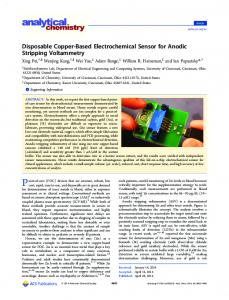
Figure 1 shows a schematic of the electrode sensor fabrication process. B. Characterization of Electrode Interface The modified gold samples were analysed using a WiTec confocal Raman microscope (alpha300) where Raman spectral imaging was performed in order to characterise the nanotubes on the surface. A Nikon 100x air objective was employed for all measurements.
METHODS
A. Preparation of Electode Interface 1) Purification and Cutting of Nanotubes
978-1-4244-5262-0/10/$26.00 © 2010 IEEE
124
ICONN 2010
Figure 1. A schematic of the step-wise fabrication of the nanotube sensor, outlining the formation of vertically aligned nanotubes followed by the peptide attachment and metal ion accumulation
clearly indicates that the nanotube array remained undisturbed and stable during further modification steps.
C. Electrochemical Analysis All electrochemical measurements were performed with a BAS-100B electrochemical analyser with a conventional three-electrode system, comprising a modified working electrode (gold), a platinum flag auxiliary electrode and an Ag/AgCl/3.0M NaCl reference electrode. All potentials were reported against this reference at room temperature. Cyclic voltammetry was conducted at a sweep rate of 100mVs-1 between -200mV and +600mV. The electrolyte consisted of 50mM ammonium acetate buffer and 50mM NaCl (1:1). Measurements were carried out at room temperature in a Teflon cell. III.
The modified gold sensor surface was successful in determining copper concentrations. Figure 3A shows the electrochemical response of Cu2+ on a GlyGlyHis modified electrode. The electrochemical response shown by the reduction of Cu2+ to Cu+ and the oxidation back to Cu2+, which is an indication that the peptide remains associated with the nanotube based sensing interface. The response with just the plain GlyGlyHis modified electrode without any prior metal ion accumulation is also shown as the initial response (Figure 3A). The electrode was also found to be stable on continuous repetitions (Figure 3B) indicating that the metal-peptide affinity is strong. The slightly diminished peak currents are caused by the slow degradation of the underlying cysteamine monolayer. This degradation takes place with the repeated oxidation and reduction that the electrode is subjected to during cyclic voltammetry.
RESULTS AND DISCUSSION
The Raman spectrum obtained, as displayed in Figure 2D, shows the characteristic nanotube peaks (G band~1590 cm-1, RBM~ 190cm-1, D band~1320cm-1 and G’ band~2700cm-1) providing chemical evidence of the presence of the nanotubes[6]. The brightness of each pixel in the three images obtained corresponds to the intensity of the peak at 1590cm-1. The plain gold slide (Figure 2A) shows the absence of nanotube modification, corresponding to zero Gband peak intensity. Figure 2B and 2C show the presence of nanotubes both before and after peptide immobilisation. This
In the absence of the peptide, with the use of just the SWNT array-modified gold surface, there was no substantial electrochemical response towards the metal ion. Thus, the peptide plays an active role in coordinating the metal ion.
125
levels between 40-60 min of accumulation. This saturation point depends not only on the concentration of the metal ion but also on the extent of nanotube coverage across the gold electrodes. Peak current is a measure of the amount of copper accumulated at each modified site. This value also increases with increase in concentration. Prior to accumulation of metal ion or at zero accumulation time, the peptide-modified nanotube array displayed a background signal. This background is the non-Faradaic current that arises due to a certain degree of roughness produced by the presence of the vertically aligned nanotube array. For the time traces observed, the background response was subtracted from peak currents obtained for each of the different accumulation times. The extent of surface coverage with nanotubes was expected to vary with each fabricated electrode. The reproducibility of a self-assembled nanotube array was examined by incubating four different gold electrodes in four different sets of nanotube suspensions, followed by peptide immobilisation. The error bars (Fig. 4) were calculated from the standard deviation of ± 2.06µA measured using the reduction peaks of the four different electrodes. Due to the high transfer of electrons across the ends of nanotubes, the SWNT arrays impart a high degree of sensitivity to such sensor systems. The fabricated electrode showed a strong electrochemical response towards Cu2+ at very low concentrations. Figure 5 shows a CV obtained after incubation of the electrode in a 1nM Cu2+ solution.
Figure 2. Raman images describing surface modification of Au surface – A) plain Au surface, B) Au self-assembled with vertically aligned nanotubes, and C) Aligned nanotubes further modified with peptide GlyGlyHis; D) Raman Spectrum of carbon nanotubes self-assembled on gold surface A)
B)
Figure 4. Top: Cyclic Voltammograms showing increased peak current with accumulation time of Cu2+ on a GlyGLyHis modified electrode. Bottom: Trend in peak current with accumulation time of Cu2+ with same electrode.
Figure 3. Top: CVs of GlyGlyHis modified NT-array electrodes before and after exposure to Cu2+ in ammonium acetate buffer solution (pH=7.0). Cu2+ was accumulated for 10min in a 0.1M copper sulphate solution. Bottom: Repeated scans of the same Cu2+ accumulated modified electrode
Fig. 4 shows the increase in peak current of the cyclic voltammogram with increase in accumulation time from 0 min to 60 min for a 1mM Cu2+ solution. The peptidenanotube arrays were observed to reach their metal saturation
Figure 5. The CV for a GlyGlyHis modified NT array subjected to 10min accumulation time in a 1nM solution
126
Nitrogen atoms from each peptide bond and the imidazole ring form a stable 4N complex with a low binding energy (816.2kJ/mol)[7]. The GlyGlyHis tripeptide was found to specifically bind to copper with minimalistic interferences from other metal ions in solution. Ni2+ ion species showed a slight but noticeable electrochemical response as indicated by previous studies[8]. The reduction peak current however, is negligible in comparison to that obtained with Cu2+. Even after 2hrs of exposure in a 100mM solution of Ni2+ the peak current rise was not observed to be significant. Figure 6 shows the slight rise in reduction peak current from 10min to 2hrs of accumulation time. This minimal interference is because of the similar binding configurations possessed by both Cu2+ and Ni2+, and their preference towards the formation of a 4N complex. For the same length of accumulation time and metal-ion concentration (100mM) for both ions (Cu2+ and Ni2+), the signal (reduction peak current) observed for Cu2+ is 2.5 times greater than that observed for Ni2+ with the GlyGlyHis modified electrode. For this reason, Ni2+ is not likely a major cause of interference in the case of in-field applications because the concentrations of the metal ions in all probability will be present in the sub-micromolar range. The electrochemical response towards Ni2+ will thus be further minimised in these lower concentration ranges.
began deteriorating rapidly. This decline in peak current observed is attributed to the weak stability of the cysteamine layer bound to the gold surface leading to the poor reusability of the overall sensor. The cysteamine layer, in addition to being unstable in water over extended periods of time, is possibly prone to degradation by constant exposure to light and air. The storage stability of the electrode was also assessed. A GlyGlyHis modified electrode was stored under dry conditions at 4oC and a CV was measured and monitored on a daily basis. The peak current decreased every 24hrs with the electrode losing all electrochemical activity after 3 days. Under wet conditions, the electrode degradation was more rapid with a drastic decrease in peak current and complete loss of electrochemistry. IV.
CONCLUSION
This mode of sensor fabrication was successful in establishing peptide-nanotube chemistry. The complexation of the copper ion with the GlyGlyHis interface demonstrated a high affinity for the ion and preferential high stable coordination geometry. The use of nanotubes was proven to be very sensitive with detection abilities in the nanomolar concentration range. The sensing methodology, with the peptide modified nanotube array, reflects the promising use of carbon nanotube arrays 1) as effective and efficient substituents for molecular wires, 2) as a platform to create multiple peptide sensor interfaces due to their potential to enhance and improve the field of sensing devices via its self-assembled alignment, high potential for miniaturisation and high transfer rate of electrons through the nanotubes. REFERENCES [1] M. S. Dresselhaus, G. Dresselhaus, and P. Avouris, "Carbon Nanotubes: Synthesis, Structure, Properties, and Applications," in Topics in applied physics. vol. 80, C. E. Ascheron, H. J. Kolsch, and W. Skolaut, Eds. Germany: Springer-Verlag Berlin Heidelberg, 2001. [2] J. Liu, A. Chou, W. Rahmat, M. N. Paddon-Row, and J. J. Gooding, "Achieving Direct Electrical Connection to Glucose Oxidase Using Aligned Single Walled Carbon Nanotube Arrays," Electroanalysis, vol. 17, pp. 38-46, 2004. [3] J. Wang, "Carbon-Nanotube based Electrochemical Biosensors: A Review," Electroanalysis, vol. 17, pp. 7-14, 2004. [4] S. Alegret and A. Merkoci, "Electrochemical Sensor Analysis," in Comprehensive analytical chemistry. vol. 49, D. Barcelo, Ed. Amsterdam, The Netherlands: Elsevier, 2007.
2+
Figure 6. Extent of observed interference from Ni on a GlyGlyHis modified NT-array for 10min and 2hr accumulation times
[5] C. S. S. R. Kumar, "Nanomaterials for Biosensors," in Nanotechnologies for the Life Sciences. vol. 8, C. S. S. R. Kumar, Ed. Weinheim: WILEY-VCH Verlag GmbH & Co. , 2007.
In order to reuse the electrode, the complexed metal ion had to be released from the peptide cavity, which was carried out by incubating the working electrode in 0.1M HClO4 for 2 minutes. A typical test for regeneration of the electrode involved obtaining the initial CV after incubation at a fixed concentration and accumulation time, followed by subsequent removal of bound metal ion and lastly recomplexation under similar conditions. After each regeneration cycle, the electrochemistry of Cu2+ disappeared, keeping the rest of the modifications intact and ready for the subsequent accumulation. The regeneration process was repeated till a decline in the peak current was observed which lasted up to four cycles, after which the response
[6] S. Saito and A. Zettl, "Carbon Nanotubes: Quantum Cylinders of Graphene," in Contemporary Concepts of Condensed Matter Science, M. L. C. E. Burstein, D.L. Mills and P.J. Stiles, Ed. Oxford, UK: Elsevier, 2008. [7] W. Yang, E. Chow, G. D. Willett, D. B. Hibbert, and J. J. Gooding, "Exploring the use of the tripeptide Gly-Gly-His as a selective recognition element for the fabrication of electrochemical copper sensors," The Analyst, vol. 128, p. 712, 2003. [8] W. Yang, E. Chow, G. D. Willett, D. B. Hibbert, and J. J. Gooding, "Exploring the use of the tripeptide Gly-Gly-His as a selective recognition element for the fabrication of electrochemical copper sensors," The Analyst, vol. 128, pp. 712-718, 2003.
127