Lepr Rev (2017) 88, 574– 582
CASE REPORT
Peripheral neuropathy is not the end but the beginning HALA I. EL GENCY*, MAHMOUD GHANEMA*, SOLIMAN A. HUSSEIN**, OMER S. ALMAGHRABY*** & WEGDAN RASHAD* *Department of Internal Medicine, Cairo University, Cairo, Egypt **Department of Dermatology Hansen Center, Cairo, Egypt ***Department of Internal Medicine, Al Azhar University, Cairo, Egypt Accepted for publication 8 November 2017 Summary Background: Diagnosing the etiology of peripheral neuropathy is a challenging clinical situation especially in a patient with multiple potential causes. This case discusses how distribution of the neuropathy and accompanying manifestations can help unmask the original culprit. Case report: A 59-year-old male patient with a 15 year history of diabetes presented with severe peripheral neuropathy affecting the upper more than lower limbs. Six months prior to this presentation he had been diagnosed with hepatitis C infection for which he received interferon, ribavirin and sofosbuvir therapy. Physical examination revealed bilateral, thickened and tender ulnar and popliteal nerves, multiple patches of vitiligo and another well defined, hypopigmented, anesthetic lesions appearing over his back and buttocks. Six slit smears were positive for Mycobacterium leprae and ultrasound of both median nerves indicated swollen and hypoechoic areas along their course with positive intraneural Doppler uptake. Conclusion: Treatment with the interferon or immunosuppressive therapy could unmask leprosy. Leprosy should be included in the differential diagnosis of severe peripheral neuropathy. Keywords: Peripheral neuropathy, cyclophosphamide
Borderline
leprosy,
Hepatitis
C,
Interferon,
Introduction Peripheral neuropathy (PN) is a clinical conundrum for most clinicians since its etiology is quite variable. It is a complication secondary to a diverse array of a large number of common diseases such as diabetes, thyroid disorders, renal failure, vitamin deficiency, viral infections Correspondence to: Wegdan Rashad, Kasr Al Ainy St. Cairo, Egypt. Post Code: 11562 (Tel./Fax: (202) 23682030; e-mail:
[email protected])
574
0305-7518/17/064053+09 $1.00
q Lepra
Peripheral neuropathy
575
and autoimmune diseases. Peripheral neuropathies are classified by the distribution pattern, which oftentimes can aid the clinician in suspecting the cause in a patient presenting with multiple potential etiologies. Diabetes is the most common cause of PN with prevalence in the Middle East ranging from 37·1% to 65·3%, compared to a prevalence of 2·4 – 8% in the general population.1 Hepatitis C is an inflammatory disease of the liver. Numerous HCV positive patients suffer neurological manifestations, ranging from cognitive impairment to peripheral neuropathy.2,3 Forty to seventy-five percent of HCV positive patients present with symptomatic PN, being more prominent with HCV associated cryoglobulinemia.4,5 Leprosy is a chronic granulomatous infectious disease caused by Mycobacterium leprae (M. leprae), affecting skin and peripheral nerves. Between 30 – 60% of patients will have nerve damage at the time of diagnosis.6 The picture of peripheral neuropathy in leprosy is distinguished by predominantly upper limb involvement. We present a case with several factors potentially causing PN in one patient and how through meticulous work up, the cause was identified. The key in this case was to notice the PN distribution and manifestations after immunosuppressive therapy.
Case Report A 59-year-old Egyptian man, with a 15 year history of diabetes was treated for hepatitis C infection with pegylated interferon, ribavirin and sofosbuvir for 3 months. Six months later he
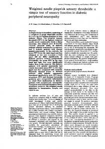
Figure 1. Areas of vitiligo on dorsum of hands.
576
H.I. El Gendy et al.
developed with gradual onset and progressive course, numbness and tingling of the upper limbs more than lower limbs. These symptoms were handicapping for the patient, impairing his daily functioning significantly. Nerve conduction studies revealed focal entrapment of both ulnar and median nerves at the wrist and elbow joints. Nerve decompression surgery on the left upper limb yielded no improvement. Follow-up nerve conduction revealed sensorymotor axonal polyneuropathy. Based on his history of HCV and positive rheumatoid factor with low C4 levels, and the possibility of cyoglobulinemia induced neuropathy, intravenous methylprednisone 1 g/day for 3 days was administered, followed by intravenous cyclophosphamide (15 mg/kg) with partial improvement of his painful neuropathy. Shortly after the second dose of cyclophosphamide, the patient demonstrated bilateral knee and ankle arthritis not accompanied by morning stiffness or other peripheral or axial joint affection.
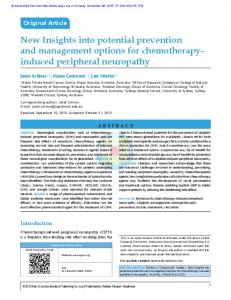
Figure 2. Areas of vitiligo over the leg.
Peripheral neuropathy
577
Figure 3. Areas of vitiligo seen over the swollen knee surrounded with erythematous borders.
The new appearance of multiple hypopigmented skin lesions were noticed affecting both extremities and trunk. He also began to develop a low-grade fever, a red painful eye, loss of limb hair and dry skin which were associated with the worsening of his neurological complaint. At this point, a revision of the diagnosis and management plan was commenced. Physical examination was otherwise unremarkable. Fundus examination was normal. Upper
Figure 4. Well-circumscribed hypopigmented lesion seen on the back.
578
H.I. El Gendy et al.
Figure 5. Well-circumscribed hypopigmented lesion seen on the buttocks.
and lower limb examination demonstrated partial left claw hand and wasting of thenar and hypothenar muscles on the left side. Distal muscle weakness (Grade 4) with peripheral neuropathy was noted in all four limbs. Bilateral asymmetrical tender thickened ulnar nerves, multiple areas of vitiligo seen over hands, knees, legs, with some surrounded by erythematous areas (Figures 1, 2, and 3) were also observed. There were three well-defined, anesthetic, hypopigmented patches on the back and buttocks (Figures 4 and 5). Laboratory investigations were unremarkable apart from positive rheumatoid factor with low C4 levels at 5·7 (NL: 10-40); cryoglobulins were negative, HCV antibodies were positive and HCV PCR negative. Ultrasonography of the median nerve at the forearm and wrist revealed thickening measuring 18 mm in the right arm and 19 mm in the left arm (NL: 10 mm), Hypoechoic, positive intraneural Doppler signs suggesting neuropathy were seen. Also both ulnar nerves were thickened and hypoechoic at the level of medial epicondyle with increased cross-sectional area. Right: 17·5 mm. Left: 26·7 mm (N:7 mm) with no Doppler signals (Figures 6, 7, 8, 9, 10 and 11).
Figure 6. Left median nerve cross-sectional area.
Peripheral neuropathy
579
Figure 7. Left median nerve positive Doppler signal.
Figure 8. Right median nerve cross-sectional area.
Six silt-skin smears for leprosy were positive (þ þ ) from his ear lobule. In the light of the clinical history and the results of the dermatological examination, a diagnosis of borderline tuberculoid leprosy with Type 1 reaction was diagnosed. The patient was maintained on a 6 month, oral regimen of dapsone (100 mg per day), rifampicin (600 mg per day) for 5 days every month and ofloxacin (400 mg per day) for 5 days every month. Oral prednisone at 0·25 mg/kg was also added for 6 months. The patient showed marked improvement and regained full functionality within 3 months, and he is currently under regular follow-up.
Figure 9. Right median nerve: positive Doppler signal.
580
H.I. El Gendy et al.
Figure 10. Right ulnar nerve cross-sectional area.
Figure 11. Left ulnar nerve cross-sectional area.
Discussion Leprosy is a chronic infectious condition caused by Mycobacterium leprae (M. leprae). It affects mainly skin and peripheral nerves and presents a wide spectrum of clinical manifestations, which are dependent on the interaction between M. leprae and host, and are related to the degree of immunity to the bacillus.7 The most frequently affected nerves are the ulnar, radial, median, tibial and peroneal nerves.6 Nerve involvement in leprosy affects sensory, motor and autonomic fibres. The destructive capability of granulomatous inflammation which is demonstrated in tuberculoid leprosy is well known and has often been accepted as the basic explanation for nerve injury in tuberculoid and borderline tuberculoid leprosy patients.7,8 Chronic hepatitis C virus (HCV) infection exhibits a wide range of extra hepatic complications, affecting various organs in the human body. Mechanisms proposed to explain the neurologic manifestation of the virus include the vascular deposition of HCV RNA containing cryoglobulins, direct viral invasion and perivascular inflammation.9 The clinical onset of peripheral neuropathy is often subacute with distal, symmetric, sensory or sensorimotor polyneuropathy, and less frequently, asymmetrical. The most common symptoms are sensory loss, paresthesia, numbness, cramps, burning feet, and
Peripheral neuropathy
581
9
tingling. Typically, a patient with diabetic neuropathy would also demonstrate a picture of diabetic retinopathy since the pathogenesis of microvascular complications is quite similar, however what was perplexing in this patient was that the peripheral neuropathy was accompanied with a normal fundus examination. This finding made diabetic neuropathy as the primary cause of his symptoms unlikely. Also, diabetes affects long nerve tracts early, hence diabetic PN has the predisposition to start in lower limbs.10,11 In the case described the patient probably had undiagnosed leprosy and the use of pegylated interferon unmasked it through the ability of IFN-b to induce IL-10. This is a mechanism by which type I IFNs inhibit type II IFN-induced host defense pathways, thereby contributing to the pathogenesis of bacterial infections in humans and may trigger the reversal reaction. This is an immune-mediated reaction, being precipitated by interferon therapy as a result of the T helper 1 response becoming induced.12 Moreover, it was found that patients with leprosy show an imbalance in Th17 and Treg populations. The reduction in Treg suppressor activity is associated with higher Th17 cell activity. Cyclophosphamide also has a great effect on the phenotype of T cells, especially switching the Treg/Th17 balance to Th17 phenotype, so it could unmask leprosy by this immunomodulation.13 How to differentiate leprosy from these neuropathies? In general, in polyneuropathies the sensory loss is classically in a glove and stocking pattern and this can be explained by the ‘dying back phenomenon’. They produce a length dependent pattern of nerve involvement beginning in the toes and ascending to involve shorter and shorter nerves as the disease progresses. Polyneuropathy due to Hansen’s disease (HD) has certain characteristics as opposed to other neuropathies; HD affects cooler areas of the body, hence the neuritis is not truly distal; for example, the first affected area is over the shin and dorsal part of the foot and not over the toes. Similarly, the first effect in upper limb in HD is over the dorsal aspect of forearm, sparing the hands.14
Conclusion A patient presenting with severe peripheral neuropathy should be examined carefully. A peripheral neuropathy affecting upper limbs mainly accompanied by nerve thickening and dermatological manifestations should trigger the astute clinician to suspect leprosy. Leprosy can also be unmasked by immunosuppressant drugs, and so a vigilant eye should be put on patients receiving immunosuppression who develop neurological manifestations.
Contributorship The authors would like to thank Dr Rasmia ElGohary for conducting the ultransonographic aspects of the case. All authors declare no financial support and no conflict of interest.
References 1
Jambart S, Ammache Z, Haddad F et al. Prevalence of painful diabetic peripheral neuropathy among patients with diabetes mellitus in the Middle East region. J Int Med Res, 2011; 39: 366 –377.
582 2 3 4 5 6 7 8 9 10 11 12 13 14
H.I. El Gendy et al.
Adinolfi LE, Nevola R, Lus G et al. Chronic hepatitis C virus infection and neurological and psychiatric disorders: an overview. World J1377 Gastroenterol, 2015; 21: 2269–2280. Shilu M, Muhammed F, Sara M et al. Hepatitis C virus and neurological damage. World J Hepatol, 2016; 8: 545–556. Migliaresi S, Di Iorio G, Ammendola A et al. Peripheral nervous system involvement in HCV-related mixed cryoglobulinemia. Reumatismo, 2001; 53: 26 –32. Nemni R, Sanvito L, Quattrini A et al. Peripheral neuropathy in hepatitis C virus infection with and without cryoglobulinaemia. J Neurol Neurosurg Psychiatry, 2003; 74: 1267– 1271. Sumit K, Ajay K, Neha S et al. Nerve damage in leprosy: An electrophysiological evaluation of ulnar and median nerves in patients with clinical neural deficits: A pilot study. Indian Dermatol Online J, 2013; 4: 97– 100. Mendonc¸a VA, Costa RD, Brito-Melo GE et al. Immunology of leprosy. An Bras Dermatol, 2008; 83: 343 –350. Lockwood DN, Suneetha L, Sagili KD et al. Cytokine and protein markers of leprosy reactions in skin and nerves: baseline results for the North Indian INFIR cohort. PLoS Negl Trop Dis, 2011; 12: 1327. Adinolfi LE, Nevola R, Lus G et al. Chronic hepatitis C virus infection and neurological and psychiatric disorders: an overview. World J1377 Gastroenterol, 2015; 21: 2269–2280. Salwa SI, Ayman SA. Explorative study on diabetic neuropathy among Type 2 diabetic patients in university Sains Malaysia Hospital. Diabet Metab Syndr Clin Res Rev, 2012; 6: 167–172. Sharma VK, Josi MV, Vishnoi AA. Interrelation of retinopathy with peripheral neuropathy in diabetes mellitus. J Clin Ophthalmol Res, 2016; 4: 83–87. Teles R, Graeber T, Modlin R. Type I IFN suppresses Type II IFN triggered antimicrobial responses in humans. J Immunol, 2013; 190: 56. Lopes VAP, Lourenc¸o DMR, Guariento A et al. Borderline tuberculoid leprosy in childhood onset systemic lupus erythematosus patient. Lupus, 2015; 24: 1448–1451. Madhusudan M. Leprous neuritis: A diagnostic dilemma. Indian J Dermatol Venerol and Leprol, 1999; 65: 59– 65.