Peritonitis in Peritoneal Dialysis Patients: The Case for Rapid Diagnosis, Targeted Treatment, and Monitoring to Improve Outcomes Chakera and colleagues have provided an excellent review on the latest developments in microbiological diagnostic techniques for peritonitis in peritoneal dialysis. Despite being a cost-effective, home-based treatment option for patients with end-stage renal disease, peritoneal dialysis use is declining in many countries due to the concerns clinicians and patients have regarding peritonitis infection. To restore confidence, better diagnostics are required to enable appropriate treatment to be started earlier, alongside improved understanding of the biology of peritonitis. I hope this paper will spark discussion and debate among clinicians. Samantha Warne Editor
Authors:
*Aron Chakera,1,2,3 Kieran T. Mulroney,2,3 Hui Juin Shak,2,3 Amanda L. McGuire,2,3 Matthias Eberl,4 Nicholas Topley2,3,5 1. Renal Unit, Sir Charles Gairdner Hospital, Hospital Avenue, Nedlands, Australia 2. Translational Renal Research Group, Harry Perkins Institute of Medical Research; QEII Medical Centre, Nedlands, Australia 3. Centre for Medical Research, The University of Western Australia, Perth, Australia 4. Division of Infection and Immunity, School of Medicine; Systems Immunity Research Institute, Cardiff University, Cardiff, UK 5. Wales Kidney Research Unit, Division of Infection and Immunity, School of Medicine, Cardiff University, Cardiff, UK *Correspondence to
[email protected]
Disclosure:
The authors have declared no conflicts of interest.
Received:
26.02.18
Accepted:
08.05.18
Keywords:
Culture-independent microbiology, immunology, infection, mesothelial cell biology, peritoneal dialysis (PD), peritonitis, signalling.
Citation:
EMJ Nephrol. 2018;6[1]:56-64.
Abstract Peritoneal dialysis (PD) is a cost-effective, home-based treatment option for patients with end-stage renal disease; however, PD is declining in many countries. A major reason for this is peritonitis, which commonly leads to technique failure and has led to negative perceptions of PD by clinicians and patients. To restore confidence in PD, better diagnostics are required to enable appropriate treatment to be started earlier; this needs to be coupled with improved understanding of the biology of peritonitis. Advances in culture-independent microbiological methods, in particular the use of bacterial flow cytometry and immune fingerprinting techniques, can enable organism
56
NEPHROLOGY • July 2018
EMJ
EUROPEAN MEDICAL JOURNAL
detection and antimicrobial susceptibility testing to be performed in as little as 3 hours after samples are received. At the same time, improved understanding of peritoneal mesothelial cell responses to infection is providing insights into pathways that may be targeted to dampen deleterious elementsof the host immune response, promote healing, and preserve membrane function.
INTRODUCTION Worldwide, >2.5 million people have end-stage renal disease and are receiving renal replacement therapy.1 Over 250,000 patients with end-stage renal disease are treated with peritoneal dialysis (PD) worldwide,2 which equates to ˜10% of all dialysis patients, but this figure can be as high as 60–70% in some countries where a PD first strategy is employed.3,4 PD is cost-effective, offers a better quality of life compared to haemodialysis, and, in some settings, may be the only available treatment option. The annual global growth rate of PD is estimated to be ˜8%, which is higher than for haemodialysis (˜6–7%).2 However, the proportion of dialysis patients treated with PD is declining in developed countries, despite PD being associated with superior survival in the first few years, better quality of life, and lower treatment costs; this is, therefore, of great concern.5-8 PD uses a catheter placed into the abdomen, with instillation of dialysis solutions of varying composition to enable fluid and toxin removal; as a result, a major complication of PD is the development of a peritonitis infection. Peritonitis is the single largest cause of patients failing on PD; approximately half of all PD technique failures are due to peritonitis, with peritoneal infection also strongly associated with mortality.9 Fear of peritonitis is a major reason for patients and clinicians not choosing PD.10 Gram-positive cocci, such as Staphylococcus epidermidis and other staphylococcal species, are the most frequent cause of PD-associated peritonitis worldwide, with Gram-negative organisms accounting for 20–25% of cases and fungal infections ~4%.11,12 Despite the use of broad spectrum antibiotics when patients present with peritonitis, many develop relapsing or recurrent life-threatening infections. Even when treatment is successful, deleterious changes may occur in peritoneal membrane function, which ultimately lead to inadequate solute or fluid removal and technique failure.
Creative Commons Attribution-Non Commercial 4.0
The treatment of, and outcomes from, peritonitis are highly variable between countries and even within medical centres in the same country. This is despite the publication of treatment guidelines.13 This suggests that despite decades of research and clinical experience, there remains concerns regarding guideline content and that there is a lack of consensus about the management of peritonitis.14-16 A major barrier to improving the treatment of patients with peritonitis is the use of traditional, culture-based diagnostic microbiology to confirm the presence of infection. These techniques are slow, with cultures usually taking 1–3 days to become positive, which can cause delays in diagnosis. In addition, many organisms are either difficult or impossible to culture,17 with reported culturenegative rates of up to 20% in some centres.11 As a result, clinicians commence empirical antimicrobial therapy based on historical profiles and published guidelines rather than patientspecific laboratory evidence. Even when cultures are positive, definitive antimicrobial susceptibility results that enable the tailoring of antibiotic treatment to the most effective regimen often require a further 1–3 days. This delay likely contributes to the increased mortality and morbidity of patients on PD and the emergence of drug-resistant microbes.18,19 Reducing the time taken for clinicians to receive results that guide effective therapeutic decision-making is therefore critical to achieving better outcomes for patients. A better understanding of the molecular pathways that control infection (susceptibility, initiation, severity, recovery, and/or relapse) should enable their manipulation to improve outcomes and reduce peritoneal membrane damage. New advances in culture-independent diagnostic methods and knowledge of mesothelial cells and peritoneal responses to infection provide hope that much-needed improvements in peritonitis outcomes are in sight.
July 2018 • NEPHROLOGY
57
Sensitivity and specificity
Time to result
• • •
• • •
Detect all true positives Identify true negatives Minimal false positives and negatives
Currently: 20% culture negative
Clinically relevant timeframes ID and AST profile 4 hours total
Currently: 2-5 days Features of an ideal test
Ease of performance • • •
Accessibility • • • •
No specialist training Minimal sample preparation Minimal biosafety concerns
Currently: Specialist staff in labs
Comprehensive laboratory test Rapid screening for ED testing Point of care for rural and remote areas Acceptable cost-per-test
Currently: Laboratory test only
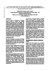
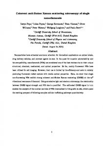
Figure 1: Features of an ideal diagnostic test for kidney disease. AST: antimicrobial susceptibility testing; ED: emergency department; ID: identification of infecting organism.
ADVANCING CULTURE-INDEPENDENT MICROBIOLOGY A number of culture-independent laboratory methods are now available that promise faster, more sensitive, and more specific aetiological diagnoses across a broad spectrum of pathogen and specimen types.20 Examples include nucleic acid-based approaches to detect bacteriaspecific DNA or RNA, and protein-based assays, such as matrix assisted laser desorption/ionisation time-of-flight (MALDI-TOF) mass spectrometry (MS), which identifies organism-unique protein signatures.21,22 These techniques are relatively rapid, leading to faster reporting times and the detection of organisms that may be difficult to culture.23 Each approach, however, has significant limitations. They do not distinguish viable from nonviable organisms, the number of defined genetic targets for nucleic acidbased detection are currently limited, and the interpretation of results can be complicated in polymicrobial infections contaminating species and/or the presence of commensal bacteria.23,24 In addition, even if specific resistance genes are detected by polymerase chain reaction (PCR), expression of these genes may vary and multiple genes may be required to yield functional resistance in vivo.25,26
58
NEPHROLOGY • July 2018
To date, neither bacterial nucleic acid nor protein-based detection techniques are routinely used for analysis of samples from patients with suspected PD peritonitis.27 Major limitations of these techniques for the analysis of PD samples are the high bacterial concentrations required for adequate sensitivity during protein detection28,29 and the poor accuracy of nucleic acid detection, which has unacceptably high false negative rates, thought to be due to the presence of inhibitors in dialysate (Figure 1).30 Bacterially derived DNA fragments in PD effluent show some value as a prognostic marker for relapsing peritonitis episodes, but as the presence of bacterial DNA does not directly correlate with live organisms capable of causing infection, the clinical applicability is limited.31 23S ribosomal (r)RNA PCR and sequencing has been applied to the problem; however, the authors concluded that this method was best reserved as an adjunctive tool to traditional culture techniques given the lack of specificity when applied as a diagnostic test.32 While not an exhaustive set of examples, this illustrates the complexity faced when attempting to use nucleic acid-based technology in a sample as complicated as PD effluent.
EMJ
EUROPEAN MEDICAL JOURNAL
Hydrodynamic
A
Acoustic
Focussed by fluid
Focussed by sound waves
Large particles
Small particles
SYTO® 9 (530/30 nm)
B
106 105 104 103 102
103
104
105
106
SYTO® 9 (530/30 nm)
Greater resolution for microbial particles
106 105 104 103 102
106 105 104 103 102
103
104
105
106
104
105
106
Forward scatter Candida glabrata
SYTO® 9 (530/30 nm)
SYTO® 9 (530/30 nm)
Forward scatter Klebsiella pneumoniae
103
Forward scatter In silico overlay
106 105 104 103 102
103
104
105
106
Forward scatter In vitro mixture
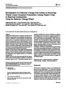
Figure 2: A) Acoustic versus standard hydrodynamic focussing for flow cytometry. B) Clear separation between bacterial and fungal pathogens in peritoneal dialysis dialysate as assessed by acoustic flow cytometry. SYTO®, Thermo Fisher Scientific, Waltham, Massachusetts, USA.
ADVANCES IN FLOW CYTOMETRY ENABLE DIRECT VISUALISATION OF BACTERIA AND FUNGI While flow cytometry (FC) has been extensively used to detect eukaryotic cells, the smaller size of bacteria coupled with variability in cell wall structures has limited its use for bacterial detection.33 However, recent advances in hardware design, including the use of
Creative Commons Attribution-Non Commercial 4.0
acoustic-focussing technology (Figure 2A), have greatly improved the effective resolution capable for small particles. Reliable detection of biological particles as small as 250 nm is now considered routine.34 Coupling this technology with DNA-intercalating and protein-binding fluorescent dyes improves resolution further and makes accurate identification of bacteria or fungi directly from clinical samples possible (Figure 2B). The quantitative nature of particle characterisation by FC also permits direct
July 2018 • NEPHROLOGY
59
enumeration of bacterial counts in dialysate providing information on inoculum dose, which may be an important feature in influencing clinical outcomes. One unique aspect of this approach is the separation of bacterial detection from identification of their species (or Gram type), which may prove challenging for clinicians and microbiologists who have traditionally decided upon treatment options based on this knowledge. While this has implications for epidemiological data collection (if a traditional culture is not also conducted),35 this approach can provide answers where traditional culture techniques have failed, for example in cases of culturenegative peritonitis, or where recent exposure to antimicrobial agents might impact culture. Furthermore, sample preparation for this technique can require as little as 25 minutes of manual handling, followed by 10 minutes for data acquisition and processing, representing a potential gain of >18 hours compared to traditional culture-based methods.36
CULTURE-INDEPENDENT ANTIMICROBIAL SUSCEPTIBILITY TESTING Most antimicrobial agents used in the treatment of PD peritonitis have bacterial (or fungal) cell
lysis as their final mechanism of action. As FC detection of this mechanism works by identifying cell shape, size, and wall integrity, the effects of antibiotics on cells can be analysed and antimicrobial susceptibility profiles determined ex vivo in samples. This method, termed flow-assisted antimicrobial sensitivity testing (FAST), has demonstrated a strong positive correlation (r2: 0.81; p