The Journal of Immunology
Persistence and Function of Central and Effector Memory CD4ⴙ T Cells following Infection with a Gastrointestinal Helminth1 Colby Zaph,* Kathryn A. Rook,* Michael Goldschmidt,* Markus Mohrs,† Phillip Scott,* and David Artis2* Immunity in the gastrointestinal tract is important for resistance to many pathogens, but the memory T cells that mediate such immunity are poorly characterized. In this study, we show that following sterile cure of a primary infection with the gastrointestinal parasite Trichuris muris, memory CD4ⴙ T cells persist in the draining mesenteric lymph node and protect mice against reinfection. The memory CD4ⴙ T cells that developed were a heterogeneous population, consisting of both CD62Lhigh central memory T cells (TCM) and CD62Llow effector memory T cells (TEM) that were competent to produce the Th type 2 effector cytokine, IL-4. Unlike memory T cells that develop following exposure to several other pathogens, both CD4ⴙ TCM and TEM populations persisted in the absence of chronic infection, and, critically, both populations were able to transfer protective immunity to naive recipients. CD62LhighCD4ⴙ TCM were not apparent early after infection, but emerged following clearance of primary infection, suggesting that they may be derived from CD4ⴙ TEM. Consistent with this theory, transfer of CD62LlowCD4ⴙ TEM into naive recipients resulted in the development of a population of protective CD62LhighCD4ⴙ TCM. Taken together, these studies show that distinct subsets of memory CD4ⴙ T cells develop after infection with Trichuris, persist in the GALT, and mediate protective immunity to rechallenge. The Journal of Immunology, 2006, 177: 511–518.
M
ucosal surfaces, such as the respiratory and gastrointestinal (GI)3 tracts, are primary entry points for many infectious agents, and studies with viral, bacterial, and parasitic pathogens have shown that protective immunity to rechallenge develops at these sites (1– 6). For instance, re-exposure to the GI nematode Trichuris muris leads to rapid immune-mediated expulsion of a secondary infection (7). In common with other GI helminths, immunity to a primary infection with Trichuris is dependent upon CD4⫹ Th type 2 (Th2) cells that develop in the GALT, produce IL-4 and IL-13, and mediate physiological changes in the GI tract (including alterations in epithelial cell turnover, goblet cell hyperplasia, and expression of resistin-like molecule (RELM)) associated with clearance of the worms and sterile immunity (8 –12). However, whereas many of the factors that
*Department of Pathobiology, University of Pennsylvania, Philadelphia, PA 19104; and †Trudeau Institute, Saranac Lake, NY 12983 Received for publication December 2, 2005. Accepted for publication March 30, 2006. The costs of publication of this article were defrayed in part by the payment of page charges. This article must therefore be hereby marked advertisement in accordance with 18 U.S.C. Section 1734 solely to indicate this fact. 1 This work was supported by National Institutes of Health Grants AI61570 (to D.A.) and AI35914 (to P.S.); Pilot Feasibility Program of the National Institute of Diabetes and Digestive and Kidney Diseases Center Grant DK50306 (to D.A.); the Crohn’s and Colitis Foundation of America’s William and Shelby Modell Family Foundation Research Award (to D.A.); and the Irvington Institute for Immunological Research (to C.Z.). 2 Address correspondence and reprint requests to Dr. David Artis, University of Pennsylvania, School of Veterinary Medicine, Department of Pathobiology, 3800 Spruce Street, Philadelphia, PA 19104; E-mail address:
[email protected] or Dr. Phillip Scott, University of Pennsylvania, School of Veterinary Medicine, Department of Pathobiology, 3800 Spruce Street, Philadelphia, PA 19104; E-mail address:
[email protected] 3 Abbreviations used in this paper: GI, gastrointestinal; Th2, Th type 2; RELM, resistin-like molecule; TCM, central memory T cell; TEM, effector memory T cell; TEFF, effector T cell; mLN, mesenteric lymph node; pos, positive; DC, dendritic cell.
Copyright © 2006 by The American Association of Immunologists, Inc.
orchestrate immunity to a primary infection with GI pathogens such as Trichuris are well defined, those that regulate T cell memory and immunity to rechallenge have not been analyzed. Memory T cells are heterogeneous and have been separated into at least two distinct subsets based upon phenotype, function, and migratory pattern (4, 13–15). Central memory T cells (TCM) express high levels of CD62L and can migrate through secondary lymphoid tissues, whereas effector memory T cells (TEM) express low levels of CD62L and accumulate at extralymphoid sites. Although memory T cell subsets have been characterized most extensively using models of CD8⫹ T cell memory (16 –19), the development and maintenance of memory CD4⫹ T cells is less well understood. We recently found that following infection with the protozoan parasite Leishmania major, no CD4⫹ effector T cells (TEFF) or TEM could be detected once the parasites were eliminated. However, Leishmania-reactive CD4⫹ TCM developed, persisted in the absence of chronic infection, and mediated immunity to rechallenge (20). In contrast, memory CD4⫹ T cells that persist following clearance of viral infection in sites draining the lung are enriched for TEM, as measured by both surface phenotype and cytokine production (21, 22). Thus, the mechanisms associated with the development and persistence of memory CD4⫹ T cell responses following exposure to different pathogens remain unclear. In this study, we functionally characterize for the first time the CD4⫹ T cell memory response that develops following exposure to the intestinal helminth parasite, Trichuris. Unlike memory CD4⫹ T cells that develop following infection with several other pathogens, sterile immunity to Trichuris is characterized by the persistence of both CD4⫹ TCM and TEM. In addition, both Trichuris-responsive CD4⫹ TCM and TEM are efficient at conferring resistance to secondary Trichuris infection. Lastly, these results demonstrate that in addition to expanding the TEFF pool, 0022-1767/06/$02.00
CD4⫹ T CELL MEMORY IN THE GI TRACT
512 CD62Llow TEM can also repopulate the CD62Lhigh TCM population, thereby replenishing the pathogen-specific TCM. Taken together, these studies show that distinct subsets of memory CD4⫹ T cells develop after infection, persist in the GALT, and mediate protective immunity to rechallenge.
Materials and Methods
were further separated based on expression of CD62L by MACS columns (Miltenyi Biotec) with 95–98% purity of CD62Lhigh and CD62Llow fractions. There were no phenotypic or functional differences in the cells isolated from animals that were between 60 and 120 days postinfection. CD4⫹ T cells were stained with CFSE (Molecular Probes) as described previously (25, 26). A total of 10 ⫻ 106 CFSE-labeled CD4⫹ T cells was transferred via the retro-orbital plexus into naive congenic recipients. Mice were infected 24 h or 3 wk later with Trichuris.
Animals
Flow cytometry and intracellular cytokine staining
BALB/cByJ mice were obtained from The Jackson Laboratory. BALB/c Thy1.1 mice were originally obtained from Dr. L. Turka (University of Pennsylvania, Philadelphia, PA). BALB/c eGFP/IL-4 reporter mice were generated as described previously (23). Animals were maintained in a specific pathogen-free environment at the University of Pennsylvania and tested negative for pathogens in routine screening. All experiments were conducted following the guidelines of the University of Pennsylvania Institutional Animal Care and Use Committee.
Cells were stained with fluorochrome-conjugated mAbs against CD4 (RM4-5), CD44 (IM7), CD62L (MEL14), IL-4 (11B11), and Thy1.1 (OX-7) or isotype-specific control Abs (eBioscience) before acquisition on a FACSCalibur flow cytometer (BD Pharmingen). Briefly, cells were isolated from the draining mLN and spleen and were analyzed for expression of surface markers directly ex vivo without further activation or for intracellular cytokines following 4 h of pharmacologic stimulation with PMA (50 ng/ml) and ionomycin (500 ng/ml) in the presence of Brefeldin A (10 g/ml). Cells were washed in staining buffer (PBS containing 0.1% BSA and 0.1% sodium azide) and incubated with Fc block (50 g/ml 2.4G2 and 500 g/ml rat Ig) before incubation with specific fluorochrome-conjugated mAbs. Cells were washed in staining buffer and fixed in 2% paraformaldehyde (Electron Microscopy Services). For intracellular staining, cells were permeabilized with 0.5% saponin before staining. Analysis was conducted using CellQuest Pro software (version 5.1; BD Biosciences).
Parasites, Ags, and infections T. muris was maintained in genetically susceptible or immunocompromised animals. Between days 35 and 42 postinfection, adult worms were isolated and cultured in RPMI 1640 containing 500 U/ml penicillin and 500 g/ml streptomycin for 24 h. Trichuris excretory-secretory Ag was isolated at 4 h, dialyzed, sterile filtered, and protein concentrations were determined by Bradford assay. Ag preparations were then used in lymphocyte restimulations (50 g/ml). Deposited eggs were collected after 24 h of culture, washed three times in sterile water, incubated at room temperature for 6 wk, and stored at 4°C. Mice were infected on day 0 with 150 –200 embryonated eggs, and parasite burdens were assessed on various days postinfection.
Abs and in vivo depletions mAbs were prepared from ammonium sulfate precipitation of hybridoma culture supernatants or ascites and dialyzed extensively in PBS. For CD8⫹ T cell depletions, 500 g of anti-CD8 mAb (H35) was administered i.p. 24 – 48 h before sacrifice, which routinely depleted ⬎98% of CD8⫹ lymphocytes. For CD4⫹ T cell depletions during infection, 1 mg of anti-CD4 mAb (GK1.5) was given i.p. on days 0, 1, 3, 6, and 9 postinfection and depleted ⬎98% of CD4⫹ T cells. Control mice received equivalent amounts of purified rat IgG (Sigma-Aldrich).
Cell culture and cytokine analysis At necropsy, the mesenteric lymph node (mLN) was harvested, and singlecell suspensions were prepared in DMEM supplemented with 10% heatinactivated FBS, 2 mM glutamine, 100 U/ml penicillin, 100 g/ml streptomycin, 25 mM HEPES, and 5 ⫻ 10⫺5 M 2-ME. Cells were plated at 5 ⫻ 106/ml in 24-well culture plates in medium alone or in the presence of T. muris excretory-secretory Ag (50 g/ml). In addition, 2.5 g/ml anti-IL4R␣ mAb (clone M1; BD Pharmingen) was added to cultures to enhance detection of IL-4. Cell-free supernatants were harvested after 48 h, and cytokine analysis was conducted by sandwich ELISA using paired mAb to detect IL-4 (11B11 and BVD6-24G2.3), IL-5 (TRFK-5 and TRFK-4), and IL-13 (R&D Systems) as described previously (24).
Analysis of goblet cell and RELM responses Segments of cecum were removed, washed in sterile PBS, and fixed for 24 h in 4% paraformaldehyde. Tissues were processed routinely and paraffin embedded using standard histological techniques. For detection of intestinal goblet cells, 5-m sections were cut and stained with Alcian blue-periodic acid-Schiffs reagent. Isolation of proteins from fecal samples was performed as described previously (8). Equal amounts of protein (20 g) were analyzed by SDS-PAGE and immunoblotted for RELM with a polyclonal rabbit anti-murine RELM Ab (PeproTech). Relative band intensities were quantified using ImageJ software (National Institutes of Health, 具http://rsb.info.nih.gov/ij/典).
CD4⫹ T cell purification and adoptive transfer Naive or immune (between 60 and 120 days postinfection) BALB/c Thy1.1, Thy1.2, or eGFP/IL-4 reporter mice were depleted of CD8⫹ T cells by injection with anti-CD8 mAb 1 and 3 days before sacrifice (⬎98% effective). Cells were isolated from draining mLN and spleen, and RBC were lysed in hypotonic solution. In some experiments, CD4⫹ T cells were purified using a T cell purification column (R&D Systems) according to the manufacturer’s recommendations. In some experiments, CD4⫹ T cells
Statistics Results represent the mean ⫾ SD of individual animals. Statistical significance was determined by Student’s t test (when comparing two groups) or ANOVA with a post hoc test (when comparing more than two groups).
Results
Long-term Trichuris-specific CD4⫹ Th2 memory in the absence of persistent infection Following sterile cure of a primary infection with Trichuris, mice exhibit enhanced resistance to reinfection (7, 27). Although CD4⫹ T cells are critical for primary worm expulsion (28), it is not known whether memory CD4⫹ T cells develop during primary infection nor whether these cells are required for resistance to reinfection. To investigate this, we examined the immune response of immune mice (⬎60 days postprimary infection) before reinfection as well as the kinetics of the response to challenge. Before reinfection, cells isolated from immune mice did not produce detectable levels of IFN-␥ or IL-13 (data not shown), but secreted significantly higher levels of Ag-specific IL-4 and IL-5 than cells from control mice following in vitro restimulation (Fig. 1A). Rechallenge of immune mice with Trichuris resulted in accelerated worm expulsion (Fig. 1B), undetectable levels of Trichuris-specific IFN-␥ (data not shown), but early increased production of IL-4, IL-5, and IL-13 (Fig. 1C). Associated with elevated expression of type 2 cytokines, rechallenged immune mice exhibited enhanced goblet cell hyperplasia (Fig. 1D; 1°, 160 ⫾ 12 goblet cells/20 crypts; 2°, 425 ⫾ 20 goblet cells/20 crypts) and rapid expression and secretion of RELM (Fig. 1E), a goblet cell-specific molecule associated with expulsion of helminth infections (8). Both the rapid expulsion and enhanced immune response were lost when CD4⫹ T cells were depleted from immune mice (Fig. 1, B–E). Furthermore, we found that CD4⫹ T cells are not only required, but are also sufficient to transfer immunity to naive mice. Transfer of CD4⫹ T cells from immune, but not naive, mice resulted in increased goblet cell hyperplasia (Fig. 1F; naive cells, 225 ⫾ 15 goblet cells/20 crypts; immune cells, 645 ⫾ 32 goblet cells/20 crypts), expression of RELM (Fig. 1G), and rapid clearance of worms from the GI tract (Fig. 1H). Consistent with the spontaneous type 2 cytokine production by cells isolated from mice infected for 60 days (Fig. 1A), we also observed increased RELM expression in unchallenged mice that received cells from immune animals (Fig. 1G). Thus, long-term mucosal immunity
The Journal of Immunology
513 against Trichuris infection is dependent upon memory CD4⫹ T cells that persist in the absence of chronic infection and mediate rapid protective immunity. Rapid expansion and effector differentiation of Trichuris-specific memory CD4⫹ T cells following rechallenge To visualize the proliferation and function of Trichuris-responsive memory CD4⫹ T cells following rechallenge, CFSE-labeled CD4⫹ T cells from naive or immune mice were adoptively transferred into congenic naive recipients that were subsequently infected with Trichuris. At day 12 postinfection, CD4⫹ T cells from immune mice responded more robustly to infection by proliferating, demonstrating that Trichuris-specific memory CD4⫹ T cells have the capability to expand rapidly following re-exposure to infection (Fig. 2A). Phenotypic analysis revealed that the Trichurisresponsive CD4⫹ T cells (CFSEdim) were predominantly CD62Llow, a characteristic of TEFF or TEM (Fig. 2B, left panels). Furthermore, consistent with the expanded population of Trichuris-specific CD62Llow cells, there was an increased frequency of effector cytokine-positive (IL-4pos) cells in the mLN of mice receiving CD4⫹ T cells from immune donors (Fig. 2B, right panels). Accelerated IL-4 production was associated with the enhanced goblet cell responses and rapid worm expulsion observed following adoptive transfer of Trichuris-responsive memory CD4⫹ T cells (Fig. 1, F–H). CD4⫹ TCM and TEM persist after worm expulsion
⫹
FIGURE 1. CD4 T cells mediate immunity to Trichuris. A, Cells isolated from the mLN of naive or long-term infected (day 60) BALB/c mice were restimulated in vitro with medium alone (Med) or Trichuris Ag, and supernatants were analyzed for IL-4 and IL-5 by ELISA. Results are presented as mean ⫾ SD of four individual mice from one representative experiment of two. B–E, Naive (1°) or immune (2°) mice were infected with Trichuris. Some immune mice were treated with anti-CD4 mAb (GK1.5) on days 0, 1, 3, 6 and 9 postinfection (2° ␣-CD4). B, Worm burdens were determined at day 12 postinfection, and results are presented as mean ⫾ SD of four individual mice from one representative experiment of three. C, Supernatants were analyzed for production of IL-4, IL-5, and IL-13 by ELISA following in vitro restimulation of mLN cells isolated at day 12 postinfection with medium (Med) or Trichuris Ag. Results are presented as mean ⫾ SD of three individual mice from one representative experiment of three. D, Representative cecal sections from groups of three infected (1°) or rechallenged immune (2°) mice ⫾ anti-CD4 mAb taken on day 12 postinfection were stained for goblet cells. Results are representative of three independent experiments. E, RELM expression was examined in pooled fecal pellets (20 g of total protein) from groups of three infected (1°), rechallenged immune (2°), or CD4⫹ T cell-depleted rechallenged immune (2° anti-CD4) mice at days 6, 8, 10, and 12 postinfection and are representative of three independent experiments. Numbers below bands represent relative band intensities. F–H, CD4⫹ T cells (10 ⫻ 106) isolated from the mLN and spleen of naive or immune (day 60 –120 postinfection) BALB/c mice were transferred into naive hosts, infected with Trichuris 24 h later, and analyzed on day 12 postchallenge. F, Representative cecal sections from groups of three mice receiving cells from naive (naive cells) or immune (immune cells) mice on day 12 postinfection were stained for goblet cells. G, RELM expression was analyzed by immunoblotting of protein isolated from pooled fecal pellets (20 g of total protein) from each group of three mice collected at day 12 postinfection and are representative of three independent experiments. Numbers below bands represent relative band intensities. H, Worm burdens were analyzed at day 12 postinfection, and results are presented as mean ⫾ SD of three individual mice from one representative experiment of three. ND, Not detected. ⴱ, p ⬍ 0.01 between naive mice and mice infected with Trichuris for 60 days (A), 1°, 2°, and 2° ⫹ anti-CD4 (␣-CD4) (B and C), and mice receiving naive or immune cells (H).
The congenic adoptive transfer system described above allows tracking of memory CD4⫹ T cells in vivo; however, identification
FIGURE 2. Trichuris-specific memory CD4⫹ T cells rapidly expand and produce IL-4 following rechallenge. CD4⫹ T cells were isolated from the mLN and spleen of naive or immune BALB/c Thy1.1 mice, labeled with CFSE, and 10 ⫻ 106 cells were transferred into naive congenic recipients that were infected with Trichuris 24 h later. A, Proliferation of CD4⫹Thy1.1⫹ cells in the mLN on day 12 postinfection was visualized by flow cytometry. Numbers represent percentage of donor CD4⫹ T cells that diluted CFSE. B, Expression of CD62L on donor CD4⫹ T cells in the mLN on day 12 postinfection was analyzed by flow cytometry directly ex vivo without additional activation, whereas cytokine production was determined after a 4-h pharmacologic stimulation. Numbers in bold represent percentage of donor CD4⫹ T cells that diluted CFSE, whereas numbers in upper and lower left corners represent percentage of proliferated cells (CFSEdim) that express high or low levels of CD62L (left panels) or IL-4 (right panels). Results are from one animal of three per group and are representative of three independent experiments. ⴱ, p ⬍ 0.01 between mice receiving naive cells or immune cells.
514 of Trichuris-specific memory T cells requires a secondary infection and does not allow identification and characterization of persistent Trichuris-specific memory CD4⫹ T cells before rechallenge. To directly examine the nature of the memory CD4⫹ T cells that develop and persist following Trichuris infection, we used bicistronic cytokine reporter mice (23). With this reporter system, expression of eGFP identifies cells that have previously been stimulated to express IL-4 and reflects the potential of cells to produce IL-4 (23, 29). Furthermore, this system allows for the detection of IL-4 competent cells without additional antigenic or pharmacologic stimulation. Following a primary infection of BALB/c reporter mice with Trichuris, ⬎95% of the eGFP/IL-4⫹ cells were CD4⫹ T cells (data not shown). There was a significant increase in the frequency of eGFP/IL-4pos CD4⫹ T cells in the mLN during the peak of the primary response, with a 2-fold increase by day 6 postinfection and a 4-fold increase by day 17 (Fig. 3A). These cytokine-positive cells displayed an effector phenotype (CD62Llow or CD44high), demonstrating the development of a primary effector Th2 response after infection with Trichuris (Fig. 3B). Following pathogen clearance (beyond day 17 postinfection), the frequency of eGFP/IL-4pos CD4⫹ T cells did not decrease over time (Fig. 3A, day 35 and day 45), demonstrating that a population of CD4⫹ TEM was maintained in the absence of persistent infection. Furthermore, the absolute number of eGFP/IL-4pos CD4⫹ cells (total, CD62Lhigh and CD62Llow populations) reflected the increased frequencies observed at all time points postinfection, with signifi-
FIGURE 3. Central and effector memory CD4⫹ T cells persist in the mLN after pathogen clearance. A, BALB/c eGFP/IL-4 reporter mice were infected with Trichuris, and on days 6, 17, 35, and 45 postinfection cells isolated from the mLN were analyzed for expression of CD4. The mean frequency ⫾SD of eGFP/IL-4pos CD4⫹ cells from four animals per time point is shown. B, Cells isolated in A were analyzed by flow cytometry for expression of CD62L and CD44, and numbers represent frequency of eGFP/IL-4pos cells expressing high or low levels of CD62L or CD44 from one representative animal of four per time point. C, Absolute numbers of eGFP/IL-4pos CD4⫹ cells and eGFP/IL-4pos CD4⫹CD62Lhigh and eGFP/ IL-4pos CD4⫹CD62Llow subsets following infection with Trichuris. Results are representative of two independent experiments. ⴱ, p ⬍ 0.01 between naive (day 0) and infected mice.
CD4⫹ T CELL MEMORY IN THE GI TRACT cantly higher numbers of cells in the mLN of infected animals compared with naive mice (Fig. 3C). Despite the lack of parasites, it is not possible to conclusively determine whether Trichuris Ag persists after sterile cure. Therefore, we refer to a population of effector cytokine-positive CD4⫹ T cells that persists following sterile cure of infection as TEM. The majority of persistent eGFP/ IL-4pos CD4⫹ T cells expressed high levels of CD44 or CD62L, characteristic of TEM and consistent with the maintenance of an Ag-specific effector response (Fig. 1A). We also observed the emergence of a population of cells competent to produce IL-4 expressing high levels of CD62L (Fig. 3B, day 35 and day 45, middle panels). Therefore, IL-4-competent cells with the phenotype of either TCM (CD62Lhigh) or TEM (CD62Llow) persisted following sterile cure of Trichuris infection. Central or effector memory CD4⫹ T cells can mediate immunity to rechallenge Several previous studies on memory CD8⫹ T cells have shown that TCM provide more effective and rapid immunity than TEM (16, 18, 19). In contrast, CD4⫹ TEFF or TEM confer more rapid protective immunity to L. major infection than TCM (20, 30, 31). To determine whether the CD4⫹ TCM or TEM populations— defined by expression of CD62L—that develop following Trichuris infection differed in their ability to mediate protective immunity, purified cells from wild-type immune donors were separated into CD62Lhigh and CD62Llow fractions, labeled with CFSE, and transferred into naive congenic recipients that were subsequently infected with Trichuris. Examination of the activated (CD44high) CD62Lhigh vs CD62Llow donor cells in the mLN 12 days after infection revealed that there was equivalent proliferation of both subsets (Fig. 4A, histograms). The proliferation of both subsets of memory CD4⫹ T cells was associated with Th2 cytokine-dependent physiological changes in the GI tract, because both populations induced goblet cells (Fig. 4B), RELM production (Fig. 4C, inset), and mediated rapid worm expulsion (Fig. 4C). Thus, unlike
FIGURE 4. Central or effector memory CD4⫹ T cells mediate immunity to rechallenge. Cells isolated from the mLN and spleen of immune BALB/c Thy1.1 mice were separated based on high or low expression of CD62L. Purified fractions were labeled with CFSE, transferred into naive congenic recipients, and 24 h later the recipient mice were infected with Trichuris. A, Proliferation of CD44highCD4⫹Thy1.1⫹ cells in the mLN on day 12 postinfection was analyzed by flow cytometry. Numbers on histograms represent percentage of donor CD44highCD4⫹Thy1.1⫹ cells that proliferated and are from one representative animal of three from three independent experiments. B, Goblet cells were quantified by direct counting of tissue sections from mice on day 12 postinfection. Data represent mean ⫾ SD number of goblet cells per 20 crypts from groups of three animals and is representative of three experiments. C, Worm burdens were determined microscopically at day 12 postinfection, and results are presented as mean ⫾ SD of three individual mice from one representative experiment of three. Inset, RELM expression in pooled fecal pellets from groups of three mice receiving CD62Lhigh (High) or CD62Llow (Low) cells was analyzed at day 12 postinfection by immunoblotting (20 g of total protein). Numbers refer to relative band intensities compared with mice receiving no cells (1.0; data not shown). Results are representative of three independent experiments. ⴱ, p ⬍ 0.01, between mice receiving no cells, mice receiving CD62Lhigh, or mice receiving CD62Llow cells.
The Journal of Immunology CD4⫹ T cell memory in other infectious diseases, both TCM and TEM persist after sterile cure of Trichuris and can mediate protective immunity against secondary infection in the gut. Relationship between CD4⫹ TCM and TEM Based on the data presented here, two phenotypically distinct populations of mucosal memory CD4⫹ T cells develop after infection with Trichuris and exhibit the characteristics of either TCM or TEM. CD62Llow IL-4-competent TEFF or TEM are evident throughout primary infection, whereas it appears that eGFP/IL-4pos CD62Lhigh TCM arise after pathogen clearance (Fig. 3B, central panel). Studies with CD8⫹ T cells have demonstrated that TCM are derived from TEM, whereas the origin and interrelationship of memory CD4⫹ T cell subsets is unknown (18). To address the relationship between cytokine-competent CD4⫹ TCM (CD62Lhigh) and TEM (CD62Llow) that develop following Trichuris infection, equal numbers (10 ⫻ 106 cells) of CD62Lhigh or CD62LlowCD4⫹ T cells were purified from immune eGFP/IL-4 reporter mice (⬎60 days postinfection). Analysis of CD62Lhigh cells revealed that only a small frequency of the population was eGFP/IL-4pos (Fig. 5A, lower left panel, 1.4%), consistent with the results presented above (Fig. 3B). Upon transfer into congenic recipients, CD62Lhigh cells gave rise to both CD62Lhigh and CD62Llow populations after challenge with Trichuris (Fig. 5A, middle histogram). The cells retaining high expression levels of CD62L remained predominantly eGFP/IL-4neg (Fig. 5A, upper right dot plot, 1.5%), suggesting that effector cytokine expression does not develop in the CD62Lhigh cells directly. In contrast, a significant proportion of CD62Llow cells derived from CD62Lhigh donors expressed eGFP/IL-4 (Fig. 5A, lower right dot plot, 42%) demonstrating that upon secondary infection, CD62Lhigh TCM can efficiently give rise to cytokinepositive CD62Llow TEFF. Therefore, CD62Lhigh TCM that develop during Trichuris infection appear to mediate protective immunity primarily by differentiating into IL-4-expressing, CD62Llow effector Th2 cells. In contrast to the CD62Lhigh population, a significant proportion of the donor CD62Llow TEM isolated from immune eGFP/IL-4
FIGURE 5. Relationship between central and effector memory CD4⫹ T cells. CD4⫹ T cells isolated from the mLN and spleen of immune BALB/c eGFP/IL-4 reporter mice were separated into CD62Lhigh (A) or CD62Llow (B) fractions, and 10 ⫻ 106 cells of each fraction were transferred into congenic recipients. Twenty-four hours later, the recipients were infected with Trichuris. Before transfer and on day 12 postinfection, donor CD4⫹Thy1.2⫹ cells were analyzed by flow cytometry for eGFP/IL-4 and CD62L expression. Histograms are gated on donor Thy1.2⫹ cells, and dot plots on the right side are gated on either CD62Lhigh or CD62Llow subpopulations. Results presented are from one individual animal of three and are representative of three independent experiments.
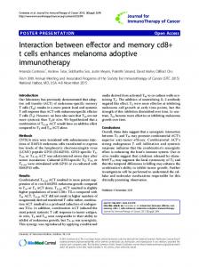
515 reporter mice were eGFP/IL-4pos before transfer (Fig. 5B, lower left panel, 15%). Upon transfer and rechallenge, there was significant expansion of CD62Llow cells that expressed eGFP/IL-4 (Fig. 5B, lower right dot plot, 65%). Highly purified CD62Llow cells also gave rise to a population of CD62Lhigh cells in which there was a diminished frequency of cells competent to express IL-4 (Fig. 5B, upper right dot plot, 16%). Therefore, CD4⫹CD62Lhigh TCM can arise from CD62Llow TEM following secondary challenge, suggesting that CD4⫹ TEM can replenish the TCM pool following rechallenge with Trichuris. CD4⫹ TEM transition to TCM in the absence of infection To test whether the development of CD4⫹CD62Lhigh TCM from CD62Llow TEM was dependent upon rechallenge or could occur in the absence of infection, equal numbers (10 ⫻ 106) of purified CD62Lhigh and CD62LlowCD4⫹ T cells from immune mice were transferred into naive recipients in the absence of infection (Fig. 6, left panels). Three weeks later, CFSEbright donor CD4⫹ T cells in the mLN cells were analyzed for expression of CD62L. Following transfer, persistent CD62LhighCD4⫹ T cells maintained expression of CD62L (Fig. 6, upper right panel). In contrast, donor CD62LlowCD4⫹ T cells that persisted after transfer did not remain uniformly CD62Llow, because a significant proportion expressing high levels of CD62L emerged (Fig. 6, lower right histogram). This increase could be the result of either increased death of CD62LlowCD4⫹ T cells or a selective outgrowth of contaminating CD62Lhigh T cells. In the absence of MHC class II tetramers or TCR transgenic T cells for helminth parasites such as Trichuris, at present we cannot definitively say that the loss of CD62Llow T cells was not a factor in the appearance of the CD62Lhigh population. However, absolute numbers of recovered CD62Lhigh and CD62Llow cells from the mLN following transfer of purified CD62Llow cells were similar (CD62Lhigh, 1.9 ⫻ 104; CD62Llow, 1.3 ⫻ 104), suggesting that this is not due to selective survival. Furthermore, analysis of CFSE-labeled cells shows that this transition occurred independent of proliferation (Fig. 6, dot plots), suggesting that the CD62Lhigh TCM that arise following transfer of CD62Llow TEM are not the result of expansion of a small number of contaminating CD62Lhigh TCM in the donor CD62Llow TEM population. Rather, these data indicate that TEM can directly convert into TCM. Thus, similar to memory CD8⫹ T cells, the transition from CD62LlowCD4⫹ TEM to CD62LhighCD4⫹ TCM does not require infection and may be a natural step in the development of long-lived memory CD4⫹ T cells. Taken together, the results presented in this study support a novel model of memory CD4⫹ T cell development and maintenance at mucosal sites (Fig. 7), in which infection results in the
FIGURE 6. CD4⫹ TEM transition to TCM in the absence of infection. CD4⫹ T cells isolated from the mLN and spleen of immune BALB/c Thy1.1 mice were separated into CD62Lhigh or CD62Llow fractions (input), labeled with CFSE, and 10 ⫻ 106 cells were transferred into congenic recipients. Before transfer and on day 21 posttransfer, donor CD4⫹Thy1.1⫹ cells in the mLN were analyzed by flow cytometry for CD62L expression and dilution of CFSE. Results presented are from one individual animal of three and are representative of two independent experiments.
516
FIGURE 7. Model of CD4⫹ T cell memory in the GALT. During a primary infection, a robust Th2 effector (TEFF) cell population develops that mediates protective immunity. Following sterile cure, TEM that are CD62Llow and IL-4pos and TCM that are CD62Lhigh and IL-4pos persist. Upon rechallenge, TEM rapidly proliferate and expand the TEFF pool (1). Persistent CD62Llow TEM also can transition into CD62Lhigh TCM following clearance of a primary or secondary infection (2), although CD62Lhigh TCM may also arise before sterile cure (dotted line). Following secondary infection, these TCM can also rapidly give rise to TEFF and contribute to the rapid immune response associated with long-term immunity to infection (3).
persistence of TCM and TEM in the GALT after pathogen clearance. Moreover, these memory cell populations appear to have the potential to repopulate central and effector memory cell pools in vivo.
Discussion In this report, we show that long-term immunity to Trichuris is dependent upon memory CD4⫹ T cells and identify three important aspects of CD4⫹ T cell memory in the GI tract. First, both CD4⫹ TCM and TEM develop and persist after sterile cure of Trichuris infection. Second, both CD4⫹ TCM and TEM that arise following infection exhibit the ability to mediate protective immunity to rechallenge. Finally, similar to memory CD8⫹ T cells, CD4⫹ TCM can derive from TEM following sterile cure of infection. Previous studies of CD4⫹ and CD8⫹ T cell memory following infection with L. major, Listeria monocytogenes, or lymphocytic choriomeningitis virus have suggested that IFN-␥-producing TEFF or TEM require persistent infection to be sustained, whereas TCM can persist in the absence of chronic infection (18, 20, 30, 31). In contrast, the majority of virus-specific memory CD4⫹ T cells that are maintained in sites draining the lung in the absence of Sendai virus infection have an activated phenotype (CD44high, CD62Llow) and rapidly express IFN-␥, characteristics of TEM (21, 22). Therefore, the identification of a population of Trichuris-specific IL-4expressing CD62LlowCD4⫹ T cells—a phenotype consistent with a TEM population—that persists after sterile cure of primary infection is more consistent with the composition of memory CD4⫹ T cells observed in the lung following clearance of viral infection than other infections. It is important to note that although mice that have cleared a primary Trichuris infection have no persistent parasites, it is not possible to conclude that there is no residual Ag that remains following sterile cure of infection. Nevertheless, upon rechallenge, this persistent TEM population is able to proliferate and rapidly expand the TEFF pool (Fig. 7 and Ref. 1). The disparity between the persistence and function of TCM and TEM in different infection models may be influenced by the cytokine polarization of the CD4⫹ T cell memory responses. Trichuris induces a strong type 2 response at the mucosal site of infection, whereas infection with viral, bacterial, or protozoan pathogens results primarily in potent type 1 memory responses associated with
CD4⫹ T CELL MEMORY IN THE GI TRACT the production of IFN-␥ (32–34). Previous studies have demonstrated that IFN-␥-producing T cells are short-lived in vivo (35, 36), whereas Th2 cells are more amenable to surviving in adoptive hosts (37) and are more resistant to activation-induced cell death than Th1 cells (38, 39). Therefore, maintenance of Trichuris-specific TEM in the GALT may reflect the differences in the survival of Th1 and Th2 memory cells in vivo. Supporting this theory, in vitro- or in vivo-generated TCR transgenic CD4⫹ Th2 cells can persist for several weeks in vivo in the absence of Ag and respond rapidly to secondary stimulation by producing effector cytokines such as IL-4 (37, 40, 41). In addition, a previous study demonstrated that adoptive transfer of effector CD4⫹ T cells isolated from mice infected with Trichuris were able to persist for more than 40 days and mediate protective immunity to rechallenge (42). IL-4-competent CD4⫹ T cells with the characteristics of TEM also persisted in the absence of chronic infection with another GI helminth parasite, Nippostrongylus brasiliensis, and mediated protective immunity to rechallenge (23). Given that virus-responsive memory CD4⫹ T cells that express IFN-␥ can persist in lung-draining LN, commitment to distinct Th cell subsets cannot be the only explanation for the differences in persistence of memory T cells. In addition to intrinsic mechanisms that may differentially regulate the persistence and function of memory T cells, tissue-specific regulation of T cell memory may contribute to the development of Trichuris-specific memory cells. For instance, microbial and environmental stimuli and/or the presence of specialized APC populations in the gut may influence the persistence and function of memory CD4⫹ T cells following exposure to Trichuris. Certainly, activation of T cells by mucosal dendritic cells (DC) results in cytokine expression and homing phenotypes that are distinct from T cells primed by peripheral DCs (43– 47). Therefore, it is possible that priming of CD4⫹ T cells by DCs in the gut and other mucosal sites such as the lung will also affect the ontogeny, survival, and function of memory T cells and the mechanisms that regulate their function upon re-exposure to infection. Furthermore, microbial stimuli from both normal and pathogenic gut flora, coupled with environmental Ags, may also constitute unique signals that affect the quality of the memory responses in the gut (48). In addition to the persistent CD62Llow TEM, Trichuris-responsive CD62LhighCD4⫹ TCM developed after infection, persisted after sterile cure, and could mediate immunity to rechallenge in the GI tract. CD4⫹ TCM develop after clearance of primary or secondary Trichuris infection, express high levels of CD62L, are able to express IL-4, and appear to derive from CD62Llow TEM cells (Fig. 7 and Ref. 2), although it is possible that they arise early following primary infection (Fig. 7, dotted line). Following rechallenge, these CD62Lhigh TCM can give rise to an IL-4-expressing CD62Llow population (Fig. 7 and Ref. 3) and contribute to the TEFF pool. Previous studies with CD4⫹ T cells have suggested that commitment to effector function is limited to TEM but not TCM, whereas CD8⫹ TCM can produce effector cytokines such as IFN-␥ and express effector molecules such as perforin (18, 20, 49 –51). Results presented in this study demonstrate that commitment to effector cytokine expression in TCM populations is not restricted to CD8⫹ T cells and support the contention that common regulatory pathways may exist in the memory CD4⫹ and CD8⫹ T cell compartments. A question that arises is why maintaining both persistent Trichuris-responsive CD4⫹ TEM and TCM would be advantageous to the host. One possibility is that having a subset of CD4⫹ TEM repopulating the TCM pool provides an intrinsic pathway to protect the repertoire of memory responses while allowing a rapid, but flexible response. Maintaining CD62Lhigh LN-homing memory T
The Journal of Immunology cells allows licensing of a subset of Trichuris-responsive memory T cells to traffic through peripheral LNs, which is primarily a CD62L-dependent phenomenon, thereby facilitating recirculation to additional sites, more extensive immunological surveillance, and allowing memory cells to encounter additional survival signals that may be present at optimal concentrations in extra-GALT sites. At the same time, persistent TEM facilitate rapid immune responses upon re-exposure to infection. Recent studies have provided compelling evidence for the importance of Th2 responses in secondary immunity to helminth infection in humans (52, 53). Jackson et al. (53) demonstrated that increased Th2 cytokine responses immediately before deworming had a significant negative effect on the probability of reinfection several months later. However, human infections tend to be repeated low-dose infections, whereas the data presented in this report are from a single inoculation of parasites. Nevertheless, the demonstration here that persistent of Trichuris-specific memory Th2 cells can mediate rapid immunity to rechallenge, reinforces the idea that long-term antihelminth immunity—via immunization or prophylactic treatment of infection—is an attainable goal. Together, these results provide a model of mucosal CD4⫹ T cell memory comprised of distinct memory T cell subsets. A population of persistent, cytokine-competent TEM provides a potent effector response to control infection in the GI tract. In addition, TCM develop in the GALT, exhibiting the ability to differentiate into effector CD4⫹ T cells and mediate protective immunity. These results identify novel aspects of CD4⫹ T cell memory in the GI tract and provide a framework to investigate the factors that regulate the maintenance of distinct memory CD4⫹ T cell populations at mucosal sites.
Acknowledgments We thank K. Joyce for technical assistance; S. L. Colpitts, P. M. Gray, C. A. Hunter, E. J. Pearce, and E. J. Wherry for critical reading of the manuscript; members of the Department of Pathobiology for helpful discussions; and PeproTech for the anti-RELM Ab.
Disclosures The authors have no financial conflict of interest.
References 1. Kim, S. K., K. S. Schluns, and L. Lefrancois. 1999. Induction and visualization of mucosal memory CD8 T cells following systemic virus infection. J. Immunol. 163: 4125– 4132. 2. Lepage, A. C., D. Buzoni-Gatel, D. T. Bout, and L. H. Kasper. 1998. Gut-derived intraepithelial lymphocytes induce long term immunity against Toxoplasma gondii. J. Immunol. 161: 4902– 4908. 3. Marzo, A. L., V. Vezys, K. Williams, D. F. Tough, and L. Lefrancois. 2002. Tissue-level regulation of Th1 and Th2 primary and memory CD4 T cells in response to Listeria infection. J. Immunol. 168: 4504 – 4510. 4. Masopust, D., V. Vezys, A. L. Marzo, and L. Lefrancois. 2001. Preferential localization of effector memory cells in nonlymphoid tissue. Science 291: 2413–2417. 5. Pope, C., S. K. Kim, A. Marzo, D. Masopust, K. Williams, J. Jiang, H. Shen, and L. Lefrancois. 2001. Organ-specific regulation of the CD8 T cell response to Listeria monocytogenes infection. J. Immunol. 166: 3402–3409. 6. Woodland, D. L. 2003. Cell-mediated immunity to respiratory virus infections. Curr. Opin. Immunol. 15: 430 – 435. 7. Wakelin, D. 1967. Acquired immunity to Trichuris muris in the albino laboratory mouse. Parasitology 57: 515–524. 8. Artis, D., M. L. Wang, S. A. Keilbaugh, W. He, M. Brenes, G. P. Swain, P. A. Knight, D. D. Donaldson, M. A. Lazar, H. R. P. Miller, et al. 2004. RELM/ FIZZ2 is a goblet cell-specific immune-effector molecule in the gastrointestinal tract. Proc. Natl. Acad. Sci. USA 101: 13596 –13600. 9. Bancroft, A. J., A. N. McKenzie, and R. K. Grencis. 1998. A critical role for IL-13 in resistance to intestinal nematode infection. J. Immunol. 160: 3453–3461. 10. Cliffe, L. J., N. E. Humphreys, T. E. Lane, C. S. Potten, C. Booth, and R. K. Grencis. 2005. Accelerated intestinal epithelial cell turnover: a new mechanism of parasite expulsion. Science 308: 1463–1465. 11. Else, K. J., F. D. Finkelman, C. R. Maliszewski, and R. K. Grencis. 1994. Cytokine-mediated regulation of chronic intestinal helminth infection. J. Exp. Med. 179: 347–351.
517 12. Finkelman, F. D., T. Shea-Donohue, J. Goldhill, C. A. Sullivan, S. C. Morris, K. B. Madden, W. C. Gause, and J. F. Urban, Jr. 1997. Cytokine regulation of host defense against parasitic gastrointestinal nematodes: lessons from studies with rodent models. Annu. Rev. Immunol. 15: 505–533. 13. Bunce, C., and E. B. Bell. 1997. CD45RC isoforms define two types of CD4 memory T cells, one of which depends on persisting antigen. J. Exp. Med. 185: 767–776. 14. Sallusto, F., D. Lenig, R. Forster, M. Lipp, and A. Lanzavecchia. 1999. Two subsets of memory T lymphocytes with distinct homing potentials and effector functions. Nature 401: 708 –712. 15. Reinhardt, R. L., A. Khoruts, R. Merica, T. Zell, and M. K. Jenkins. 2001. Visualizing the generation of memory CD4 T cells in the whole body. Nature 410: 101–105. 16. Lang, K. S., M. Recher, A. A. Navarini, N. L. Harris, M. Lohning, T. Junt, H. C. Probst, H. Hengartner, and R. M. Zinkernagel. 2005. Inverse correlation between IL-7 receptor expression and CD8 T cell exhaustion during persistent antigen stimulation. Eur. J. Immunol. 35: 738 –745. 17. Marzo, A. L., K. D. Klonowski, A. Le Bon, P. Borrow, D. F. Tough, and L. Lefrancois. 2005. Initial T cell frequency dictates memory CD8⫹ T cell lineage commitment. Nat. Immunol. 6: 793–799. 18. Wherry, E. J., V. Teichgraber, T. C. Becker, D. Masopust, S. M. Kaech, R. Antia, U. H. von Andrian, and R. Ahmed. 2003. Lineage relationship and protective immunity of memory CD8 T cell subsets. Nat. Immunol. 4: 225–234. 19. Wherry, E. J., D. L. Barber, S. M. Kaech, J. N. Blattman, and R. Ahmed. 2004. Antigen-independent memory CD8 T cells do not develop during chronic viral infection. Proc. Natl. Acad. Sci. USA 101: 16004 –16009. 20. Zaph, C., J. Uzonna, S. M. Beverley, and P. Scott. 2004. Central memory T cells mediate long-term immunity to Leishmania major in the absence of persistent parasites. Nat. Med. 10: 1104 –1110. 21. Cauley, L. S., T. Cookenham, T. B. Miller, P. S. Adams, K. M. Vignali, D. A. Vignali, and D. L. Woodland. 2002. Cutting edge: virus-specific CD4⫹ memory T cells in nonlymphoid tissues express a highly activated phenotype. J. Immunol. 169: 6655– 6658. 22. Roman, E., E. Miller, A. Harmsen, J. Wiley, U. H. Von Andrian, G. Huston, and S. L. Swain. 2002. CD4 effector T cell subsets in the response to influenza: heterogeneity, migration, and function. J. Exp. Med. 196: 957–968. 23. Mohrs, M., K. Shinkai, K. Mohrs, and R. M. Locksley. 2001. Analysis of type 2 immunity in vivo with a bicistronic IL-4 reporter. Immunity 15: 303–311. 24. Artis, D., A. Villarino, M. Silverman, W. He, E. M. Thornton, S. Mu, S. Summer, T. M. Covey, E. Huang, H. Yoshida, et al. 2004. The IL-27 receptor (WSX-1) is an inhibitor of innate and adaptive elements of type 2 immunity. J. Immunol. 173: 5626 –5634. 25. Zaph, C., and P. Scott. 2003. Th1 cell-mediated resistance to cutaneous infection with Leishmania major is independent of P- and E-selectins. J. Immunol. 171: 4726 – 4732. 26. Lyons, A. B., and C. R. Parish. 1994. Determination of lymphocyte division by flow cytometry. J. Immunol. Methods 171: 131–137. 27. Cliffe, L. J., and R. K. Grencis. 2004. The Trichuris muris system: a paradigm of resistance and susceptibility to intestinal nematode infection. Adv. Parasitol. 57: 255–307. 28. Koyama, K., H. Tamauchi, and Y. Ito. 1995. The role of CD4⫹ and CD8⫹ T cells in protective immunity to the murine nematode parasite Trichuris muris. Parasite Immunol. 17: 161–165. 29. Voehringer, D., K. Shinkai, and R. M. Locksley. 2004. Type 2 immunity reflects orchestrated recruitment of cells committed to IL-4 production. Immunity 20: 267–277. 30. Uzonna, J. E., G. Wei, D. Yurkowski, and P. Bretscher. 2001. Immune elimination of Leishmania major in mice: implications for immune memory, vaccination, and reactivation disease. J. Immunol. 167: 6967– 6974. 31. Belkaid, Y., C. A. Piccirillo, S. Mendez, E. M. Shevach, and D. L. Sacks. 2002. CD4⫹CD25⫹ regulatory T cells control Leishmania major persistence and immunity. Nature 420: 502–507. 32. Pamer, E. G. 2004. Immune responses to Listeria monocytogenes. Nat. Rev. Immunol. 4: 812– 823. 33. Sacks, D., and N. Noben-Trauth. 2002. The immunology of susceptibility and resistance to Leishmania major in mice. Nat. Rev. Immunol. 2: 845– 858. 34. Wherry, E. J., and R. Ahmed. 2004. Memory CD8 T-cell differentiation during viral infection. J. Virol. 78: 5535–5545. 35. Hayashi, N., D. Liu, B. Min, S. Z. Ben-Sasson, and W. E. Paul. 2002. Antigen challenge leads to in vivo activation and elimination of highly polarized TH1 memory T cells. Proc. Natl. Acad. Sci. USA 99: 6187– 6191. 36. Wu, C. Y., J. R. Kirman, M. J. Rotte, D. F. Davey, S. P. Perfetto, E. G. Rhee, B. L. Freidag, B. J. Hill, D. C. Douek, and R. A. Seder. 2002. Distinct lineages of TH1 cells have differential capacities for memory cell generation in vivo. Nat. Immunol. 3: 852– 858. 37. Swain, S. L., H. Hu, and G. Huston. 1999. Class II-independent generation of CD4 memory T cells from effectors. Science 286: 1381–1383. 38. Roberts, A. I., S. Devadas, X. Zhang, L. Zhang, A. Keegan, K. Greeneltch, J. Solomon, L. Wei, J. Das, E. Sun, et al. 2003. The role of activation-induced cell death in the differentiation of T-helper-cell subsets. Immunol. Res. 28: 285–293. 39. Zhang, X., T. Brunner, L. Carter, R. W. Dutton, P. Rogers, L. Bradley, T. Sato, J. C. Reed, D. Green, and S. L. Swain. 1997. Unequal death in T helper cell (Th)1 and Th2 effectors: Th1, but not Th2, effectors undergo rapid Fas/FasL-mediated apoptosis. J. Exp. Med. 185: 1837–1849. 40. Harbertson, J., E. Biederman, K. E. Bennett, R. M. Kondrack, and L. M. Bradley. 2002. Withdrawal of stimulation may initiate the transition of effector to memory CD4 cells. J. Immunol. 168: 1095–1102.
518 41. Hu, H., G. Huston, D. Duso, N. Lepak, E. Roman, and S. L. Swain. 2001. CD4⫹ T cell effectors can become memory cells with high efficiency and without further division. Nat. Immunol. 2: 705–710. 42. Lee, T. D., D. Wakelin, and R. K. Grencis. 1983. Cellular mechanisms of immunity to the nematode Trichuris muris. Int. J. Parasitol. 13: 349 –353. 43. Campbell, D. J., and E. C. Butcher. 2002. Rapid acquisition of tissue-specific homing phenotypes by CD4⫹ T cells activated in cutaneous or mucosal lymphoid tissues. J. Exp. Med. 195: 135–141. 44. Lambrecht, B. N., and H. Hammad. 2003. Taking our breath away: dendritic cells in the pathogenesis of asthma. Nat. Rev. Immunol. 3: 994 –1003. 45. Lanzavecchia, A., and F. Sallusto. 2001. Regulation of T cell immunity by dendritic cells. Cell 106: 263–266. 46. Mora, J. R., M. R. Bono, N. Manjunath, W. Weninger, L. L. Cavanagh, M. Rosemblatt, and U. H. Von Andrian. 2003. Selective imprinting of gut-homing T cells by Peyer’s patch dendritic cells. Nature 424: 88 –93. 47. Mora, J. R., G. Cheng, D. Picarella, M. Briskin, N. Buchanan, and U. H. von Andrian. 2005. Reciprocal and dynamic control of CD8 T cell homing by dendritic cells from skin- and gut-associated lymphoid tissues. J. Exp. Med. 201: 303–316.
CD4⫹ T CELL MEMORY IN THE GI TRACT 48. Nagler-Anderson, C. 2001. Man the barrier! Strategic defences in the intestinal mucosa. Nat. Rev. Immunol. 1: 59 – 67. 49. Barber, D. L., E. J. Wherry, and R. Ahmed. 2003. Cutting edge: rapid in vivo killing by memory CD8 T cells. J. Immunol. 171: 27–31. 50. Kaech, S. M., E. J. Wherry, and R. Ahmed. 2002. Effector and memory T-cell differentiation: implications for vaccine development. Nat. Rev. Immunol. 2: 251–262. 51. Sallusto, F., J. Geginat, and A. Lanzavecchia. 2004. Central memory and effector memory T cell subsets: function, generation, and maintenance. Annu. Rev. Immunol. 22: 745–763. 52. Turner, J. D., H. Faulkner, J. Kamgno, F. Cormont, J. Van Snick, K. J. Else, R. K. Grencis, J. M. Behnke, M. Boussinesq, and J. E. Bradley. 2003. Th2 cytokines are associated with reduced worm burdens in a human intestinal helminth infection. J. Infect. Dis. 188: 1768 –1775. 53. Jackson, J. A., J. D. Turner, L. Rentoul, H. Faulkner, J. M. Behnke, M. Hoyle, R. K. Grencis, K. J. Else, J. Kamgno, M. Boussinesq, and J. E. Bradley. 2004. T helper cell type 2 responsiveness predicts future susceptibility to gastrointestinal nematodes in humans. J. Infect. Dis. 190: 1804 –1811.