JOURNAL OF CLINICAL MICROBIOLOGY, July 1996, p. 1739–1744 0095-1137/96/$04.0010 Copyright q 1996, American Society for Microbiology
Vol. 34, No. 7
Persistence, Replacement, and Microevolution of Cryptococcus neoformans Strains in Recurrent Meningitis in AIDS Patients DEREK SULLIVAN,1 KEN HAYNES,2 GARY MORAN,1 DIARMUID SHANLEY,1 1 AND DAVID COLEMAN * School of Dental Science and Dublin Dental Hospital, Department of Oral Medicine and Pathology, Trinity College, University of Dublin, Dublin 2, Republic of Ireland,1 and Department of Infectious Diseases and Bacteriology, Royal Postgraduate Medical School, Hammersmith Hospital, London W12 0NN, United Kingdom2 Received 1 February 1996/Returned for modification 27 February 1996/Accepted 25 April 1996
Six separate human immunodeficiency virus-positive patients with cryptococcal meningitis were each found to have been infected with a unique strain of Cryptococcus neoformans on the basis of genomic DNA fingerprinting analysis with the microsatellite sequence-containing oligonucleotide probe (GGAT)4 and by random amplification of polymorphic DNA. Two patients (A and B) experienced a recurrent episode of infection. Between 12 and 16 single-colony isolates recovered from primary isolation media (>50% of C. neoformans colonies recovered) from cerebrospinal fluid specimens were fingerprinted from both patients during each episode. The fingerprints of both isolate collections from patient B were very similar, although minor polymorphisms were evident in both sets of profiles. The fingerprints of the isolate collection from the initial episode of infection in patient A were also identical to each other, apart from minor polymorphisms, but they were clearly different from the corresponding profiles of the isolate collection from the recurrent episode, the latter of which were completely identical, apart from minor polymorphisms in a single isolate. Furthermore, prolonged storage and in vitro subculture of the isolates did not alter the fingerprint profiles. These results provided convincing evidence that patients A and B were each infected with a single C. neoformans strain during each episode of infection and that in patient B, the same strain persisted and caused both episodes, while in patient A, a different strain was responsible for each episode. The prevalence of polymorphisms in multiple single-colony isolates from both patients also suggested that C. neoformans populations may undergo microevolution. ogy, including electrophoretic karyotyping (17, 19), PCR fingerprinting (13), random amplified polymorphic DNA (RAPD) analysis (2, 8), multilocus enzyme typing (1, 2), allelic variation of the URA5 locus (3, 5), and DNA fingerprinting with (i) genomic DNA probes (5, 18, 22, 23, 26, 27), (ii) mitochondrial DNA probes (25), and (iii) oligonucleotide probes homologous to microsatellite sequences (8). The general consensus from many of these studies is that C. neoformans strains exhibit considerable genetic heterogeneity and that recurrent infections are apparently due to the persistence of the original infecting strain (17, 22, 23). However, a recent study by Haynes et al. (8) indicated, on the basis of oligonucleotide and RAPD fingerprint analysis of genomic DNA, that in two patients (from five examined in total), recurrent infections may have been due to reinfection with a novel strain. In addition, this study indicated that one patient was coinfected with more than one strain during a single episode of infection. These data have aroused some controversy (4), and their clinical implications are of sufficient importance to warrant the analysis of additional cases of recurrent C. neoformans infection in order to confirm unequivocally that reinfection with novel strains may occur. The purpose of this study was to corroborate these earlier findings by analyzing multiple single-colony isolates recovered from patients during two recurrent episodes of meningitis and to determine the extent of genetic diversity within phenotypically homogeneous populations of C. neoformans recovered from the same clinical specimen.
Cryptococcus neoformans is an encapsulated basidiomycetous yeast species which occurs naturally in the environment and is frequently associated with pigeon droppings and soil contaminated with avian guano (11). Under most circumstances, inhalation of this organism fails to cause symptomatic infection (14). However, in 5 to 10% of individuals with AIDS, severe life-threatening disease in the form of meningoencephalitis can occur (6). The vast majority of the isolates responsible for these infections are C. neoformans var. neoformans serotype A (10). Because of the perceived importance of these organisms as human pathogens, the species has been subjected to intense study during the last decade. In this regard, the recent development and application of techniques designed to differentiate between individual isolates are of particular relevance because of the high incidence of recurrent cryptococcal infections in AIDS patients once antifungal therapy has ceased (20). In order to design effective antifungal drug treatment regimens, it is important to determine if recurrent disease is the result of reinfection with the original strain or infection with a novel strain. Unfortunately, individual strains of C. neoformans are morphologically and physiologically indistinguishable, and isolates of C. neoformans var. neoformans can only be divided into three serotypes (serotypes A, D, and A-D) (9). Consequently, techniques used in the epidemiological analysis of these organisms have concentrated on detecting genetic differences between individual isolates. To date, a wide variety of molecular typing systems have been applied to C. neoformans epidemiol-
MATERIALS AND METHODS C. neoformans isolates. C. neoformans isolates were recovered from six human immunodeficiency virus-infected individuals with cryptococcal meningitis at The Chelsea and Westminster Hospital, London, United Kingdom, between January 1994 and April 1995. In the case of four of these patients, C. neoformans isolates were recovered from a single episode of infection. The remaining two patients,
* Corresponding author. Mailing address: University of Dublin, School of Dental Science, Dental School Office, Trinity College, Dublin 2, Republic of Ireland. Phone: 353 1 6082015. Fax: 353 1 6799294. Electronic mail address:
[email protected]. 1739
1740
SULLIVAN ET AL.
both of whom were homosexual males with AIDS, experienced two successive episodes of meningitis. These two patients, termed A and B, respectively, presented with the classical symptoms of cryptococcal meningitis for the first time in November 1994, and in both cases, C. neoformans was isolated by plating of aliquots of cerebrospinal fluid (CSF) on Sabouraud’s agar. After treatment with 0.6 to 0.7 mg of amphotericin B kg21 day21 for 2 to 4 weeks, both patients made a complete clinical recovery. Thereafter, each patient received 200 to 400 mg of fluconazole day21 on an ongoing basis. However, clinical symptoms of meningitis reappeared in patient A in April 1995 and in patient B in March 1995, and in both cases, C. neoformans was again isolated from CSF samples. The time intervals between the initial and recurrent isolation of C. neoformans from patients A and B were 158 and 112 days, respectively. No additional specimens were taken from either patient during the symptom-free period between episodes of meningitis. Isolates were recovered on Sabouraud’s agar after incubation at 378C for 48 h and subcultured on fresh media prior to identification with the API ID 32C Yeast Identification System (bioMe´rieux, Marcy l’Etoile, France) and urea assimilation. Each isolate was stored in Protect cryo-storage vials (STC, Heywood, Lancashire, United Kingdom) at 2208C prior to detailed analysis. The CSF specimens from the initial and recurrent episodes of meningitis for both patients A and B each yielded between 10 and 30 C. neoformans CFU on primary isolation media. In the case of both episodes of infection in patient A and in the recurrent episode of infection in patient B, 20 individual well-separated C. neoformans colonies from the primary isolation plates were selected at random and stored; 16 of these from each isolate collection were subjected to further detailed analysis. In the case of the initial episode of infection in patient B, only 12 C. neoformans CFU were recovered on primary isolation media, all of which were stored and subjected to detailed analysis. The isolate collections from the initial episodes of infection in patients A and B were labelled AI1–20 and BI1–12, respectively, while those from the recurrent episodes were labelled AII1–20 and BII1–20, respectively. Fluconazole susceptibility testing. The susceptibility of C. neoformans clinical isolates to fluconazole was determined by broth microdilution in RPMI 1640 medium (15). Isolates were grown in 96-well microtiter plates (Corning) incubated at 358C for 72 h with agitation (16). An end point of 80% growth inhibition (IC80) was determined for each isolate by measuring the A405 with an automated microplate reader (Spectra I; SLT-Labinstruments, Salzburg, Austria). DNA fingerprinting. C. neoformans total cellular DNA was purified as described previously (22). Restriction fragments generated by digesting total cellular DNA samples to completion with the restriction enzyme EcoRI (Promega Corp., Madison, Wis.) were separated by electrophoresis through 0.8% (wt/vol) agarose gels and transferred onto nylon membrane filters (MSI, Westboro, Mass.) according to the method of Southern (21). The oligonucleotide probe (GGAT)4 was end labelled with [g-32P]dATP (Amersham International Plc., Little Chalfont, Buckinghamshire, United Kingdom [.5,000 Ci mmol21]) and T4 polynucleotide kinase (Promega Corp.) according to the manufacturer’s instructions. Labelled (GGAT)4 was hybridized to the digested DNA as described previously (24). RAPD. PCRs were performed in a final volume of 25 ml containing 10 mM Tris-HCl (pH 8.3); 50 mM KCl; 3.0 mM MgCl2, 200 mM (each) dATP, dCTP, dTTP, and dGTP (Promega Corp.); 20 pM primer; 0.5 U of Taq DNA polymerase (Promega Corp.); and approximately 10 ng of C. neoformans total genomic DNA. The following oligonucleotide primers were used: 1, 59GCGATCCCCA39; 2, 59(GATA4)39; and 3, 59AACGCGCAAC39. Amplification reactions were performed in a Perkin-Elmer Cetus DNA thermal cycler under the following conditions: 948C for 5 min, 368C for 5 min, and 728C for 5 min for 4 cycles followed by 30 cycles at 948C for 1 min, 368C for 1 min, and 728C for 2 min. This was followed by an incubation period of 728C for 10 min. After amplification, 20-ml aliquots of the reaction mixtures were electrophoresed in 1.5% (wt/vol) agarose gels, and the amplified products were visualized under UV light after being stained with ethidium bromide.
RESULTS Oligonucleotide fingerprinting of C. neoformans isolates. Total cellular DNA was purified from a single isolate of C. neoformans recovered in each case from CSF, skin, and blood samples obtained from two patients with meningitis and from CSF cultures recovered from another four patients suffering from meningitis. Aliquots of DNA from these isolates were digested to completion with EcoRI, and the resulting fragments were separated by agarose gel electrophoresis and transferred to nylon membrane filters prior to hybridization analysis with the 32P-labelled oligonucleotide probe (GGAT)4. The fingerprints generated from isolates recovered from different anatomical sites in the same patient were found to be indistinguishable. However, the fingerprints generated from isolates recovered from separate patients were each found to be very
J. CLIN. MICROBIOL.
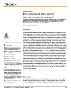
FIG. 1. Autoradiogram of EcoRI-digested total cellular DNA from singlecolony isolates of C. neoformans recovered from CSF specimens from patients A and B during two successive episodes of meningitis and hybridized with the oligonucleotide probe (GGAT)4. Lanes 1 and 2 and 3 and 4 show fingerprint profiles generated from two single-colony isolates recovered from the same CSF sample during the initial and recurrent episodes of infection, respectively. (A) Lanes 1 to 4 show profiles generated from isolates AI1, AI2, AII1, and AII2, respectively. (B) Lanes 1 to 4 show profiles generated from isolates BI1, BI2, BII1, and BII2, respectively. The relative positions of molecular size standards are indicated to the right.
different (data not shown), indicating that each patient was infected with a unique strain of C. neoformans. Total cellular DNA was purified from three single-colony isolates from the initial and recurrent isolate collections recovered from two AIDS patients (A and B), each of whom experienced two successive episodes of meningitis, and DNA fingerprints were generated as described above. In the case of patient A, the hybridization profiles obtained with the three isolates recovered during the initial episode of meningitis (data for isolates AI1 and AI2 are shown in Fig. 1A, lanes 1 and 2) were identical to each other but were clearly significantly different from the corresponding profiles obtained with the three isolates (also identical to each other) recovered during the recurrent episode of infection (data for isolates AII1 and AII2 are shown in Fig. 1A, lanes 3 and 4). In contrast, the hybridization patterns obtained with both sets of isolates recovered from patient B were found to be very similar, but some band differences were evident. For example, there was a hybridization band with a size of approximately 3 kb, which was present in the profiles of the three isolates from the initial episode of infection (data for isolates BI1 and BI2 are shown in Fig. 1B, lanes 1 and 2) but was absent in the corresponding profiles of the three isolates from the recurrent episode (data for isolates BII1 and BII2 are shown in Fig. 1B, lanes 3 and 4). Direct visual analysis of ethidium bromide-stained agarose gels containing separated EcoRI-generated fragments of genomic DNA from the isolates concerned showed that the restriction fragment length polymorphism patterns of the recurrent isolates from patient B lacked a heavily stained band, also approximately 3 kb in size, that was present in the restriction fragment length polymorphism patterns of the three isolates from the initial episode of infection (data not shown). Furthermore, an additional hybridization band with a size of approximately 2.8 kb was present in the profile of isolate BII1 which was not present in the profile of isolate BII2 (Fig. 1B, lanes 3 and 4).
VOL. 34, 1996
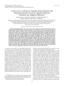
FIG. 2. RAPD products from single-colony isolates of C. neoformans recovered from CSF specimens from patients A and B during two successive episodes of meningitis and amplified with oligonucleotide primer 1 (59GCGATCCCC A39). Lanes 1 to 3 (isolates AI1 to 3) and 8 to 10 (isolates BI1 to 3) show RAPD profiles generated from three single-colony isolates recovered from the same CSF sample from patients A and B, respectively, during the initial episodes of infection. Lanes 4 to 6 (isolates AII1 to 3) and 11 to 13 (isolates BII1 to 3) show RAPD profiles generated from three single-colony isolates recovered from the same CSF sample from patients A and B, respectively, during recurrent episodes of infection. Lane 14, 100-bp DNA ladder size standards (Promega).
The fingerprint profiles of the isolates described above were found to be stable after repeated subculture (minimum of eight times) of the isolates over a 6-month period. During this time, total cellular DNA was prepared from each isolate on three separate occasions, and in each instance, fingerprint profiles identical to those shown in Fig. 1 were obtained with (GGAT)4. RAPD analysis of recurrent isolates. The RAPD profiles generated with primer 1 and target DNA from three singlecolony isolates from the same clinical specimen examined from the initial episode of infection in patient A (AI1, AI2, and AI3) were identical to each other (Fig. 2, lanes 1 to 3). Similarly, the corresponding profiles obtained with the target DNA from an additional three single-colony isolates from the same clinical specimen recovered during the recurrent episode of infection (AII1, AII2, and AII3 [Fig. 2, lanes 4 to 6]) were also found to be identical to each other. However, the patterns obtained from both sets of isolates were totally distinct. In contrast, the RAPD patterns generated with the three isolates examined from both the initial and recurrent episodes of meningitis in patient B (BI1, BI2, BI3, BII1, BII2, and BII3) were all found to be identical (Fig. 2, lanes 8 to 13). Additional RAPD experiments with the same target DNA as that used in the experiments described above and primers 2 and 3 also generated distinct profiles for the three AI and the three AII isolates and were unable to discriminate between the three BI and the three BII isolates. RAPD fingerprint profiles were also found to be reproducible after repeated subculture of the isolates with target DNA prepared on three separate occasions.
MICROEVOLUTION OF C. NEOFORMANS
1741
Analysis of multiple single-colony isolates. In order to investigate the possibility that patients A and B may have been infected with more than one strain of C. neoformans during both episodes of meningitis, total cellular DNA was prepared from an additional 13 single-colony isolates (9 in the case of the initial episode of infection in patient B) recovered, in each case, from the same clinical specimens as the isolates used in the experiments described above, and then hybridization fingerprints were generated with (GGAT)4. For both patients, only a single fingerprint pattern was found for each group of single-colony isolates examined. As in the results described above, the patterns of the 13 additional AI isolates differed considerably from those of the 13 additional AII isolates, while those of the 9 additional BI isolates were almost identical to those of the 13 additional BII isolates. However, whereas the AII isolates all yielded identical fingerprint patterns (fingerprints from 14 isolates are shown in Fig. 3a), the corresponding profiles of the AI, BI, and BII isolate collections, respectively, although essentially homogeneous, showed numerous subtle but distinct minor polymorphisms. An example of the profiles obtained with the BII isolate collection is shown in Fig. 3b. RAPD fingerprints of the additional single-colony isolates from each isolate collection from patients A and B were also generated with primer 2 and primer 3, respectively. The results confirmed the findings obtained in the experiments described above with three single-colony isolates from each isolate collection. However, minor polymorphisms were evident in some of the RAPD profiles of all four single-colony isolate collections, including one single-colony isolate from the recurrent episode of infection in patient A (Fig. 4). Fluconazole susceptibility of C. neoformans isolates. The fluconazole susceptibilities of isolates 1 to 3 from the initial and recurrent isolate collections for both patients A and B were determined by broth microdilution. Isolates AI1 to 3, BI1 to 3, and BII1 to 3 yielded an IC80 of 32 mg ml21, whereas isolates AII1 to 3 yielded an IC80 of 8 mg ml21. DISCUSSION Only a small number of studies have focused on the relationship between isolates of C. neoformans from successive episodes of meningitis in individual patients (8, 17, 22, 23). Some of these studies have indicated that relapse of cryptococcal meningitis is due to the persistence of the originally infecting strain (17, 22, 23), whereas another study has shown that in two separate individuals, recurrence of infection was apparently due to reinfection with a novel strain (8). In addition, the latter study provided evidence that one patient was infected with more than one C. neoformans strain during a single episode of meningitis. The present study was undertaken to confirm these findings by analysis of multiple C. neoformans single-colony isolates recovered from the same clinical specimen obtained from two patients during each of two successive episodes of disease. Fingerprint analysis of genomic DNA with the microsatellite sequence-containing oligonucleotide probe (GGAT)4 and RAPD analysis with three separate oligonucleotide primers showed that the six patients included in the study were each infected by different C. neoformans strains. This is in agreement with earlier studies which described the genetic heterogeneity of serotype A C. neoformans strains (3, 7, 8, 17, 26, 27). In addition, in the case of the two patients from which C. neoformans isolates were recovered from specimens taken from different anatomical sites during the same episode of meningitis, all of the isolates from the same individual yielded the same fingerprint pattern. Two of the six patients under study (A and B) suffered
1742
SULLIVAN ET AL.
J. CLIN. MICROBIOL.
FIG. 3. Autoradiograms of EcoRI-digested total cellular DNA from C. neoformans single-colony isolates recovered from the same CSF specimen in the case of patients A and B, respectively, during the recurrent episode of meningitis after hybridization analysis with the oligonucleotide probe (GGAT)4. (a) Profiles shown in lanes 1 to 14 were from single-colony isolates AII1 to AII14, respectively. (b) Profiles shown in lanes 1 to 16 were from single-colony isolates BII1 to BII16, respectively. The arrow and arrowhead shown to the left of panel b indicate the relative positions of polymorphic hybridization bands with sizes of approximately 2.6 and 2.9 kb present in isolate profiles shown in lanes 1, 4, 6, 7, 8, 9, 14, and 15 and lanes 10, 11, 12, and 15, respectively.
FIG. 4. Amplified RAPD products from single-colony isolates of C. neoformans recovered from CSF specimens from patient A during the initial and recurrent episodes of infection. (a) Profiles shown in lanes 1 to 7 were from single-colony isolates (AI5 to AI11) from the same specimen recovered during the initial episode of infection. (b) Profiles shown in lanes 1 to 7 were from single-colony isolates (AII1 to AII7) from the same specimen recovered during the recurrent episode of infection. The positions of the molecular size reference markers indicated to the right are in kilobase pairs. The arrowhead and arrow shown to the left of panel a indicate the relative positions of polymorphic bands with sizes of approximately 0.7 and 0.9 kb present in isolate profiles shown in lanes 1, 2, 4, 5, and 7 and 4, 5, 6, and 7, respectively. The arrowhead to the left of panel b indicates the position of a single polymorphic band with a size of approximately 0.45 kb present only in lane 1.
relapses in infection within 6 months of clinical resolution of the symptoms after therapy. Preliminary experiments were performed with three single-colony isolates recovered from CSF specimens from patients A and B during each episode of disease by both DNA fingerprinting techniques (Fig. 1 and 2). The results showed that the (GGAT)4-generated hybridization patterns obtained with the three single-colony isolates from the initial episode of meningitis in patient A, although identical to each other, were significantly different from the corresponding profiles of the three single-colony isolates recovered during the recurrent episode of disease in the same patient. In fact, the two sets of profiles, each of which contained $15 clearly resolved hybridization bands, shared no bands in common, strongly suggesting that patient A was infected with unrelated C. neoformans strains during each episode of meningitis. In contrast, the hybridization patterns obtained with the three single-colony isolates from both the initial and recurrent episodes of infection in patient B were very similar, with the majority (;75%) of bands shared in common. These results suggested that the same strain of C. neoformans was responsible for both episodes of disease. This also suggested that the recurrent episode of infection was due to persistence of the strain responsible for the initial episode in patient B. However, the possibility that patient B was reinfected with the same strain from an environmental source cannot be discounted. Fluconazole susceptibility data obtained from three singlecolony isolates from each episode of meningitis in both patients A and B showed that isolates from the recurrent episode in patient A had a fourfold lower (8 mg ml21) fluconazole IC80 than that obtained with isolates from the initial episode (32 mg ml21). In contrast, all six isolates tested from patient B yielded
VOL. 34, 1996
the same fluconazole IC80 (32 mg ml21). These results strengthen the conclusions derived by fingerprinting analysis that the recurrent episode of meningitis in patient A was caused by a novel strain, while both episodes in patient B were caused by the same strain. On the basis of the results described above, it was still possible that both patients A and B were infected with two or more strains of C. neoformans during each episode of disease and that this was not reflected in the single-colony isolates tested from primary isolation plates. If two strains were present during a particular episode of infection in different relative abundance, the chance of detecting a particular strain would directly reflect the number of single-colony isolates tested from the primary isolation plates. A strain present in low abundance relative to a second strain during a particular episode of disease in a given patient might not be detected if only a few colonies were sampled. A change in the relative abundance of the two strains during a subsequent episode of disease, assuming that both strains persisted between episodes, could lead to the conclusion that the recurrent episode was due to a novel strain. To unequivocally determine the presence of multiple strains during a specific episode of infection would require analysis of all of the C. neoformans colonies recovered on primary isolation. However, from a routine perspective, this would clearly not be practical for logistical reasons. To improve the chances of detecting the presence of more than one C. neoformans strain from each of the specimens obtained during both episodes of infection in patient A and from the recurrent episode of infection in patient B, a total of 16 singlecolony isolates from the primary isolation plates were analyzed (i.e., an additional 13 single-colony isolates from each specimen). This represented .50% of the total number of C. neoformans colonies recovered from the original specimens in each case. In the case of the initial episode of infection in patient B, all 12 (i.e., 100%) of the C. neoformans CFU recovered on primary isolation were analyzed. Fingerprinting analysis of the additional single-colony isolates from the initial and recurrent specimens from patients A and B confirmed the preliminary findings described above with each of three singlecolony isolates from each episode of infection. The very close similarity of the fingerprint patterns obtained with the initial and recurrent isolate collections from patient B makes it unlikely that this patient was infected by more than one strain of C. neoformans during each episode of meningitis and suggests that both disease episodes were caused by the same strain. Furthermore, although the fingerprint patterns obtained with the single-colony isolate collections from the initial and recurrent episodes of disease in patient A were totally different from each other, the profiles obtained with the 16 isolates comprising each collection were remarkably homogeneous, apart from minor polymorphisms evident in patterns from isolates from the initial episode. These findings and the confirmatory data obtained by RAPD analysis provided strong evidence that patient A was infected with a single strain of C. neoformans during each episode of disease and that a different strain was responsible for each episode. As far as we are aware, the present work and another recent report from our laboratories (8) are the only studies which have indicated that recurrent episodes of meningitis in the same patient can be caused by different strains of C. neoformans. The only other published studies on this subject indicate that recurrent episodes of meningitis in the same patient are due to persistence of the same C. neoformans strain responsible for the initial episode of disease (17, 22, 23). There are several possible reasons for the disparity between our results and those of previous studies, including different patient man-
MICROEVOLUTION OF C. NEOFORMANS
1743
agement and treatment regimens. However, the use of different fingerprinting techniques in the various studies to distinguish between isolates and their relative discriminatory powers is probably more important. Each of the fingerprinting probes employed to date recognizes different genetic markers and is likely to have a different discriminatory ability. Failure to detect minor polymorphisms in the single-colony isolate collection from the recurrent episode of infection in patient A by fingerprinting with the (GGAT)4 probe was surprising, given that they were readily detectable in both isolate collections from patient B and in the initial isolate collection from patient A (Fig. 4a, lane 1). However, minor polymorphisms were evident in the RAPD profiles of one of the 16 single-colony isolates from the recurrent episode of meningitis in patient A. It is possible that some strains of C. neoformans are inherently more unstable or genetically pleomorphic and thus are more prone to generate polymorphisms than others. In order to investigate whether the polymorphisms were due to genetic instability in vitro, three of the single-colony isolates from each isolate collection recovered from patients A and B were subcultured at least eight times over a 6-month period, and fingerprinting experiments were repeated on three separate occasions with freshly prepared total cellular DNA. The results demonstrated that the fingerprint profiles of the singlecolony isolates were reproducible. Furthermore, storage of isolates for a period of at least 6 months at 2208C did not detectably affect the fingerprint profiles. All of these results demonstrated that the generation of polymorphisms was not due to genetic instability in vitro; the generation of polymorphisms is more likely to be a response to unfavorable environmental conditions in vivo. Similar polymorphisms in Candida albicans DNA fingerprint profiles have recently been described and have been attributed to the occurrence of microevolution (12). It has been suggested that C. neoformans strains only rarely reproduce sexually and that clinical populations of the organism are usually clonal in origin (5, 14). In the absence of genetic exchange through sexual reproduction, microevolution could conceivably confer a selective advantage under unfavorable environmental conditions. ACKNOWLEDGMENTS This work was supported by the Irish Health Research Board Opportunistic Infection in AIDS Unit Programme grant. We thank all of the Medical Microbiology staff from Chelsea and Westminster Hospital, in particular, Matthew Readwin, Bob Wooley, and Mark Green, who helped in the collection of the C. neoformans isolates. REFERENCES 1. Brandt, M. E., S. L. Bragg, and R. W. Pinner. 1993. Multilocus enzyme typing of Cryptococcus neoformans. J. Clin. Microbiol. 31:2819–2823. 2. Brandt, M. E., L. C. Hutwagner, R. J. Kuykendall, R. W. Pinner, and The Cryptococcal Disease Active Surveillance Group. 1995. Comparison of multilocus enzyme electrophoresis and random amplified polymorphic DNA analysis for molecular subtyping of Cryptococcus neoformans. J. Clin. Microbiol. 33:1890–1895. 3. Casadevall, A., L. F. Freundlich, L. Marsh, and M. D. Scharff. 1992. Extensive allelic variation in Cryptococcus neoformans. J. Clin. Microbiol. 30:1080– 1084. 4. Casadevall, A., and E. D. Spitzer. 1995. Involvement of multiple Cryptococcus neoformans strains in a single episode of cryptococcosis and reinfection with novel strains in recurrent infection demonstrated by random amplification of polymorphic DNA and DNA fingerprinting. J. Clin. Microbiol. 33: 1682–1683. (Letter.) 5. Chen, F., B. P. Currie, L.-C. Chen, S. G. Spitzer, E. D. Spitzer, and A. Casadevall. 1995. Genetic relatedness of Cryptococcus neoformans clinical isolates grouped with the repetitive DNA probe CNRE-1. J. Clin. Microbiol. 33:2818–2822. 6. Chuck, S. L., and M. A. Sande. 1989. Infections with Cryptococcus neofor-
1744
7.
8.
9. 10.
11. 12.
13.
14. 15.
SULLIVAN ET AL.
mans in the acquired immune deficiency syndrome. N. Engl. J. Med. 321: 794–799. Currie, B. P., L. F. Freundlich, and A. Casadevall. 1994. Restriction fragment length polymorphism analysis of Cryptococcus neoformans isolates from environmental (pigeon excreta) and clinical sources in New York City. J. Clin. Microbiol. 32:1188–1192. Haynes, K. A., D. J. Sullivan, D. C. Coleman, J. C. K. Clarke, R. Emilianus, C. Atkinson, and K. J. Cann. 1995. Involvement of multiple Cryptococcus neoformans strains in a single episode of cryptococcosis and reinfection with novel strains in recurrent infection demonstrated by random amplification of polymorphic DNA and DNA fingerprinting. J. Clin. Microbiol. 33:99–102. Kwon-Chung, K. J., and J. E. Bennett. 1984. Epidemiological differences between the two varieties of Cryptococcus neoformans. Am. J. Epidemiol. 120:123–130. Kwon-Chung, K. J., A. Varma, and D. H. Howard. 1990. Ecology of Cryptococcus neoformans and prevalence of its two varieties in AIDS and nonAIDS-associated cryptococcosis, p. 103–113. In H. Vanden Bossche, D. W. R. Mackenzie, G. Cauwenbergh, J. Van Cutsem, E. Drouhet, and B. Dupont (ed.), Mycoses in AIDS patients. Plenum Press, New York. Levitz, S. M. 1991. The ecology of Cryptococcus neoformans and the epidemiology of cryptococcosis. Rev. Infect. Dis. 13:1163–1169. Lockhart, S. R., J. J. Fritch, A. Sturdevant Meier, K. Schro¨ppel, T. Srikantha, R. Galask, and D. R. Soll. 1995. Colonizing populations of Candida albicans are clonal in origin but undergo microevolution through C1 reorganization as demonstrated by DNA fingerprinting and C1 sequencing. J. Clin. Microbiol. 33:1501–1509. Meyer, W., T. G. Mitchell, E. Z. Freedman, and R. Vilgalys. 1993. Hybridization probes for conventional DNA fingerprinting used as single primers in the polymerase chain reaction to distinguish strains of Cryptococcus neoformans. J. Clin. Microbiol. 31:2274–2280. Mitchell, T. G., and J. R. Perfect. 1995. Cryptococcosis in the era of AIDS— 100 years after the discovery of Cryptococcus neoformans. Clin. Microbiol. Rev. 8:515–548. National Committee for Clinical Laboratory Standards. 1992. Reference method for broth dilution antifungal susceptibility testing of yeasts. Proposed guideline M27-P. National Committee for Clinical Laboratory Standards, Villanova, Pa.
J. CLIN. MICROBIOL. 16. Odds, F. C., T. De Backer, G. Dams, L. Vranckx, and F. Westenborghs. 1995. Oxygen as limiting nutrient for growth of Cryptococcus neoformans. J. Clin. Microbiol. 33:995–997. 17. Perfect, J. R., N. Ketabchi, G. M. Cox, C. W. Ingram, and C. L. Beiser. 1993. Karyotyping of Cryptococcus neoformans as an epidemiological tool. J. Clin. Microbiol. 31:3305–3309. 18. Polacheck, I., G. Lebens, and J. B. Hicks. 1992. Development of DNA probes for early diagnosis and epidemiological study of cryptococcosis in AIDS patients. J. Clin. Microbiol. 30:925–930. 19. Polacheck, I., and G. A. Lebens. 1989. Electrophoretic karyotype of the pathogenic yeast Cryptococcus neoformans. J. Gen. Microbiol. 135:65–71. 20. Powderly, W. G. 1992. Therapy for cryptococcal meningitis in patients with AIDS. Clin. Infect. Dis. 14:554–559. 21. Sambrook, J., E. F. Fritsch, and T. Maniatis. 1989. Molecular cloning: a laboratory manual, 2nd ed. Cold Spring Harbor Laboratory Press, Cold Spring Harbor, N.Y. 22. Spitzer, E. D., and S. G. Spitzer. 1992. Use of a dispersed repetitive DNA element to distinguish clinical isolates of Cryptococcus neoformans. J. Clin. Microbiol. 30:1094–1097. 23. Spitzer, E. D., S. G. Spitzer, L. F. Freundlich, and A. Casadevall. 1993. Persistence of initial infection in recurrent Cryptococcus neoformans meningitis. Lancet 341:595–596. 24. Sullivan, D., D. Bennett, M. Henman, P. Harwood, S. Flint, F. Mulcahy, D. Shanley, and D. Coleman. 1993. Oligonucleotide fingerprinting of isolates of Candida species other than C. albicans and of atypical Candida species from human immunodeficiency virus-positive and AIDS patients. J. Clin. Microbiol. 31:2124–2133. 25. Varma, A., and K. J. Kwon-Chung. 1989. Restriction fragment polymorphism in mitochondrial DNA of Cryptococcus neoformans. J. Gen. Microbiol. 135:3353–3362. 26. Varma, A., and K. J. Kwon-Chung. 1992. DNA probe for strain typing of Cryptococcus neoformans. J. Clin. Microbiol. 30:2960–2967. 27. Varma, A., D. Swinne, F. Staib, J. E. Bennett, and K. J. Kwon-Chung. 1995. Diversity of DNA fingerprints in Cryptococcus neoformans. J. Clin. Microbiol. 33:1807–1814.