FAN LI,1'2 HUI ZHUANG,2'3 SOTIRIOS KOLIVAS,' STEPHEN A. LOCARNINI,3. AND DAVID ...... Ching, K. E. Fry, G. R. Reyes, D. W. Bradley, and M. Carl. 1992.
JOURNAL OF CLINICAL MICROBIOLOGY, Sept. 1994, p. 2060-2066
Vol. 32, No. 9
0095-1137/94/$04.00+0 Copyright © 1994, American Society for Microbiology
Persistent and Transient Antibody Responses to Hepatitis E Virus Detected by Western Immunoblot Using Open Reading Frame 2 and 3 and Glutathione S-Transferase Fusion Proteins FAN LI,1'2 HUI ZHUANG,2'3 SOTIRIOS KOLIVAS,' STEPHEN A. LOCARNINI,3 AND DAVID A. ANDERSON'* Macfarlane Burnet Centre for Medical Research' and Victorian Infectious Diseases Reference Laboratories, Fairfield Hospital,3 Melboume, Australia, and Department of Epidemiolog, Beijing Medical University, Beijing, People's Republic of China Received 14 February 1994/Returned for modification 28 April 1994/Accepted 24 May 1994
Recombinant proteins containing amino acid sequences from open reading frame (ORF) 2 and ORF3 of a Chinese strain of hepatitis E virus (HEV) were constructed as fusions with glutathione S-transferase (GST). Stable fusion proteins were separated by sodium dodecyl sulfate-polyacrylamide gel electrophoresis, the proteins were transferred to nitrocellulose membranes, and the immobilized proteins were probed with sera from hepatitis E patients from various regions or from rhesus monkeys (Macaca mulatta) experimentally infected with the Chinese strain of HEV. Immunoglobulin G-class antibodies were detected with chemiluminescence and anti-human immunoglobulin G conjugated to horseradish peroxidase. Anti-ORF3 antibodies were detected in most patients and monkeys within 17 days of exposure, but this humoral response declined with time and was usually undetectable by approximately 100 days. Anti-ORF2.1 antibody was usually detected as early as anti-ORF3 but persisted in all animals and many patients, whereas reactivity to the larger GST-ORF2.2 fusion protein was more transient, even though all sequences present in GST-ORF2.1 are present in GST-ORF2.2. Rechallenge of these monkeys with HEV suggested that immunity to reinfection was incomplete, as levels of anti-ORF2.1 (but not anti-ORF2.2) were boosted after each rechallenge. The results demonstrate that the carboxy-terminal region of HEV ORF2 contains epitopes which are recognized by convalescent-phase antibody and are likely to be associated with limited immunity to infection, but these epitopes may be masked when larger portions of ORF2 are expressed as recombinant proteins.
Hepatitis E virus (HEV), which is responsible for epidemic and sporadic hepatitis E, has a genome of single-stranded, polyadenylated RNA of positive polarity which encodes three open reading frames (ORFs). The particle morphology and genome organization of HEV are consistent with those of the Caliciviridae family (3, 12, 19), but more detailed analysis of the genome sequences places this classification in some doubt (15). ORF2 and ORF3 are partially overlapping in different reading frames and are thought to be structural proteins of the virus, while ORF1 encodes replicative proteins, most likely as a polyprotein, and overlaps ORF3 by one nucleotide. The cloning and sequencing of the genomes of Burmese and Mexican strains of HEV (9, 16, 17) have allowed the development of convenient assays for the detection of antibody to HEV, largely replacing the use of immunoelectron microscopy for this purpose. Two immunodominant epitopes (406.3-2, 406.4-2) encoded by ORF2 and ORF3, respectively, were identified by immunoscreening of the cDNA clones of HEV (18). More recently, the deduced amino acid sequences from the three ORFs were used to synthesize overlapping peptides to study linear B-cell epitopes. Reactivity with antibodies was localized to epitopes contained within 12 discrete regions of the polyprotein encoded by ORF1, to 3 discrete regions for ORF2, and to 1 discrete region for ORF3 (10, 11). Solid-phase enzyme immunoassays (EIAs) have been developed to detect anti-HEV immunoglobulin G (IgG) and IgM by
using synthesized polypeptides or recombinant proteins from ORF2 and/or ORF3 of the Burmese and Mexican strains of HEV (5). Western blot (immunoblot) assays have also been reported (8, 14). These assays have proven useful for epidemiological studies during outbreaks of non-A, non-B hepatitis, but it has been observed that both IgM and IgG antibody titers wane rapidly following infection. As a result, it has proven difficult to interpret serological surveys in both areas where hepatitis is endemic and areas where it is not endemic, and the relationship between antibody reactivity and immunity to reinfection remains unclear. In addition, although cross-protection between Mexican and Burmese strains of HEV has been demonstrated (3), little is known of antigenic variation between these and other geographically distinct isolates of HEV. Large epidemics as well as sporadic cases of hepatitis E have been reported in China (4, 20). The full-length cDNA sequence of a strain of HEV isolated from the epidemic in Xinjiang, China, has been determined, with 93.9% nucleotide sequence identity with the genome of the Burmese strain (2). Preliminary studies with EIAs using sequences derived from the Mexican and Burmese strains have shown inconsistent reactivity in patients and animals infected with this strain, suggesting antigenic differences between strains and possible differences in the responses of humans and rhesus macaques to infection. We report here the development of a Western blot assay for antibody to HEV utilizing fusion proteins containing sequences from ORF2 and ORF3 of a Chinese strain of HEV and examine the appearance and dynamics of anti-HEV antibodies. The results show that antibody reactivity to ORF3
* Corresponding author. Mailing address: Hepatitis Research Unit, Macfarlane Burnet Centre for Medical Research, P.O. Box 254, Fairfield 3078, Melbourne, Victoria, Australia. Phone: 61 3 280 2239. Fax: 61 3 280 2561.
2060
and the carboxy-terminal part of ORF2 can be detected in patients and experimental animals and that the antibody to ORF2 persists in animals and coincides with limited immunity, representing a significant advance in understanding of the immune responses to HEV infection.
with 50 p.1 of mineral oil. The amplifications were carried out in a thermocycler (DNA thermal cycler 480; Perkin-Elmer Cetus) with the following cycling conditions: 94°C denaturation for 1 min, 55°C annealing for 1 min, and 72°C extension for 2 min. Processing was done for a total of 40 cycles followed by a final extension at 72°C for 7 min. Construction of plasmids for expression. The plasmids and cloning strategy used in this study are shown in Fig. 1. PCR
2061
PCR Product
3Wbp
I=
EcoRl
MATERUILS AND METHODS Reverse transcriptase PCR (RT-PCR). (i) Primers. Two pairs of primers were designed to isolate the complete sequence of ORF3 and the carboxy-terminal one-third of the 3' end of ORF2 (ORF2.1). These primers were modified to contain EcoRI restriction sites to facilitate cloning. The nucleotide sequences of the primers, with the HEV-specific sequences underlined, are as follows: ORF3, 5'-GTGAAT TCATGAATAACATGTC`1lllGC-3' (forward) and 5'-GTG AATTCTTAGCGGCGCGGCCCCAGCT-3' (reverse);
ORF2.1,5'-GTGAATTCCAGCTGTTCTACTCCCGTCC-3' (forward) and 5'-GTGAA1TCGGCACTFCGfllAlITGAT GTT-3' (reverse). A third primer pair was designed to amplify approximately two-thirds of ORF2, terminating 27 nucleotides from the carboxy terminus (ORF2.0). This PCR product did not contain modified restriction enzyme sites and instead was cloned by using the 3' A overhang (see below). The sequences for ORF2.0 are 5'-TCGTAGACCTACCACAGCTG-3' (forward) and 5'-TCTTAAGGCGCTGAAGCTCA-3' (reverse). (ii) Extraction of RNA from bile. Bile specimens were collected directly from the gallbladder of an infected rhesus monkey at days 7 to 14 after inoculation (12a). A 10-,ul volume of bile was mixed with 25 RI of nuclease-free water and 465 IlI of GIT (4 M guanidine thiocyanate, 25 mM sodium citrate, 0.5% Sarkosyl, 0.1 M 2-mercaptoethanol). A 50-,ul volume of 3 M sodium acetate (pH 5.2) was added to the mixture, and then extraction was done with 450 ,ul of phenol-chloroform (5:1). After separation by centrifugation, the upper phase was transferred to a fresh tube containing 2 ,ul of tRNA (10 mg/ml) per ml. The RNA was precipitated in 750 ,ul of ice-cold isopropanol at -20°C for a minimum of 2 h and then pelleted at 13,000 rpm in an Eppendorf Microfuge for 15 min, washed with 80% ethanol, and repelleted as described above. The dried pellet was resuspended in 10 ,ul of nuclease-free water and incubated at 65°C for 10 min. (iii) RT-PCR. Prior to PCR amplification, the RNA was reverse transcribed into cDNA. To each RNA sample, 10 ,ul of RT master mixture (4 ,ul of 25 mM MgCl2, 2.0 ,ul of 10 x RT buffer [500 mM Tris, pH 8.3; 500 mM KCI; 5 mM spermidine; 100 mM dithiothreitol], 2.0 p1 of 10 mM deoxynucleoside triphosphate [dNTP] mixture, 1.0 pI of reverse primer [200 ng/pul], 0.5 pul of rRNAsin [Promega], 0.3 ,u1 of RT [avian myeloblastosis virus; Promega]) was added. After a 60-min incubation at 42°C, the reaction mixture was boiled for 5 min and quenched on ice for 5 min. For the PCR, 30 ,ul of PCR master mixture (19.1 plI of nuclease-free water, 3.6 ,ul of 25 mM MgCl2, 3.0 ,u1 of 10x Taq buffer [500 mM KCI; 100 mM Tris, pH 9.0; 1% Triton X-100], 3.0 ,ul of 2 mM dNTP mixture, 1.0 ,u1 of forward primer [200 ng/ul], 0.3 plI of AmpliTaq DNA polymerase [Promega]) was mixed with the cDNA and overlaid
~ ~GSTIORF2.
PERSISTENT ANTIBODY TO HEV
VOL. 32, 1994
Kpnl,amHl
Kpn1
D_"H
IcA \
pGEXI
I
1
Lgton
BaniHi
sCIon. Into
EcoRl
GX-AC2.2 |
Expression
t G3sT-ORF3
GST-ORF2.2
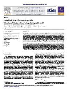
FIG. 1. Diagram of the cloning strategy for the recombinant HEVGST fusion proteins used in this study, as described in Materials and Methods. The cDNA fragments of 1,658, 830, and 380 bp were generated after RT-PCR of HEV RNA extracted from the bile of an experimentally infected monkey. All DNA fragments were purified by agarose gel electrophoresis and Prep-A-Gene at each step.
products were separated on a 1.0% agarose gel. Fragments of about 380 (ORF3), 830 (ORF2.1), and 1,658 (ORF2.0) bp were recovered by cutting the bands out of the gel. The DNA was purified with the Prep-A-Gene kit (Bio-Rad, Richmond, Calif.). Fragments ORF3 and ORF2.1 were digested with EcoRI, and the restriction fragments (370 or 820 bp) were separated on a gel and purified as described above. Approximately 6 ng of ORF3 or 16 ng of ORF2.1 DNA was ligated with 10 ng of pGEX1 vector (EcoRI cut and dephosphorylated) (Pharmacia Biotech) to give the plasmids pGEX1-AC3 and pGEX1-AC2.1, respectively, which were transformed into Escherichia coli NM522 (Pharmacia Biotech) with a Gene Pulser (Bio-Rad) and plated on L agar plus ampicillin. Plasmids were prepared from transformants by minipreparation and after EcoRI digestion were examined for inserts of the expected sizes. Fragment ORF2.0 (20 ng) was ligated with 50 ng of dephosphorylated vector pCR (Invitrogen, San Diego, Calif.) by utilizing the T overhangs on the plasmid and A overhangs on the PCR product to give the plasmid pCRAC2.0. After transformation and selection of recombinants, purified plasmids were subjected to partial EcoRI digestion and the resulting fragment of 1,670 bp was purified and ligated into pGEX1 as described above to give plasmid pGEX1AC2.0, representing the carboxyl two-thirds of ORF2 but with a 3' truncation of 27 nucleotides. To add the C-terminal sequences, this plasmid and pGEX1-AC2.1 were digested to
2062
LI ET AL.
completion with KpnI and BamHI. The restriction fragment of 1,390 bp from pGEX1-AC2.0 was then ligated with the fragment from pGEX1-AC2.1 containing the pGEX1 sequence together with the ORF2 sequences downstream of the KpnI site, to generate plasmid pGEX1-AC2.2, representing approximately two-thirds of ORF2 with the authentic C terminus. Selected colonies from each transformant were then induced with isopropylthio-13-D-galactoside (IPTG) and examined for expression of fusion proteins of the expected sizes. Production of GST fusion proteins. The transformants of pGEX1-AC3, pGEX1-AC2.1 and pGEX1-AC2.2, and pGEX1 (as a control) were grown overnight in L-broth medium containing 50 ,ug of ampicillin per ml. A 0.5-,u sample of the overnight culture was inoculated into 200 ml of L-broth medium plus ampicillin and incubated for 6 h. IPTG (200 p1 of 100 mM stock) was added, and the culture was grown overnight. Cells were pelleted at 3,500 rpm for 15 min in a bench centrifuge. The pellet was washed in 10 mM Tris (pH 7.5) and spun as before. For each gram of packed cells, 3 [lI of lysis buffer (50 mM Tris [pH 8.0], 1 mM EDTA, 100 mM NaCl), 4 plI of 100 mM phenylmethylsulfonyl fluoride, and 80 pul of lysozyme (10 mg/ml) were added. The suspension was incubated at room temperature for 30 min and then at 37°C for an additional 30 min. The solution was sonicated for 3 min and centrifuged at 2,800 x g for 15 min. The pellet (containing most of the fusion proteins) was washed with ice-cold 1 M NaCi-10 mM Tris (pH 7.5). The protein pellet was resuspended in 3 ml of 10 mM Tris (pH 7.5) and stored in aliquots at -20°C. SDS-PAGE. Each of the fusion proteins (GST-ORF3, GSTORF2.1, and GST-ORF2.2) was resuspended in 50 mM Tris (pH 6.8)-2% sodium dodecyl sulfate (SDS)-5% 2-mercaptoethanol-8% glycerol-0.01% bromophenol blue. In each experiment, unfused GST (prepared as described above) was mixed with the fusion proteins before electrophoresis. The protein mixture was heated at 100°C for 5 min and loaded on SDSpolyacrylamide gel electrophoresis (PAGE) gels in a mini-gel apparatus (Bio-Rad) with a separating gel of 10% acrylamide0.267% bisacrylamide by the discontinuous buffer system of Laemmli (15 mM Tris [pH 6.8]), 333 mM Tris [pH 8.8]. The gels were electrophoresed at 75 V for 20 min and then at 150 V until the dye reached the bottom of the gel. The proteins were transferred to nitrocellulose membranes (Hybond-C Extra; Amersham) on a semi-dry transfer cell (Bio-Rad) in transfer buffer (0.15 M glycine, 0.025 M Tris [pH 8.5], 20% methanol). Biotinylated marker proteins (Bio-Rad) were run in reference lanes. Western blot. The nitrocellulose membranes were blocked with 0.1 M Tris (pH 7.5)-0.1 M NaCl-0.3% Tween-20 (TBST) containing 3% casein and 0.01% sodium azide at room temperature for 1 h. Test sera were diluted 1:500 in TBST plus 1% casein and preabsorbed with 1% (wt/vol) GST lysate suspension at 37°C for 1.5 h. The diluted sera were incubated at 35°C with agitation for 1 h with the membrane, using the MiniProtean Multi Screen apparatus (Bio-Rad). The membranes were washed three times for 5 min in TBST, incubated with horseradish peroxidase (HRPO)-conjugated rabbit anti-human IgG (diluted 1:5,000; Dako Co., Glostrup, Denmark) at 35°C for 1 h, and then washed as before. Biotinylated markers were detected by using HRPO-conjugated streptavidin (diluted 1:10,000; Amersham). Enzyme complexes were detected by enhanced chemiluminescence (ECL) (Amersham). The membranes were soaked in ECL reagents for 1 min, then placed between acetate sheets, and exposed to Hyperfilm ECL (Amersham) for 30 s to 2 min at room temperature. Test sera for Western blot. (i) Human sera. Sixty-six serum
J. CLIN. MICROBIOL.
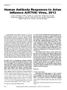
specimens were collected from acute-phase patients in Xinjiang, China, during the hepatitis E epidemic of 1988 (14), and convalescent-phase serum specimens were also collected from 19 of these patients between 3 and 9 months after the onset of disease. Negative control serum specimens were from 19 healthy controls and patients with other acute viral illnesses (10 patients with cytomegalovirus and 10 with Epstein-Barr virus) or other forms of acute viral hepatitis in Australia (10 patients with acute hepatitis A [positive for anti-hepatitis A virus IgM; Abbott Diagnostics], 18 with acute hepatitis B [positive for anti-hepatitis B core IgM; Abbott Diagnostics], and 58 with hepatitis C [positive by second-generation hepatitis C virus ETA; Abbott Diagnostics]. (ii) Macaque sera. Serum specimens were collected approximately twice weekly from seven rhesus monkeys (M27, M31, M32, M33, M34, M35, and M36) experimentally infected with HEV (12a). Samples were stored at -20°C. RESULTS Expression of HEV-GST fusion proteins. Clones of putative HEV structural proteins were constructed in the highly efficient pGEX expression system, giving rise to proteins fused with GST (normal molecular mass, 26 kDa). The clones studied represent the complete sequence of ORF3 (GSTORF3) and partially overlapping sequences within ORF2 (GST-ORF2.1 and GST-ORF2.2), as shown in Fig. 1. After induction with IPTG, bacterial lysates were found to contain unique proteins of 39.5, 57, and 86 kDa (results not shown), consistent with the predicted molecular masses of these translated sequences. It should be noted that pGEX1-AC2.2 contains all the sequences within pGEX1-AC2.1 (that is, the carboxy-terminal one-third of ORF2) with an N-terminal extension of 294 amino acids. Detection of anti-HEV IgG by Western blot. To select conditions for a sensitive diagnostic assay and to examine the dynamics of antibody responses to each fusion protein, Western blots were conducted to detect anti-HEV IgG in sera from acute-phase hepatitis patients and from experimentally infected monkeys. (i) Detection of anti-HEV IgG in sera from humans with acute hepatitis. Sera from well-documented cases of acute hepatitis A, B, C, and E and from controls were assayed by Western blot (Fig. 2; Table 1). The results show that none of 10 hepatitis A and 18 hepatitis B serum specimens contained anti-ORF3 or anti-ORF2.1 IgG, but 3 of 58 hepatitis C serum samples were weakly reactive with GST-ORF2.1 and one of these was also reactive with GST-ORF3. None of the serum samples from healthy controls or cytomegalovirus- and Epstein-Barr virus-infected patients were reactive. Sixty-one of 66 (92%) acute-phase hepatitis E serum specimens from Xinjiang, People's Republic of China, contained anti-HEV IgG that bound specifically to the GST-ORF2.1 band but not to the negative control (GST). Of these, 55 were also positive for anti-ORF3, and one serum sample was reactive with ORF3 but not with ORF2.1. Nine of 19 serum specimens collected between 3 and 9 months after the onset of hepatitis E were reactive against ORF2.1, but only 3 of these were reactive with ORF3. When tested by a commercial EIA based on ORF3 of the Burmese and Mexican strains, only one of the convalescent-phase serum specimens was reactive (results not shown), and this specimen was reactive against both GST-ORF2.1 and GST-ORF3. Both fusion proteins were therefore immunoreactive by Western blot and resulted in the sensitive detection of antiHEV IgG in epidemic cases of hepatitis E in China, with a high
PERSISTENT ANTIBODY TO HEV
VOL. 32, 1994
66KDa -
45KDa
-
31 KDm
-
m 4
-.
-
GST-ORF2.1
-
p-
M4-
GST-ORF3
-
66KD:a-
DGST-ORF2.1 -6 WI} 45KOa 6
Qe*
--
GST-ORF3
31 KDa
IWMW1s
10 xl1213lols16l7]8192o212223 22 33 44 53 66 77 8S 99 10 11 12 1314 17 1819 2021 2223
1
GST
1516
level of specificity indicated by the low rate of reactivity in other forms of viral hepatitis and other controls. Some hepatitis E samples strongly positive for anti-ORF2.1 or anti-ORF3 were also reactive with degradation products of the respective fusion proteins (as indicated in Fig. 2 through 5), but no serum showed reactivity with the control GST protein band. (ii) Detection of anti-HEV IgG in serial serum specimens from rhesus monkeys. Rhesus monkeys were inoculated intravenously with a Chinese strain of HEV and displayed an increase in alanine transaminase (ALT) at 7 to 26 days, indicating acute hepatitis (12a). Serial blood samples were tested for anti-HEV IgG by Western blot, and the results for one monkey (M27) are shown in Fig. 3. In this monkey, both anti-GST-ORF2.1 and anti-GST-ORF3 antibodies were barely detectable at day 16 but strongly positive at day 34, with anti-GST-ORF2.1 continuing to increase throughout the study period, whereas anti-GST-ORF3 reached a peak at 71 days and then declined. Degradation products of both fusion proteins were sometimes detected in Western blots, as indicated in Fig. 3 and subsequent figures. All seven monkeys seroconverted, since anti-ORF2.1 was detected at day 14 to 21 (results not shown), immediately after ALT elevation. Anti-ORF3 antibody was first detected at the TABLE 1. Detection of anti-HEV IgG by Western blotting No. (%) positive Infection
Total (%)a
n
GST-ORF2.1
None (healthy controls) Cytomegalovirus Epstein-Barr virus Hepatitis A Hepatitis B Hepatitis C Hepatitis E Acute phaseb Convalescent phasec
GST-ORF3
19
0
0
0
10 10 10 18 58
0 0 0 0 3 (5)
0 0 0 0 1(2)
0 0 0 0 3 (5)
66 19
61(92) 9 (47)
56 (85) 3 (16)
62 (94) 9 (47)
a Positive for GST-ORF2.1 and/or GST-ORF3. b Less than 3 months after onset of disease. Three to 9 months after onset of disease.
-0
_--GST MW 9 13 16 34 71 92 131
FIG. 2. Western blot of human acute-phase hepatitis sera against recombinant GST-ORF2.1 and GST-ORF3 proteins. Approximately equal masses of each recombinant protein and unfused GST were mixed and electrophoresed on a 10% acrylamide gel, the proteins were transferred to nitrocellulose, and the membrane was blocked with casein. The membrane was then incubated with a 1:500 dilution of patient sera or a 1:10,000 dilution of HRPO-streptavidin (marker lane), and immune complexes were detected by using HRPO-conjugated anti-human IgG and enhanced chemiluminescence. Lanes: MW, biotinylated molecular mass markers (sizes indicated on the left); 1 to 10, acute-phase hepatitis A specimens; 11 to 18, acute-phase hepatitis B specimens; 19 to 23, acute-phase hepatitis E specimens. The positions of reactive degradation products of GST-ORF2.1 and GSTORF3 (closed and open circles, respectively), GST, and each fusion protein are indicated on the right.
'
2063
Days Postinoculation FIG. 3. Western blot of GST-ORF2.1 and GST-ORF3 with sera from a rhesus macaque experimentally infected with HEV. The animal was inoculated intravenously with a fecal suspension from a firstpassage monkey, and serum samples were collected at the days indicated. The sera were diluted 1:500 and incubated with the membrane as described for Fig. 2. Lane MW, biotinylated molecular mass markers (sizes indicated on the left). Circles are as defined in the legend to Fig. 2.
same time or 7 days after first detection of anti-ORF2.1 for four monkeys but was weakly detectable in only one serum sample from each of two monkeys and in no samples from the remaining monkey. Anti-ORF2.1 therefore appears to be a more consistent marker of infection in macaques, as was also observed with samples from humans (Table 1). Two monkeys were rechallenged at days 53, 107, and 316 and a third monkey was rechallenged at days 107 and 316 after initial infection (12a). We have previously reported (12a) that all monkeys showed biochemical evidence of hepatitis after primary inoculation and less severe hepatitis after the first rechallenge, but only one (M32) of two monkeys appeared to be infected after the second rechallenge, suggesting the acquisition of partial protective immunity after each infection. Coincident with this partial immunity, we detected antiORF2.1 antibody in each monkey (Fig. 4), which remained at high levels throughout the study and was often boosted after rechallenge. At present, we cannot determine whether the boosted reactivity results from limited viral replication or at least in part from viral antigen present in the inocula, but it should be noted that in both M31 and M32 there was no further increase in reactivity following a third rechallenge, which suggests that the increased reactivity observed previously was due to virus replication. Anti-ORF3 was detected at high levels in M31 and M32 between days 17 and approximately 60, but the levels waned thereafter. Interestingly, anti-ORF3 was boosted after the second rechallenge in monkey M32, and for M33 the only sample positive for this antibody was after rechallenge. The reactivity with GST-ORF3 closely reflects that observed when a commercial HEV EIA based on ORF3 synthetic proteins was used, in which the day 151 sample was also the only reactive sample from M33 (12a). Comparison of GST-ORF2.1 and GST-ORF2.2. The fusion protein GST-ORF2.1 contains only the carboxy-terminal onethird of the coding sequences from HEV ORF2. In order to examine whether additional sequences from ORF2 would show greater reactivity, serum specimens from monkeys M31, M32, and M33 were reacted with GST-ORF2.1 and GSTORF2.2, which has an N-terminal extension of 294 amino acids but retains the authentic C terminus of ORF2. In the acute phase of infection, the extended protein was indeed more strongly reactive, although no samples were positive for only this protein. However, we were surprised to observe that the peak reactivity of the extended protein was in fact much lower than that of GST-ORF2.1 for each of the animals. In addition,
2064
A
_~
J. CLIN. MICROBIOL.
LI ET AL.
M31 S6KD.-
45KDa31 KD-
Rechallenge
Rechallonge 107
53
M31 4-GST4ORF2.1
-
GST-ORF2.2
4-GST-ORF3
-
GST-ORF2.1
0-GS
MW 7 10 17 38 455 2 56 59 63 66 73 80 87
B
A
316
53
107
122
129 168 16
3%1
47 MW 7 1=l so437 33, 3343,:M -A (4S7
B
316
M32
1;22
1479 431S9
31(7
Ah316
M32
4 GSTO4RF2.1
-
45KD& _
97K Da
t!P1etI ttt O-GST-ORIF3
MW 7 17 21 28 38 45 63 87129
-
--
45KDa
-
31 KDa
-
S
- -.... .......
W
34 1 1
M33 66KDa
_
4OKDa31 KDa
.
C
316
1~~~~~ I if 4- QST4ORP21 - I!flfl1I ~~~~~~~GST-OAF3
p lb
_..
4-
m ------wl -
MW 7 14 17 2124 38 45
122
136 159 151
168
GST-ORF2.1
.w a_"4 .
40
151 168 355 362 16 159 3-50 360 165
107
GST-ORF2.2
66KDa-
31 KDa 4
C
3511
.0-GST
344 355 362 35 360 365
3 f6
-.
M33
-a-- GST-ORF2.2
97K3a -
66KDa
-
45KDa
-
31 KDa
-
GST-ORF2.1 .. ...*.~~~~
I "I~~~~~~I 04D
3l' j
Days Postinfection
FIG. 4. Western blot of GST-ORF2.1 and GST-ORF3 with sera from rhesus macaques M31 (A), M32 (B), and M33 (C), which were experimentally infected with HEV and subsequently rechallenged with aliquots of the original inoculum. The days of rechallenge and the days of serum collection are indicated (above and below the lanes, respectively). Lanes MW, biotinylated molecular mass markers (sizes indicated on the left). Note that reactivity to GST-ORF2.1 remains at high levels and is boosted following rechallenge, whereas reactivity to GST-ORF3 declines with time. Circles are as defined for Fig. 2.
the antibody was found to decline with time and was only weakly boosted after each rechallenge. These two proteins were also used to screen some of the sera from patients with acute hepatitis A, B, or E as described above, with no difference observed in the proportion of reactive sera (results not shown), although in most cases the signal for GST-ORF2.2 was stronger than that for GSTORF2.1, as was also seen during the acute phase in monkeys (Fig. 5). DISCUSSION A Western blot assay was developed for the detection of anti-HEV with three HEV-GST fusion proteins: GST-ORF2.1 and GST-ORF2.2, which are encoded by portions of ORF2 overlapping at the C terminus, and GST-ORF3, which represents the entire ORF3 from a Chinese strain of HEV. This assay proved to be sensitive and specific for HEV in tests with sera from patients with different types of acute hepatitis, and to
4h4 4I(
"44
4'3
437'
Days Postinfection
FIG. 5. Western blot of GST-ORF2.1 and GST-ORF2.2 with sera from infected macaques. GST and fusion proteins GST-ORF2.1 and GST-ORF2.2 were mixed in equal proportions, electrophoresed, and transferred essentially as described for Fig. 2. Serum samples are from the same animals as for Fig. 4. Lanes MW, biotinylated molecular mass markers (sizes indicated on the left). Note that reactivity to GSTORF2.1 is weaker than that to GST-ORF2.2 during the acute phase of infection but is boosted after rechallenge and subsequently remains at higher levels than that to GST-ORF2.2. Positions of reactive degradation products (closed circles) are indicated.
our knowledge it is the first assay described which detects long-lasting antibody reactivity in a high proportion of patients
and experimentally infected animals. The detection of this persistent antibody reactivity represents an important advance in the understanding of immunity to HEV infection. Many serum specimens used in this study were previously tested for anti-HEV IgG with a commercial EIA kit (12a) (unpublished observations), with overall detection rates of 69% (50 of 72) for acute-phase patients and 57% (4 of 7) for monkeys, compared with 92% for patients and 100% for monkeys by the Western blot reported here. In addition, anti-HEV IgG was detected in macaque sera by Western blot (Fig. 3 to 5) over a longer period than with the EIA (12a), and almost half of the convalescent-phase hepatitis E sera were reactive in the Western blot (Table 1). This Western blot therefore shows utility as a diagnostic assay for HEV infection. While only IgG-class antibodies were detected in this study, in
PERSISTENT ANTIBODY TO HEV
VOL. 32, 1994
preliminary studies we have found specific IgM reactivity against the fusion proteins in acute-phase hepatitis E sera. However, the reactivity is not consistent. As the basis for a diagnostic assay, we believe that detection of anti-ORF2.1 indicates exposure to HEV, with the obvious potential to detect rising antibody titers during the acute phase. Conversely, the detection of anti-ORF3 suggests acute infection, but it should also be noted that in some cases this reactivity may be the result of repeated exposure to the virus (Fig. 4B and C). The assay appears to be highly specific for hepatitis E infection, as indicated by the low rate of reactivity in control specimens including other forms of acute viral hepatitis (Table 1), with only 3 of 125 specimens (2.4%) being reactive with GST-ORF2.1 and only 1 specimen (0.8%) being reactive with GST-ORF3. We do not know whether there is any significance in this reactivity being observed for hepatitis C patients, but in view of the persistence of anti-ORF2.1 observed for monkeys and hepatitis E patients, we suggest that those hepatitis C patients who were reactive to only GST-ORF2.1 represent true positives but had past infections. The one patient reactive to both GST-ORF2.1 and GST-ORF3 may represent protracted reactivity to antigens of ORF3, as has been described in rare cases (6), but must be considered a false positive for acute infection at present. Previous reports of both EIA and Western blot assays for antibody to HEV have described similar rates of detection for acute hepatitis E (5, 7); however, the detection of past infection appears to be less efficient, with a rapid decline in antibody reactivity within weeks or months. We therefore used the sera from experimentally infected macaques to examine the dynamics of the anti-HEV response following infection. The results indicate that anti-ORF2.1 antibody is detectable just after ALT elevation, while anti-ORF3 antibody appears only in some monkeys but generally coincident with antibody to GST-ORF2.1 (Fig. 3 and 4). Sera remained strongly positive for anti-ORF2.1 for at least 12 months, while anti-ORF3 declined after day 70, approximately 1 month after ALT levels have returned to normal. This suggests that these two antibodies may have different roles in the immune response to HEV infection. Epidemiological studies have shown the preponderance of clinically apparent hepatitis E in young to middle-aged patients, while subclinical infections occur more often in young patients. Second clinical infections are a rarity (15, 16), as has also been shown in cross-protection experiments in monkeys (3) and in the rechallenge studies examined here (also see reference 12a). Thus, it seems that individuals can acquire protective immunity following exposure to HEV on one or more occasions. The studies presented here provide important information about the potential protective role of antibody to HEV epitopes. After rechallenge at day 53 and/or 107, each of the three monkeys had elevations in ALT but less pronounced than those after primary infection, suggesting partial immunity. At the times of rechallenge, anti-ORF2.1 was the major or only reactive antibody found (Fig. 4), which implies that this protein includes a highly immunogenic epitope. Coincidence of this antibody with partial protection from rechallenge (12a) further suggests that antibody to this epitope may be protective. The results of this study also have implications for the development of more convenient assays for HEV antibody by other methods. First, the reactivity of the HEV fusion proteins in the Western blot appears to be greater than that of native, synthetic proteins in an EIA (5). Accordingly, we suggest that important epitopes of HEV ORF2 are commonly hidden when the protein is expressed in recombinant systems and that the exposure of these epitopes can be achieved under the dena-
2065
turing conditions of SDS-PAGE and subsequent renaturation. We are therefore currently evaluating the EIA reactivity of GST-ORF fusion proteins after bulk purification by SDSPAGE. Second, the greater reactivity of convalescent-phase sera with GST-ORF2.1 compared with that with the larger GST-ORF2.2, despite the latter containing all the sequences included in the smaller protein, is unexpected and suggests that the inclusion of additional viral sequences may hamper the correct folding of the protein to expose the required epitopes, at least in the GST fusion system. While this has not yet been demonstrated with other expressed HEV proteins, some of which are not fusion proteins (such as the full-length ORF2 expressed in insect cells [8]) or have different fusion partners, we suggest that vaccines and diagnostic reagents should be designed with the minimal sequences necessary for correct folding of the epitope(s) found in GST-ORF2.1. In this respect, it should be noted that Purdy et al. (13) have suggested that a trpE-HEV fusion protein, approximately analogous to GST-ORF2.2, may protect macaques against challenge with HEV. The results of the present study suggest that immunization with a smaller fragment of ORF2 may be more effective, but the relative contributions of viral sequences and different fusion proteins to the correct presentation of epitopes remain to be resolved. ACKNOWLEDGMENTS This work was supported in part by the Research Fund of the Macfarlane Burnet Centre for Medical Research and the Senior Medical Staff Fund of Fairfield Hospital. We are grateful to Scott Bowden, Angeline Bartholemeusz, and Joseph Torresi for helpful discussions, to Elizabeth Grgacic and John Mills for critical reading of the manuscript, and to Anthony Price for photography. REFERENCES 1. Aye, T. T., T. Uchida, and X. Ma. 1992. Sequence comparison of the capsid region of hepatitis E viruses isolated from Myanmar and China. Microbiol. Immunol. 36:615-621. 2. Aye, T. T., T. Uchida, and X. Z. Ma. 1992. Complete nucleotide sequence of a hepatitis E virus isolated from the Xinjiang epidemic (1986-1988) of China. Nucleic Acids Res. 20:3512. 3. Bradley, D. W. 1992. Hepatitis E: epidemiology, aetiology and molecular biology. Rev. Med. Virol. 2:19-28. 4. Cao, X., X. Ma, Y. Liu, Z. Liu, X. Jin, Q. Gao, H. Dong, H. Zhuang, C. Liu, and G. Wang. 1989. Epidemiological and etiological studies on enterically transmitted non-A, non-B hepatitis in the south part of Xinjiang, China. Chin. J. Exp. Clin. Virol. 3:1-9. 5. Dawson, G. J., K. H. Chau, C. M. Cabal, P. 0. Yarbough, G. R. Reyes, and I. K. Mushahwar. 1992. Solid-phase enzyme-linked immunosorbent assay for hepatitis E virus IgG and IgM antibodies utilizing recombinant antigens and synthetic peptides. J. Virol. Methods 38:175-186. 6. Dawson, G. J., I. K. Mushahwar, K. H. Chau, and G. L. GitnicL 1992. Detection of long-lasting antibody to hepatitis E virus in a US traveller to Pakistan. Lancet 340:426-427. (Letter.) 7. Favorov, M. O., H. A. Fields, M. A. Purdy, T. L Yashina, A. G. Aleksandrov, M. J. Alter, D. M. Yarasheva, D. W. Bradley, and H. S. Margolis. 1992. Serologic identification of hepatitis E virus infections in epidemic and endemic settings. J. Med. Virol.
36:246-250. 8. He, J., A. W. Tam, P. O. Yarbough, G. R. Reyes, and M. Carl. 1993. Expression and diagnostic utility of hepatitis E virus putative structural proteins expressed in insect cells. J. Clin. Microbiol. 31:2167-2173. 9. Huang, C. C., D. Nguyen, J. Fernandez, K. Y. Yun, K. E. Fry, D. W. Bradley, A. W. Tam, and G. R. Reyes. 1992. Molecular cloning and sequencing of the Mexico isolate of hepatitis E virus (HEV). Virology 191:550-558. 10. Kaur, M., K. C. Hyams, M. A. Purdy, K. Krawczynski, W. M. Ching, K. E. Fry, G. R. Reyes, D. W. Bradley, and M. Carl. 1992.
2066
LI ET AL.
Human linear B-cell epitopes encoded by the hepatitis E virus include determinants in the RNA-dependent RNA polymerase. Proc. Natl. Acad. Sci. USA 89:3855-3858. 11. Khudyakov, Y. E., N. S. Khudyakova, H. A. Fields, D. Jue, C. Starling, M. 0. Favorov, K. Krawczynski, L. Polish, E. Mast, and H. Margolis. 1993. Epitope mapping in proteins of hepatitis E virus. Virology 194:89-96. 12. Krawczynski, K. 1993. Hepatitis E. Hepatology 17:932-941. 12a.Li, F., et al. Submitted for publication. 13. Purdy, M. A., K A. McCaustland, K. Krawczynski, J. Spelbring, G. R. Reyes, and D. W. Bradley. 1993. Preliminary evidence that a trpE-HEV fusion protein protects cynomolgus macaques against challenge with wild-type hepatitis E virus (HEV). J. Med. Virol. 41:90-94. 14. Purdy, M. A., K. A. McCaustland, K. Krawczynski, A. Tam, M. J. Beach, N. C. Tassopoulos, G. R. Reyes, and D. W. Bradley. 1992. Expression of a hepatitis E virus (HEV)-trpE fusion protein containing epitopes recognized by antibodies in sera from human cases and experimentally infected primates. Arch. Virol. 123:335349.
J. CLIN. MICROBIOL. 15. Purdy, M. A., A. W. Tam, C.-C. Huang, P. O. Yarbough, and G. R. Reyes. 1993. Hepatitis E virus: a non-enveloped member of the "alpha-like" RNA virus supergroup. Semin. Virol. 4:319-326. 16. Reyes, G. R., M. A. Purdy, J. P. Kim, K. C. Luk, L. M. Young, K. E. Fry, and D. W. Bradley. 1990. Isolation of a cDNA from the virus responsible for enterically transmitted non-A, non-B hepatitis. Science 247:1335-1339. 17. Tam, A. W., M. M. Smith, M. E. Guerra, C. C. Huang, D. W. Bradley, K. E. Fry, and G. R. Reyes. 1991. Hepatitis E virus (HEV): molecular cloning and sequencing of the full-length viral genome. Virology 185:120-131. 18. Yarbough, P. O., A. W. Tam, K. E. Fry, K. Krawczynski, K A. McCaustland, D. W. Bradley, and G. R. Reyes. 1991. Hepatitis E virus: identification of type-common epitopes. J. Virol. 65:57905797. 19. Zhuang, H. 1992. Hepatitis E and strategies for its control. Monogr. Virol. 19:126-139. 20. Zhuang, H., X. Y. Cao, C. B. Liu, and G. M. Wang. 1991. Epidemiology of hepatitis E in China. Gastroenterol. Jpn. 3:135138.