... MD, PhD,*w Jesús Rodriguez-Pascual, MD,*w Fernando López-Rıós, MD, PhD, ... MD, PhD,*w Yolanda Quijano, MD, PhD,*w Ovidio Hernando, MD, PhD,*w.
ORIGINAL ARTICLE
Phase II Trial of Target-guided Personalized Chemotherapy in First-line Metastatic Colorectal Cancer Antonio Cubillo, MD, PhD,*w Jesu´s Rodriguez-Pascual, MD,*w Fernando Lo´pez-Rı´os, MD, PhD,wz Carlos Plaza, MD, PhD,wz Elena Garcı´a, MD, PhD,y Rafael A´lvarez, MD,*w Emilio de Vicente, MD, PhD,*w Yolanda Quijano, MD, PhD,*w Ovidio Hernando, MD, PhD,*w Carmen Rubio, MD, PhD,*w Sofı´a Perea, MD,*w Gema Sanchez, MSc,* and Manuel Hidalgo, MD, PhD*wy
Purpose: The aim of this study was to investigate the feasibility and efficacy of personalizing treatment of patients with advanced untreated colorectal cancer (CRC). Patients and Methods: Patients with untreated metastatic CRC, performance status 0-1, and candidates for systemic chemotherapy were eligible. Tumor tissues were analyzed for KRAS, BRAF, and PI3K mutations and expression of topoisomerase-1 (Topo-1), excision repair cross-complementing gene 1 (ERCC1), thymidylate synthase (TS), and thymidine phosphorylase (TP). Patients with Topo-1 expression received irinotecan, whereas patients with negative Topo-1 and ERCC1 expression received oxaliplatin. Otherwise, patients received physician’s choice of treatment. If TS was positive, no fluoropyrimidine was administered and if negative, 5-flurorouracil if TP was negative, or capecitabine if TP was positive. KRAS-mutated patients were treated with bevacizumab, whereas KRAS-native received cetuximab. The primary endpoint of the study was progression-free survival (PFS). Results: A total of 74 patients were enrolled and 67 received personalized treatment including irinotecan (n = 27), oxaliplatin (n = 16), FOLFIRI (n = 12), and FOLFOX (n = 12). Thirty-eight patients received cetuximab and 29 bevacizumab. With a median follow-up time of 18.3 months (95% confidence interval [CI], 4-36), the overall median PFS was 8.3 months (95% CI, 6.9-9.7), representing a 12-month PFS rate of 36.5% (95% CI, 25-48). Overall clinical benefit, including response rate and disease stabilization, was 86% (95% CI, 73%-97%). The overall median survival was 21 months (95% CI, 11-40). Conclusions: Real-time target-guided personalized first-line treatment of patients with advanced CRC is feasible but, with the approached used, did not result in a clear improvement in PFS to warrant phase III testing. Key Words: molecular targets, personalize treatment, colon cancer
(Am J Clin Oncol 2016;39:236–242)
T
he medical treatment of patients with colorectal cancer (CRC) has greatly improved over the last few years with the introduction of effective chemotherapy and biological agents such as oxaliplatin, irinotecan, cetuximab, and bevacizumab.1 Current standard management is based on combinations of these agents with several potential equally effective
From the *Centro Integral Oncolo´gico Clara Campal; zTherapeutics Targets Laboratory; wUniversidad CEU San Pablo; and yCentro Nacional de Investigaciones Oncologicas, Madrid, Spain. The authors declare no conflicts of interest. Reprints: Antonio Cubillo, MD, PhD, Centro Integral Oncolo´gico Clara Campal, C/On˜a 10, Madrid 28050, Spain. E-mail: acubillo@ hmhospitales.com. Copyright r 2014 Wolters Kluwer Health, Inc. All rights reserved. ISSN: 0277-3732/16/3903-0236 DOI: 10.1097/COC.0000000000000045
regimens. Treatment decisions remain largely empirical. The only universally accepted biomarker is KRAS genotyping supported by the lack of efficacy of cetuximab in patients with certain activating mutations in this oncogene.2 More recent data also suggest that mutation in other genes in the RAS pathway such as NRAS, BRAF, and PI3K also confer resistant to cetuximab, but this is not yet standard of care.3–5 For all other agents, including 5-fluoropyrimidines, oxaliplatin, and irinotecan, there is no clinically accepted biomarker. However, for each one of these drugs there are putative biomarkers, with different levels of validation, in the literature.6–12 Some studies support the concept that chemotherapy used in CRC is more effective in certain subgroups of patients whose tumors express the drug’s target or different quantifiable mechanism of resitance.9 For example, tumors with high expression of thymidylate synthase (TS) are considered to be resistant to treatment with 5-fluorouracil (5-FU) and tumors with high expression of thymidine phosphorylase (TP) respond better to capecitabine.10,11 Topoisomerase I (Topo-1) expression has been associated to irinotecan activity, whereas excision repair crosscomplementing gene 1 (ERCC1) expression has been related to resistance to oxaliplatin treatment.6,12 However, recent studies in other cancer types have questioned the usefulness of these biomarkers in contrast to older studies that supported it.13,14 Despite the data available, except for KRAS mutations, this information is not used in daily clinical practice. This is because of different factors, including the lack of conclusive studies, laboratory technique limitations, and the difficulties to implement these approaches in real-time clinical practice. Our group has been interested in personalizing the treatment of CRC by applying a full panel of biomarkers and a treatment algorithm that provides a guide for treatment selection for each individual patient.15 In this work, we have tested the feasibility and the effectiveness of applying this approach to patients with advanced, untreated CRC. The results show that the approach is feasible, leading to individual patient treatment recommendation in real time. Overall, however, the biomarkers tested are not predictive enough to get the expected therapeutic results. Hypothesis generating subgroup analysis suggest that certain subgroups may derive benefit and could be of interest to explore a more focused strategy in future trials.
PATIENTS AND METHODS Eligibility Criteria Patients with stage IV colorectal adenocarcinoma and candidates for first-line systemic chemotherapy according to regular clinical practice were eligible for the study. Other eligibility criteria included measurable metastatic disease
236 | www.amjclinicaloncology.com American Journal of Clinical Oncology � Volume 39, Number 3, June 2016 Copyright r 2016 Wolters Kluwer Health, Inc. All rights reserved.
American Journal of Clinical Oncology
�
Volume 39, Number 3, June 2016
according to RECIST 1.1 criteria, life expectancy of Z6 months, availability of tumor tissue, or possibility to perform a tumor biopsy to determine therapeutic targets and adequate renal (Cr < 1.5 mg/d), liver (bilirubin level r1.5 mg/dL, aspartate aminotransferase and alanine aminotransferase levels r3.0 � the upper limit of normal), and bone marrow function (absolute neutrophil count Z1500/mL, hemoglobin level Z9.0 g/dL, and a platelet count of Z100.000/mL). Exclusion criteria included contraindication for the administration of any of the drugs used in the study including 5-FU, capecitabine, irinotecan, oxaliplatin, cetuximab, or bevacizumab. Previous adjuvant treatment was allowed. The study was approved by the local ethics committee and the Spanish Health Authorities as per European Regulations and conducted in accordance with the Declaration of Helsinki (October 2000). The trial was registered with Clinical Trials.gov identifier NCT01453257.
Pretreatment and Follow-up Assessments In addition to a medical history, physical examination, hematology, biochemistry, and tumor markers, patients were studied with computed tomography (CT) scan of the chest, abdomen, and pelvis and colonoscopy if primary tumor had not been resected. Other imaging technique (PET-CT, magnetic resonance, or abdominal US) was performed, if needed, for a better assessment of tumor burden and response to treatment. Reponses were evaluated according to the RECIST 1.1 criteria every 8 weeks by CT scan and tumor markers if available. Patients were followed up until disease progression. Toxicity was assessed biweekly and reported according to the NCI CTC ver. 4.0 throughout the study.
Biological Studies A set of 7 molecular therapeutic targets were determined in pretreatment tumor tissues including mutation analysis of KRAS, BRAF, and PI3K genes and immunohistochemical (IHC) determination of Topo-1, ERCC1, TS, and TP. These markers were determined according to well-established and published methods.6,16 IHC data was analyzed according to the
Personalized Chemotherapy in Colon Cancer
percentage of tumor cells staining (0 = 0% of tumor cells; 0.1 = 1% to 9% of tumor cells; 0.5 = 10% to 49%; 1Z50% of tumor cells) and intensity of staining (0 = no staining, 1 = mild staining, 2 = moderate staining, 3 = strong staining). These 2 parameters were multiplied to generate a combined final score. The expression of marker was considered positive if the aggregate score was Z1.15 Sections were deparaffinised in xylene and rehydrated in graded alcohol. Antigen retrieval was carried out using EDTA, pH 9.0, in a pressure chamber (Pascal; Dako Cytomation), except for TP. All tissues were immunostained using the Dako Autostainer (Dako Cytomation). The antibody incubation was 60 minutes for ERCC1 (dilution 1:100; Neomarkers; clone: 8F1) and Topo-1 (dilution 1:50; Novocastra; clone 1D6), and 30 minutes for TP (dilution 1:50; Neomarkers; clone PGF44C). Immunodetection was carried out with the Dako Envision + dual-link polymer-horseradish peroxidase (Dako Cytomation) visualization method with diaminobenzidine chromogen (DAB +) as the substrate. Sections were counterstained with hematoxylin. KRAS, BRAF, and PIK3Ca mutations were analyzed from formalin-fixed and paraffin-embedded tumors (FFPE). A hematoxylin and eosin (H&E) slide of each tumor were reviewed by a pathologist to assess the percentage of tumor cells before DNA extraction. Macrodissection of tumors was performed in samples with small percentage of tumor tissue to enrich the final amount of tumor DNA. Mutation screening was performed using polymerase chain reactions and automatic direct sequencing as previously described.16
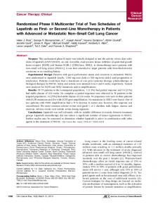
Treatment Decision-making Tree Patients with Topo-1-positive tumor received irinotecan. If Topo-1 was negative, ERCC1 status was considered. Patients with ERCC1-negative tumor received oxaliplatin. If ERCC1 expression was positive, patients received irinotecan or oxaliplatin at investigators discretion. Patients with TSnegative tumor received 5-FU if TP was negative or capecitabine if TP was positive. Patients with TS-positive tumor did
FIGURE 1. Decision-making tree for guided chemotherapy based on molecular markers expression. Patients with positive Topo-1 expression received irinotecan. If Topo-1 was negative, ERCC1 expression was considered. If ERCC1 was negative, patients received oxaliplatin and if positive, irinotecan or oxaliplatin, at their physician discretion. If TS was negative, patients received a fluoropyrimidine, either 5-fluoruracil or capecitabine depending on TP expression. If TS was positive, patients received irinotecan or oxaliplatin alone. Patients with KRAS-mutated tumor received bevacizumab, whereas patients with wild-type tumors received cetuximab.
Copyright
r
2014 Wolters Kluwer Health, Inc. All rights reserved.
www.amjclinicaloncology.com |
Copyright r 2016 Wolters Kluwer Health, Inc. All rights reserved.
237
American Journal of Clinical Oncology
Cubillo et al
not receive fluoropyrimidines in their treatment schema. In addition, patients with KRAS-mutated tumors received bevacizumab, and patients with KRAS wild-type received cetuximab (Fig. 1).
Treatment Schemas The chemotherapy regimens were administered according to their different package inserts, using conventional clinical protocols for dose, schedules, concomitant medication, and dose adjustment. Patients with responding tumors and limited anatomic disease spread were considered for locoregional treatments as per standard practice. Patients who underwent any locoregional treatment completed 6 months of chemotherapy after the procedure was completed. Maintenance treatment with bevacizumab or cetuximab plus a fluoropyrimidine, according to the chosen regimen, was recommended, but not mandatory, after completing the 6 months of treatment.
Statistical Analysis The primary endpoint of the study was progression-free survival (PFS). The strategy was considered to be effective if the proportion of patient’s PFS rate at 12 months was Z50% and “non-effective” when the PFS rate at 1 year was r35%. With these parameters, H0 was established as PFS at 1 year (%) r35% and H1 as PFS at 1 year (%) Z50%. Applying the Fleming method for phase II clinical trials, with a statistical power of 80% and an a of 0.05, 65 patients need to be included. Assuming a 10% loss rate, a total of 74 patients was to be included.
RESULTS Patients A total of 74 patients, whose principal characteristics are listed in Table 1, were enrolled in the study between January 2010 and October 2011. The median age was 67 (range, 39 to 84) years and 97% of the patients had a 0-1 ECOG performance status. Seventeen patients had rectal cancer. Most patients had only 1 or 2 metastatic lesions that were located in the liver in 70% of them.
Patient Disposition Sixty-seven patients were evaluated. Seven patients (9.5%) were not evaluable because they did not complete the first 8 weeks of treatment. Reasons for treatment discontinuation included disease progression in 45 patients, drug-related toxicity in 5 (1 infusion reaction, 1 mucositis, and 3 peripheral neuropathy), and serious unrelated events in 2 patients (1 bowel obstruction and 1 pulmonary embolism). Two patients died during the study, one because a surgical complication and another one for an unknown reason. Thirteen patients (19%) are still on treatment. Twenty-six patients (39%) received locoregional treatment (surgery, radiosurgery, or radiofrequency ablation) for their metastatic disease during the study.
�
Volume 39, Number 3, June 2016
TABLE 1. Baseline Patients Characteristics No. Patients Age (median [range]) Sex (M/F) ECOG 0 1 2 Primary tumor location Rectum Sigmoid colon Colon Tumor grade G1 G2 G3 Gx Number of metastatic lesions 1 2 >2 Metastases location (%) Liver Lung Lymph nodes Other organs
74 67 (39-84) (46/28) 31 40 3 17 12 45 13 48 6 6 46 20 8 70 30 13 13
part of their treatment. The other 28 patients with KRASmutated tumors received bevacizumab. BRAF and PI3K mutations were performed in all the samples. Three patients with BRAF-mutated KRAS-native tumors received cetuximab. The PFS of these patients was 16.5 months (95% CI, 9.9-23.2), but 2 of these patients received locoregional treatment. Seven patients, 3 with KRAS-mutated tumors, had a PI3K mutation. As expected, the PFS of patients with PI3K-mutated tumor who received cetuximab was very short, that is, 1.9 months (95% CI, 0.5-4.2). There were 40 patients with TS-positive tumor who did not receive a fluoropyrimidine in their treatment schema and 27 patients with a TS-negative tumor who received 5-FU. Topo-1 expression was positive in 33 tumors, and these patients received irinotecan-containing regimens. In addition, 6 more patients with Topo-1-negative and ERCC-1-positive tumors were treated with irinotecan-containing regimens as per physician discretion. Twenty-eight patients received oxaliplatin as part of their tailored treatment, 24 because of ERCC1negative, Topo-1-negative tumor expression and 4 with ERCC-1-positive, Topo-1-negative tumor because of investigator option.
Outcome With a median follow-up time of 18.3 (95% CI, 4-36) months, the median PFS was 8.3 (95% CI, 6.9-9.7) months,
Biomarker Assessment and Treatment Allocation Tumor material of sufficient quantity and quality was obtained in all patients to perform the full set of pre-specified markers. The results of these studies are summarized in Table 2, and a representative IHC staining example is showed in Figure 2. Median time from informed consent to treatment was 17 (95% confidence interval [CI], 4-35) days. On the basis of the results of the molecular analysis a total of 39 patients with KRAS-native tumors received cetuximab as
TABLE 2. Results of Biomarker Analysis
Topo-1 ERCC1 TS TP KRAS BRAF PI3K Positive Negative
33 34
29 38
40 27
0 67
28 39
3 64
7 60
ERCC1 indicates excision repair cross-complementing gene 1; Topo-1, topoisomerase-1; TP, thymidine phosphorylase; TS, thymidylate synthase.
238 | www.amjclinicaloncology.com Copyright r 2014 Wolters Kluwer Health, Inc. All rights reserved. Copyright r 2016 Wolters Kluwer Health, Inc. All rights reserved.
American Journal of Clinical Oncology
�
Volume 39, Number 3, June 2016
Personalized Chemotherapy in Colon Cancer
FIGURE 2. Representative IHC staining. Immunohistochemical staining for TS (A and B); ERCC1 (C and D); and Topo-1 (D and E). (A), (C), and (E) represent positive expression (score Z1), whereas (B), (D), and (F) were considered negative (score