Phosphate replicated and replaced microstructure of molluscan shells from the earliest Cambrian of China WEIMIN FENG and WEIGUO SUN Feng, W.M. and Sun, W.G. 2003. Phosphate replicated and replaced microstructure of molluscan shells from the earliest Cambrian of China. Acta Palaeontologica Polonica 48 (1): 21–30. The earliest Cambrian Meishucunian phosphoritic succession in eastern Yunnan, China, contains well−preserved mollus− can shells that offer insights into the early evolution of skeletonization. Phosphate internal moulds, phosphate replaced originally carbonate shells, and phosphate coatings show lamello−fibrillar structure, prismatic structure, and regularly fo− liated structure. The lamello−fibrillar structure appears earlier in the fossil record than laminar structures such as nacreous or foliated structures. It has been identified in fossil mollusks, which occur in China as early as the lower phosphate layer of the Zhongyicun Member of the Meishucunian. Therefore, the lamello−fibrillar structure appears to be primitive in mol− lusks. The lamello−fibrillar and prismatic aragonite is the most common shell material of molluscan skeletons in the Early Cambrian Meishucunian and equivalents around the world. Although the early molluscan microstructure is not so diverse as that of extant mollusks, it may be of use in high rank taxonomic classification as shown by the early conchiferan mol− lusks discussed here. These mollusks are characterized by the horizontal fibrillae that are layered and parallel, and thereby differ from hyoliths, in which the horizontal fibrillae appear to be in the form of the bundles of fibres that can branch or anastomose. Key words: Mollusca, small shelly fossils, microstructure, biomineralisation, Cambrian, Meishucunian Stage, China. Weimin Feng [
[email protected]] and Weiguo Sun [
[email protected]], Nanjing Institute of Geology and Paleon− tology, Academia Sinica, 39 East Beijing Road, 210008 Nanjing.
Introduction The apparently sudden appearance of abundant and diverse small skeletal fossils in rock successions near the base of the Cambrian is an expression of the “Cambrian explosion”, a long−standing enigma in both geology and biology. The most spectacular difference between the latest Precambrian and the earliest Cambrian faunas is the advent of biomineral skeletonization. For this reason, research on the biominerali− zation characters of the early small skeletal fossils may pro− vide significant information about the origin and early diver− sification of the major groups of the Metazoa. Although in the last three decades Early Cambrian small skeletal fossils have been found in basal Cambrian rocks at numerous localities throughout the world, little is known about their biomineralization and diagenesis (Bengtson and Conway Morris 1992; Lowenstam 1989; Runnegar 1989). Previous studies commonly concentrated on morphological characters of “small shelly fossils”. More and more small skeletal fossils, such as Microdictyon (see Bengtson et al. 1986; Chen et al. 1989), Wiwaxia (see Bengtson and Conway Morris 1984; Butterfield 1990), and Halkieria (see Conway Morris and Peel 1990, 1995), have been proven to be dis− persed hard parts (sclerites) covering bodies of relatively large animals. Their original taxonomic affiliation based on morphology of single sclerites appeared incorrect. Other Early Cambrian small skeletal fossils have been attributed to Acta Palaeontol. Pol. 48 (1): 21–30, 2003
many phyla, such as the Mollusca, Brachiopoda, and Hyo− litha. Qian and Bengtson (1989) in a comprehensive review attributed the Meishucunian small skeletal fossils to a cata− logue of morphological groups. Those different major groups may have their own diag− nostic characters of skeletonization, which can be used as ev− idence for identification and classification. Although most of the Early Cambrian small skeletal fossils are problematic due to incomplete preservation or unusual morphology, they may help in recognizing ancestors of later major taxa and reveal their phylogenetic relationships. Mollusks, as a good example to start with, contributed a very important part to the earliest Cambrian small skeletal fossils. Pioneering work by Runnegar (1983, 1985a, b, 1989; Runnegar and Jell 1976) on the microstructures of Cambrian mollusks and works of other researchers (Müller 1975; Mis− sarzhevsky 1989; Brasier 1990; Bengtson et al. 1990; Carter and Hall, 1990; Bengtson 1992; Li and Chen 1992; Kou− chinsky 1999, 2000b; Feng 1998; Feng et al. 2000, 2001) have provided very useful information about early molluscan biomineralization. Our investigations are based on mollus− can fossil materials from the Meishucunian in eastern Yunnan, southwestern China. The aim of this paper is to use diagenetic phosphate replication of the original mineral tis− sues to determine the original shell structure of early mol− lusks. http://app.pan.pl/acta48/app48−021.pdf
22
ACTA PALAEONTOLOGICA POLONICA 48 (1), 2003
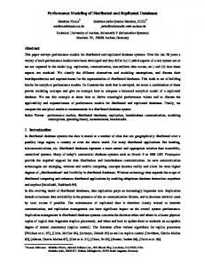
Material and methods The Meishucunian is the basalmost Cambrian chronostrati− graphic unit in the eastern Yunnan of China and corresponds, in ascending order, to Xiaowaitoushan, Zhongyicun, Dahai Members of the Dengying Formation and the Shiyantou Member of the Yuanshan Formation. Small skeletal fossils abruptly appear in abundance in the Zhongyicun Member. The fossil material dealt with in this paper was collected in 1996 and 1999 from the Zhongyicun and Dahai Members of the Early Cambrian Meishucunian at Beideng of Anning County, Baizhai of Xundian County, and Dahai and Yulu of Huize County (Fig. 1). The rock samples were carbonates and phosphorites. Most of the fossil specimens were obtained by washing weathered rock with water. Acid maceration with 5–8% acetic acid was also applied. Observations were done under SEM (JSM−6300) in addition to Figs. 2B and 5B, C, taken with Philips X−30 at Uppsala University, Sweden. This paper adopts the standardized microstructure terminology set up by Carter et al. (1990). Molluscan fossils used for the research on microstructure include the monoplacophorans Ramenta cambrina Jiang, 1982 (Luo et al. 1982), Latouchella cf. korobkovi Vostokova, 1962, Papilloconus explanatus Feng et al., 2000, Ilsanella cf. orectes (Jiang, 1982), Ilsanella? rozanovi Wang, 1994, Wat− sonella yunnanensis (He and Yang, 1982), and the para− gastropod Archaeospira ornata Yu, 1979. Watsonella is con− sidered to be a monoplacophoran based on the most complete phylogenetic analysis available (Carter et al. 2000). It is the link between the stenothecid monoplacophorans and the Bivalvia. It lacks a true pegma, which, as indicated by Pojeta and Runnegar (1974), is necessary for membership in the Rostroconchia, and its microstructure is in part much like that of Pojetaia and Fordilla (see Carter et al. 2000). All these specimens are housed in the Nanjing Institute of Geology and Palaeontology, Academia Sinica, abbreviated NIGP.
Mode of preservation Phosphatization, so common in strata near the Precambrian– Cambrian boundary around the world, played a key role in preserving information on the original shell structure (Bengt− son and Conway Morris 1992; Brasier 1990; Dzik 1994; Runnegar 1985a; Runnegar and Bengtson 1990). As a gen− eral rule, phosphatic replacement or filling occurs only in small specimens, whereas silicification affects specimens of all sizes (Yochelson 1999). Phosphate fillings of small shells can replicate very fine structures, up to 100 nm (Runnegar and Pojeta 1985). This allows determination of both the to− pography of the shell surface and its microstructure (Run− negar 1989). Crystal morphologies replicated on internal moulds were reported to be rather common in phosphate and carbonate successions of the earliest Cambrian (Runnegar
75
90
105
135
120
45
Beijing
Dahai
Yulu
China 30
26 N
Kunming 15
Baizai 25 N
Kunming Anning 24 N
102 E
103 E
104 E
Fig. 1. Location of studied exposures of the Meishucunian in the Yunnan Province.
1989). Information about early molluscan microstructures is obtained mostly from the internal moulds of conchs (Table 1). As typical for secondarily phosphatized small shelly fos− sils, the Meishucunian specimens usually show phosphoritic internal moulds, calcite−replaced original shell, and a phos− phate coating of its exterior. In acid treated materials the calcitic shell is removed and represented by a narrow empty cavity between the mould and coating. The coating then easily exfoliates. Frequently the shell itself is also phosphatized. Each of these three kinds of phosphatic structures is poten− tially a source of information for original shell microstructure. The external phosphatic coating, usually only roughly replicates the external shell morphology, but it can be some− times recovered and observed from the inside (e.g., Olemp− ska 1994; Hinz−Schallreuter 1995). It shows very fine details of the external shell surface in negative. In the case of mol− lusks these may be details of organic periostracum, if pho− sphatization was early enough. Frequently, internal moulds of cavities within the shell wall are attached to the coating. Mostly these are channels etched by endolithic cyanobacteria (e.g., Runnegar 1985b; Olempska 1986). Phosphoritic internal moulds provide additional informa− tion, in this case replicating the internal shell structure (e.g., Runnegar 1985a). In both of the above cases the information is rather reliable and relatively easy to interpret. Much more difficulty arises when the secondarily phosphatized shell wall is considered. The phosphatization, which may be late diagenetic in some cases, may replace the original organization of the shell or this
FENG AND SUN—MICROSTRUCTURE OF CAMBRIAN MOLLUSCA
23
Table 1. Distribution of microstructures in the early molluscan fossils. Fossil name Aldanella crassa Anabarella argus Anabarella plana Anabarella sp. Archaeospira ornata Ardrossania paveyi Bellerophon sp. Bemella? mirabilis Ceratoconus cf. rusticus Chancelloria eros Enigmaconus sp. Eotebenna pontifex Euphemites sp. Fordilla troyensis Watsonella varensalensis Watsonella varensalense Ilsanella cf. orectes Ilsanella sp. Ilsanella? rozanovi Lathamella symmetrica Latouchella cf. korobkovi Latouchella sp. Leptostega? corrugata Mackinnonia sp. Mackinonia davidi
Maikhanella multa Mellopegma georginensis Obtusoconus rostriptutea Obtusoconus sp. Papilloconus explanatus Pararaconus staiconum Pelagiella cf. subangulata Pelagiella deltoids Pelagiella subangulata
Microstructures Concave polygons and crossed fibres Prismatic outer shell layer Prismatic, crossed−lamellar Prismatic outer shell layer and “nacreous” inner shell layer
Horizon Lower Cambrian Lower Cambrian Lower Cambrian Lower Cambrian
Reference Kouchinsky 2000b Bengtson et al. 1990 Kouchinsky 1999 Runnegar 1983 Runnegar 1985a Horizontal and parallel fibrillae Lower Cambrian This paper Fibrous structure Lower Cambrian Bengtson et al. 1990 Shell aragonitic and predominantly aragonitic CL, cone CCL Upper Carboniferous MacClintock 1967 and irregular CCL with thin bands of irregular simple prisms Rollins 1967 Carter and Hall 1990 Prismatic Lower Cambrian Runnegar 1985a Tubercles and transverse fibres Lower Cambrian Kouchinsky 2000b Radial rays plus one central ray Lower Cambrian Kouchinsky 2000b Convex polygons Lower Cambrian Kouchinsky 2000b Inner shell layer largely calcitic, regularly foliated Lower Cambrian Runnegar 1983 Upper Carboniferous MacClintock 1967 Shell aragonitic and predominantly CL and CCL with Rollins 1967 sublayers of irregular simple prisms in the inner layer and Carter and Hall 1990 possibly with an exterior optically homogeneous layer Large tablet, imbricated nacre Lower Cambrian Runnegar and Pojeta 1992 Polygonal texture Lower Cambrian Müller 1975 Prismatic and probably aragonitic shell Lower Cambrian Runnegar 1983 Runnegar 1985a Lamello−fibrillar structure Lower Cambrian This paper Pits and polygonal texture Lower Cambrian Kouchinsky 2000b Prismatic structure Lower Cambrian This paper Prismatic, Crossed lamello−fibrillar structure Lower Cambrian Li Guoqiang 1992 Lamello−fibrillar structure Lower Cambrian This paper Prismatic outer shell layer Lower Cambrian Runnegar, 1985a Outer shell layer of prismatic structure Lower Cambrian Bengtson et al. 1990 Tubercles and concave polygonal texture Lower Cambrian Kouchinsky 2000b Bengtson et al. 1990 Outer layer of narrow fibrous prisms, a middle layer of wider Lower Cambrian J.Carter personal communi− columnar prisms with thick interprismatic organic matrices cation and in the thicker parts of the shell an inner layer with no dis− tinctive impression on steinkerns Spicular fibres Lower Cambrian Bengtson 1992 Outer columnar prismatic and inner rhombohedral (parallelo− Lower Cambrian Runnegar 1983 gram) small tablet, sheet nacreous Runnegar 1985a Prismatic structure Lower Cambrian Runnegar 1983 Runnegar 1985a Pits and polygonal texture Lower Cambrian Kouchinsky 2000b Polygonal elevations Lower Cambrian This paper Polygonal elevations Lower Cambrian Bengtson et al. 1990 Fine radial continuous fibres and shallow concave polygons Lower Cambrian Kouchinsky 2000b Inner shell layer of flattened, elongated crystallites arranged in a spiral fashion Outer radial and inner comarginal layer of fibrous crystals
Lower Cambrian
Runnegar 1985a
Lower Cambrian
Runnegar 1985 Bengtson et al. 1990 Runnegar 1983 Runnegar and Bentley 1983 Runnegar 1985a Bengtson et al. 1990 Carter 1990 Rollins 1967 Rollinns et al. 1971 Carter and Hall 1990
Pojetaia runnegari
Lower Cambrian Outermost very thin, columnar prismatic layer and middle and inner layers large tablet, imbricated nacre, probably sepa− rated with myostracal irregular simple prisms
Praematuratropis sp.
Shell aragonitic and varies from fine CCL to irregular CCL in Middle Devonian the outer layer, grading inward into cone CCL in the middle layer, and irregular CCL to cone CCL in the inner layer, with a thin, irregular simple prismatic innermost layer; inductura commarginal linear CL
http://app.pan.pl/acta48/app48−021.pdf
24
ACTA PALAEONTOLOGICA POLONICA 48 (1), 2003
Table 1 continued. Fossil name Pseudomyona queenslandica Ptomatis sp.
Microstructures Largely regularly foliated calcite, columnar prisms
Horizon Middle Cambrian
Reference Runnegar 1985a
Shell aragonitic with a thin, outer layer of nearly vertical, ir− regular simple prisms; a thick middle layer of cone CCL, and an inner layer of fine CCL and very faintly CL (?) structure, with a parietal shelf of irregular CCL Punctella maidipingensis Concentrically arranged granules Purella cf. cristata Fused needle−like sclerites Ramenta cambrina Inner lamello−fibrillar structure Retispira sp. Shell aragonitic and predominantly CL, cone CCL,irregular CCL,and fine CCL, with sublayers of irregular simple prisms
Middle Devonian
Carter and Hall 1990
Lower Cambrian Lower Cambrian Lower Cambrian Middle Devonian
Securiconus cf. costulatus Positive polygonal relief and casts of fibres Securiconus incertus Subradially oriented fibres and tightly packed spherulites sit− uated under the beak Tuarangia Largely (regularly) foliated calcite gravgaerdensis Tuarangia paparua Largely (regularly) foliated calcite
Lower Cambrian Lower Cambrian
Kouchinsky 2000b Kouchinsky 2000b This paper Rollins 1967 Carter and Hall 1990 Kouchinsky 2000b Kouchinsky 2000b
Middle Cambrian
Berg−Madsen 1987
Middle Cambrian
Watsonella sp. Watsonella yunnanensis Yochelcionella cyrano Yochelcionella sp. Yuwenia bentleyi
Lower Cambrian Lower Cambrian Lower Cambrian Lower Cambrian Lower Cambrian
MacKinnon1982 Runnegar 1985a Kouchinsky 1999 This paper Runnegar 1985a Kouchinsky 2000b Runnegar 1985a Bengtson et al. 1990
Prismatic, crossed−lamellar lamello− fibrillar structure Prismatic structure Tubercles and polygonal texture Outer shell layer of radially fibrous prismatic and inner shell layer of commarginal simple crossed lamellar
0.5 mm
b a
A B
C
100 µm
D
50 µm
10 µm
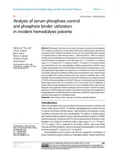
Fig. 2. Ilsanella cf. orectes (Jiang, 1982). Specimen NIGP Mo 131353 from sample yb96929−1, upper phosphatic bed of the Zhongyicun Member, Baizai Xundian in the eastern Yunnan, Meishucunian. A. Lateral view. B. Lamello−fibrillae (arrow a) approximately perpendicular to growth lines (arrow b). C. Microstructure of inner surface of external coating. D. Enlargement of place shown by arrow in C, note lamello−fibrillae.
FENG AND SUN—MICROSTRUCTURE OF CAMBRIAN MOLLUSCA
A
0.5 mm
B
C
25
10 µm
D
10 µm
10 µm
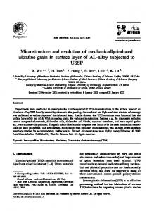
Fig. 3. Watsonella yunnanensis (He and Yang, 1982). Specimen NIGP Mo 131363 from sample yy9649−1, upper phosphatic bed of the Zhongyicun Mem− ber, Yulu of Huize in the eastern Yunnan, Meishucunian. A. Lateral view. B, C. Lamello−fibrillar structure. D. Enlargement of C.
may be completely obliterated by chaotically or perpendicu− larly growing apatite crystals. To be sure that the original structure is replicated, the matter must be sufficiently different from normal phosphate precipitation or replacement.
Description of structures Structure of the external coating.—Ilsanella cf. orectes (Jiang) is represented by cyrtoconic internal moulds and an in− complete phosphatic coating (Fig. 2A). The inner surface of the coating shows fine details of microstructure (Fig. 2C, D). Several nearly parallel lines seem to represent growth lines (Fig. 2C). In close−up, the fibrillae obviously comprise a set of rice−shaped granules (Fig. 2D). These fibrillae are generally arranged in two directions. The rice−shaped granules are phos− phatic in composition; they form the phosphatic coating. Structure on the internal mould.—Microstructures repli− cated on the internal mould have been found in the mono− placophorans Latouchella cf. korobkovi Vostokova, Ilsanella cf. orectes (Jiang), Ilsanella? rozanovi Wang, Papilloconus explanatus Feng, Ramenta cambrina Jiang, Watsonella yunnanensis (He and Yang) and paragastropod Archaeospira ornate Yu. Two microstructural morphologies, lamello− fibrillar and simple prisms, are recognized.
Lamello−fibrillae are seen on the surface of internal moulds of W. yunnanensis (Fig. 3B–D), R. cambrina (Fig. 4C, D), I. cf. orectes, and L. cf. korobkovi (Fig. 5B, C). Commonly, the fibrillae are replicated as a crossed or branched pattern on the internal mould (Figs. 3B–D, 5B, C), but parallel fibrillae are visible as well (Figs. 3D, 5C). Crossed or branched fibrillae might be diagenetic artifacts from fine phosphate grains replicating fibrillae of different lamellae and then being compressed during diagenesis. Re− gardless of the seemingly branched fibrillae, they are distrib− uted approximately along shell growth direction or perpen− dicular to growth lines (Fig. 2B). Horizontally parallel fibrillae can be also seen on the internal mould of A. ornata (Fig. 6A2). Another structural morphology replicated on the internal mould is a prism. This prismatic structure can appear in two replicated shapes. In P. explanatus Feng the prismatic preser− vation shows a positive relief. Ends of prisms resemble tuber− cles of diameter about 10 µm, very regularly exposed on the surface of the internal mould (Fig. 7B, C). This may reflect more intense weathering of the exposed prism facets than the destruction of organic material at their edges (Kouchinsky 1999). In I.? rozanovi Wang the prismatic structure is pre− served in negative relief, as shown by a series of hexagons about 12–20 µm in diameter. The hexagon periphery, 1–2 µm in width, may represent an originally organic sheath sur− http://app.pan.pl/acta48/app48−021.pdf
26
ACTA PALAEONTOLOGICA POLONICA 48 (1), 2003
0.5 mm
a
A
B
10 µm
C
10 µm
D
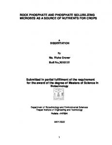
Fig. 4. Ramenta cambrina Jiang, 1982. Specimen NIGP Mo 131200 from sample yy9649−1, upper phosphatic bed of the Zhongyicun Member, Beideng of Anning in the eastern Yunnan, Meishucunian. A. Lateral view. B. Apical view. C. Enlargement of place shown by arrow a in B, note irregular polygonal con− vexity. D. Enlargement of C, note lamello−fibrillar structure.
rounding the prisms (Fig. 8C, D). The organic sheaths are very likely to have been decayed preferentially and were filled with a phosphatic matrix. A similar pattern of micro− structural preservation has been reported in the early mollus− can fossils from Siberia and Australia (Kouchinsky 2000b; Runnegar 1985a; Bengtson et al. 1990). Phosphate replaced shell wall.—Phosphate replaced shell walls have been found in various Meishucunian fossils. In a specimen of Ramenta cambrina Jiang with an internal mould, a replaced shell and a phosphatic coating, the re− placed shell wall is well exposed between the layer of the coating and the internal mould (Fig. 4C, D). The external coating replicated the surface sculpture while phosphatic re−
A
0.5 mm
B
placement preserved lamello−fibrillae, where the fibrillae ap− pear to be distributed in lamellae and arranged in two direc− tions. Fibrillae in the same lamella are generally parallel to each other, but those in the adjacent lamella are arranged in another direction, thus forming a crossed pattern in dorsal view (Fig. 4D). Phosphate replaced shell walls can be also seen in cross sections of several fractured specimens of Archaeospira ornata Yu. These shell sections comprise the outer and inner phosphate coatings and the phosphate replaced shell wall in between (Fig. 6B, C). Most of the shell wall has been filled with chaotically orientated phosphate granules, but the origi− nal fibrillae can still be recognized. They appear to be parallel to the shell surface (Fig. 6B, C).
20 µm
C
5 µm
Fig. 5. Latouchella cf. korobkovi Vostokova, 1962. Specimen NIGP Mo 131371 from yd96924−8A upper part of the Dahai Member, Dahai of Huize in the eastern Yunnan, Meishucunian. A. Lateral view. B, C. Enlargement of place shown by arrow in B, note lamello−fibrillar structure.
FENG AND SUN—MICROSTRUCTURE OF CAMBRIAN MOLLUSCA
27
0.5 mm
A1
B
A2
10 µm
C
10 µm
10 µm
Fig. 6. Archaeospira ornata Yu, 1979. A. Specimen NIGP Mo 131370 from sample yd96923−10, upper part of the Dahai Member, Dahai of Huize in the eastern Yunnan, Meishucunian. Apical view (A1); lamello−fibrillae at the apex (A2). B. Specimen NIGP Mo131370A from sample yd9649−2, upper phos− phatic bed of the Zhongyicun Member, Yulu of Huize in the eastern Yunnan, Meishucunian. Lamello−fibrillae of the shell wall. C. Specimen NIGP Mo 131365 from yd96923−10, upper part of the Dahai Member, Dahai of Huize in the eastern Yunnan, Meishucunian (note fibrous structure).
Discussion In this paper we show that phosphate coated and phosphate re− placed shell walls from Meishucunian age mollusks can pro− vide important information about original shell micro− structure. The preservation of microstructure is rather complex due to diagenesis. It may reflect fine morphology of the inner layer’s surface, or sometimes both inner and outer layers and different lamellae. This is because phosphatic granules may replicate fine structures of different layers as the inner layer is progressively destroyed. For example, prismatic and nacreous structures have been found in Anabarella sp. and Mellopegma georginensis Runnegar and Jell, 1976 (Runnegar 1983, 1985), prismatic and crossed fibrous structure in Lathamella sym− metrica Li and Chen, 1992, Anabarella plana Vostokova, 1962, Watsonella sp. (Kouchinsky 1999), Securiconus cf. costulatus Missarzhevsky, 1989, Securiconus incertus Bokova, 1985, and Pelagiella cf. subangulata (Tate, 1982) (Kouchinsky 2000b), and outer vertical fibres and inner lamello−fibrillar structure in Ramenta cambrina (Jiang, 1982) (Feng et al. 2002). The replicated morphology of fine structure may be used to indicate the orientation of crystals and the dis− tribution of organic matter between or within them prior to phosphatization (Fig. 8D). Positive and negative reliefs of
prismatic structure on many internal moulds from the Yangtze platform of China resemble those from the Siberian platform (Kouchinsky 1999: figs. 2B, 3F). Different modes of phosphatization in the Meishucunian fossils offer information on various aspects of shell structure. For example, in Ilsanella the prismatic structure is known from internal moulds (Fig. 8C, D; see also Kouchinsky 2000b: fig. 8) and lamello−fibrillar structure is known from the inner surface of the coating (Fig. 2C, D). The inner and outer layers of the shell shown prismatic and lamello−fibrillar structures, respectively. Lamello−fibrillar structure is commonly developed among Early Cambrian monoplacophorans and paragastro− pods (Table 1). However, it should be stressed that lamello− fibrillae are easily to be replicated as a crossed or branched pattern on internal moulds (Figs. 3B–D, 5B, C) due to the diagenesis. The present investigation of the phosphate re− placed shell wall of R. cambrina clarifies the real aspect of the microstructure. Lamello−fibrillar structure is similar to the crossed lamellar structure of well known extant and fossil mollusks, but it is characterized by only two orders of ar− rangement and the latter by three orders. However, crossed lamellar structure without well−defined second−order lamellae is present in the Polyplacophora (Haas 1981; Poulicek and Kreusch 1986; Carter and Hall 1990). The polyplacophorans http://app.pan.pl/acta48/app48−021.pdf
28
ACTA PALAEONTOLOGICA POLONICA 48 (1), 2003
A 0.5 mm
10 µm
C
B
Fig. 7 . Papilloconus explanatus Feng et al., 2000. Specimen NIGP Mo 131364 from sample yd96924, upper phosphatic bed of the Zhongyicun Member, Baizai of Xundian in the eastern Yunnan, Meishucunian. A. Lateral view. B. Apical view. C. Enlargement of place shown by arrow in B, note regular ar− rangement of nodules, corresponding to the end of prisms.
and aplacophorans are thought to be primitive within the Mollusca. The shell plates of the polyplacophoran seem to be more primitive than the conchiferan shell (Haas 1981). The thin periostracum−like cuticle of polyplacophorans does not serve as a crystallization surface in biomineralization, the
role usually performed by the conchiferan periostracum. Aragonitic aplacophoran and polyplacophoran spicules and polyplacophoran plates develop in crystallization chambers provided by individual cells or groups of cells below the cuti− cle. The lamello−fibrillar structure appeared earlier in the fos−
0.5 mm
A
C
B
100 µm
D
10 µm
Fig. 8. Ilsanella? rozanovi Wang, 1994. Specimen NIGP Mo 131358 from sample yd96924−8, upper phosphatic bed of the Zhongyicun Member, Baizai of Xundian in the eastern Yunnan, Meishucunian. A. Lateral view. B. Apical view. C. Enlargement of place shown by arrow in B, note cross section of prism replicated by internal mould. D. Enlargement of place shown by arrow in C.
FENG AND SUN—MICROSTRUCTURE OF CAMBRIAN MOLLUSCA
sil record than laminar structures such as nacreous or foliated structure. It has been identified in fossil mollusks in China as low as the lower phosphate bed of the Zhongyicun Member. Moreover, the nacreous structure of some extant mollusks consists of radial horizontal fibres (Mutvei 1983: fig. 4). Prisms seem to be secondary in evolutionary terms, as docu− mented by the occurrence of fibres on convex polygons (Kouchinsky 2000b: fig. 7G). Therefore, the lamello−fibrillar structure appears to be very primitive in molluscan skeletoni− zation. Lamello−fibrillar and prismatic aragonite is the most common shell materials of molluscan skeletons in the Early Cambrian Meishucunian and its equivalents around the world. While developing such microstructures the earliest conchiferan mollusks made a leap forward in mechanical quality of their skeleton, especially compared with the uni− form granular structure of Cloudina Germs, 1972 (Chen Zhe 1999), the only well known and widely distributed skeletal fossil of the latest Precambrian. Although the early mollus− can microstructures are not so diverse as in extant mollusks, they may be useful in high rank taxonomic classification. In the early conchiferan mollusks discussed here, crossed lamello−fibres are generally layered and arranged parallel to one another. In hyolith shell the fibres appear to be in the form of bundles of fibres that branch or anastomose (Kouchinsky 2000a; Feng, Mu, and Kouchinsky 2001). The difference in fibre arrangement between early mollusks and hyoliths testifies that diversification within fibrous structures occurred before the beginning of the Cambrian.
Acknowledgements We are very grateful to Prof. Jerzy Dzik (Warsaw) and Prof. Harry Mutvei (Stockholm) for their kind help in improvement of the manu− script, to Dr. Artem V. Kouchinsky (Uppsala) for beneficial discus− sions, to Prof. Joseph G. Carter (Chapel Hill) for providing valuable references and comments, and to anonymous referees for their critical remarks and constructive suggestions. This project is supported by grants from the Chinese Major Basic Research Projects of Ministry of Science and Technology (G2000077700, CAS−KZCX2−116) and from the Chinese National Sience Fundation (DST 95−Special−01).
References Bengtson, S. and Conway Morris, S. 1984. A comparative study of Lower Cambrian Halkieria and middle Cambrian Wiwaxia. Lethaia 17: 307–329. Bengtson, S., Mattews, S. C., and Missarzhevsky, V. V. 1986. The Cambrian netlike fossil Microdictyon. In: A. Hoffman and M.H. Nitecki (eds.), Problematic Fossil Taxa, H197–H115. Oxford University Press, New York. Bengtson, S., Conway Morris, S., Cooper, B. J., Jell, P. A., and Runnegar, B. N. 1990. Early Cambrian fossils from south Australia. Memoirs of the Associations of Australasian Palaeontologists 9: 1–364. Bengtson, S. and Conway Morris, S. 1992. Early radiation of biomineralizing phyla. In: J.H. Lipps and P.W. Signor (eds.), Origin and Early Evolution of the Metazoa, H447–H482. Plenum Press, New York.
29
Bengtson, S. 1992. The cap−shaped Cambrian fossil Maikhanella and the rela− tionship between Coeloscleritophora and mollusks. Lethaia 25: 401–420. Berg−Madsen, V. 1987. Tuarangia from Bornholm (Denmark) and similari− ties in Baltoscandian and Australasian late Middle Cambrian faunas. Alcheringa 11: 245–259. Bokova, A.R. 1985. The oldest complex of organisms in the Cambrian of the western Anabar region [in Russian]. In: V. Homentovskij, A.A. Terleev, and S.S. Bragin (eds.), Stratigrafiâ Poznego Dokembriâ i Rannego Kembriâ Paleozoâ Sibiri, 13–28. Nauka. Novosibirsk. Brasier, M.D. 1990. Phosphogenic events and skeletal preservation across the Precambrian–Cambrian boundary interval. Geological Society Spe− cial Publication 52: 289–303. Butterfield, N.J. 1990. A reassessment of the enigmatic Burgess Shale fossil Wiwaxia corrugata (Matthew) and its relationship to the polychaete Canadia spinosa Walcott. Paleobiology 16: 287–303. Carter, J.G. 1990, Evolutionary significance of shell microstructure in the Palaeotaxodonta, Pteriomorphia, and Isofilibranchia (Bivalvia: Mollusca). In: J.G. Carter (ed.), Skeletal Biomineralization, Patterns, Processes, and Evolutionary Trends, H135–H296. Van Nostrand Reinhold, Co., NY. Carter, J.G. and Hall, R.M. 1990. Part 3, Polyplacophora, Scaphopoda, Archaeogastropoda and Paragastropoda (Mollusca). In: J.G. Carter (ed.), Skeletal Biomineralization: Patterns Processes and Evolutionary Trends, H1–H101. Van Nostrand Reinhold, New York. Carter, J.G., Bandel, K., Buffrenil, V. de, Carlson, S.J., Castanet, J., Cren− shaw, M.A., Dalingwater, T.E., Francillon−Vieillot, H., Crerandie, T., Menier, F.J., Mutvei, H., Ricqles, A. de, Sire, J.Y., Smith, A.B., Wendt, J., Williams, A., and Zylberberg, L. 1990. Glossary of skeletal bio− mineralization, 609–671. In: J.G. Carter (ed.), Skeletal Biominerali− zation: Patterns Processes and Evolutionary Trends, H1–H101. Van Nostrand Reinhold, New York. Carter, J.G., Campbell, D.C., and Campbell, M.R. 2000. Cladistic perspec− tives on early bivalve evolution. In: E.M. Harper, J.D. Taylor, and J.A. Crame (eds.), The Evolutionary Biology of the Bivalvia, H47–H79. Spe− cial Publications 177, Geological Society, London. Chen, J.Y., Hou, X.G., and Lu, H.Zh. 1989. Early Cambrian netted scale− bearing worm−like sea animal. Acta Palaeontologica Sinica 28: 1–16. Chen, Zh. 1999. Late Sinian Metazoan Tubular Fossils from Western Hubei and Southern Shaanxi, China. 128 pp. Ph.D. thesis. Nanjing Institute of Geology and Palaeontology, Academia Sinica, Nanjing. Conway Morris, S. and Peel, J.S. 1990. Articulated halkieriids from the Lower Cambrian of north Greenland. Nature 345: 802–805. Conway Morris, S. and Peel, J.S. 1995. Articulated halkieriids from the Lower Cambrian of North Greenland and their role in early protostome evolution. Philosophical Transactions of the Royal Society London B 347: 305–358. Dzik, J. 1994. Evolution of “small shelly fossils” assemblages of the early Paleozoic. Acta Palaeontologica Polonica 39: 27–313. Feng, W.M. 1998. Skeletonization Characters of Early Molluscs and Their Evolutionary Significance. 151 pp. Ph.D. thesis. Nanjing Institute of Geology and Palaeontology, Academia Sinica, Nanjing. Feng, W.M., Sun, W.G., and Qian, Y. 2000. Earliest Cambrian mono− placophora in northeastern Yunnan with some new genera and species. Acta Micropalaeontologica Sinica 17: 365–377. Feng, W.M., Sun, W.G., and Qian, Y. 2001. Skeletalization characters, clas− sification and evolutionary significance of Early Cambrian Mono− placophoran maikhanellids. Acta Palaeotologica Sinica 40: 195–213. Feng, W.M., Mu, X.N., and Kouchinsky, A.V. 2001. Hyolith−type micro− structure in a mollusc−like fossil from the Early Cambrian of Yunnan, China. Lethaia 34: 305–309. Feng, W.M., Mu, X.N., Sun, W.G., and Qian, Y. 2002. Microstructure of Ramenta cambrina from the Early Cambrian Meishucunian. Alcherin− ga 26 (1): 9–17. Germs, G.B. 1972. New shelly fossils from the Nama Group, South West Africa. American Journal of Science 272: 752–761. Haas, W. 1981. Evolution of calcareous hardparts in primitive molluscs. Malacologia 21: 403–418. He, T.G. and Yang, X.H. 1982. Lower Cambrian Meishucun Stage of the http://app.pan.pl/acta48/app48−021.pdf
30 western Yangtze stratigraphic rehion and its small shelly fossils. Bulle− tin of the Chengdu Institute of Geological and Mineral Research 1982 (3): 69–95. Hinz−Schallreuter, I. 1995. Muscheln (Pelecypoda) aus dem Mittelkambrium von Bornholm. Geschiebekunde aktuell 11: 71–84. Kouchinsky, A.V. 1999. Shell microstructures of the Early Cambrian Ana− barella and Watsonella as new evidence on the origin of the Rostro− conchia. Lethaia 32: 173–180. Kouchinsky, A.V. 2000a. Skeletal microstructures of hyoliths from the Early Cambrian of Siberia. Alcheringa 24: 65–81. Kouchinsky, A.V. 2000b. Shell microstructure in Early Cambrian mollusks. Acta Palaeontologica Polonica 45: 119–150. Li, G.X. and Chen, J.Y. 1992. Early Cambrian cap−shaped lathamellids, their microstructures and systematics. Acta Palaeontologica Sinica 31: 459–471. Lowenstam, H.A. 1989. On Biomineralization. 300 pp. Oxford University Press, New York. Luo, H.L., Jiang, Z.W., Wu, X.Ch., Song, X.L., and Lin, Q.Y. 1982. The Sinian–Cambrian Boundary in Eastern Yunnan, China. 265 pp. Pub− lishing House, Yunnan. MacKinnon, D.I. 1982. Tuarangia paparua n. gen. and n. sp., a late Middle Cambrian pelecypod from New Zealand. Journal of Paleontology 56: 589–598. MacClintock, C. 1967. Shell structure of patelloid and bellerophontoid gas− tropods (Mollusca). Peabody Museum of Natural History, Yale Univer− sity, Bulletin 22: 1–140. Missarzhevsky, V.V. 1989. Oldest skeletal fossils and stratigraphy of Pre− cambrian and Cambrian boundary beds [in Russian]. Trudy geolo− gičeskogo Instituta Akademii Nauk SSSR 443: 1–237. Müller, K.J. 1975. Heraultia varensalensis Cobbold (Crustacea) aus dem Unteren Kambrium, der alteste Fall von Geschlechtsdimorphismus. Palaontologische Zeitschrift 49: 168–180. Mutvei, H. 1983. Flexible nacre in the nautiloid Isorthoceras, with remarks on the evolution of cephalopod nacre. Lethaia 16: 233–240. Olempska, E. 1986. Endolithic microorganisms in Ordovician ostracod valves. Acta Palaeontologica Polonica 31: 229–236. Olempska, E. 1994. Ostracods of the Mójcza Limestone in Ordovician car− bonate platform ecosystem of the Holy Cross Mountains. Palaeonto− logia Polonica 53: 129–212. Pojeta, J., Jr., and Runnegar, B. 1974. Fordilla troyensis and the early history of pelecypod mollusks. American Scientist 62: 706–711. Poulicek, M. and Kreusch, B. 1986. Evolutionary trends in skeletal struc−
ACTA PALAEONTOLOGICA POLONICA 48 (1), 2003 tures of Polyplacophora. Proceedings of the 8th International Mala− cological Congress, Budapest 1983: 207–212. Qian, Y. and Bengtson, S. 1989. Palaeontology and biostratigraphy of the Early Cambrian Meishucunian Stage in Yunnan Province, South China. Fossils and Strata 24: 1–156. Rollins, H.B. 1967. The Phylogeny and Functional Morphology of the Knightitinae, Carinaropsinae and Praematuratropidae (Gastropoda, Bellerophontacea). 196 pp. Ph.D. thesis. Columbia University, New York. Rollins, H.B., Eldredge, N., and Spiller, J. 1971. Gastropoda and Mono− placophora of the Solsville Member (Middle Devonian, Marcellus For− mation) in the Chenango Valley, New York State. Bulletin of the Ameri− can Museum of Natural History 144: 129–170. Runnegar, B. 1983. Molluscan phylogeny revisited. Memoirs of the Associ− ation of Australasian Palaeontologists 1: 121–144. Runnegar, B. 1985a. Shell microstructures of Cambrian molluscs replicated by phosphate. Alcheringa 9: 245–257. Runnegar, B. 1985b. Early Cambrian endolithic algae. Alcheringa 9: 179–182. Runnegar, B. 1989. The evolution of mineral skeletons. In: R.E. Crick (ed.), Origin, Evolution and Modern Aspects of Biomineralization in Plants and Animals, H75–H94. Plenum Press, New York. Runnegar, B. and Bengtson, S. 1990. Origin of hard parts—Early skeletal fossils. In: D.E.G. Briggs and P.R. Crowther (eds.), Palaeobiology. A Synthesis, 24–29. Blackwell, Oxford. Runnegar, B. and Bentley, C. 1983. Anatomy, ecology and affinities of the Australian Early Cambrian bivalve Pojetaia runnegari Jell. Journal of Paleontology 57: 73–92. Runnegar, B. and Jell, P.A. 1976. Australian Middle Cambrian mollusks and their bearing on early molluscan evolution. Alcheringa 1: 109–138. Runnegar, B. and Pojeta, J., Jr. 1985. Origin and diversification of the Mollusca. In: E.R. Trueman and M.R. Clarke (eds.), The Mollusca, 10, H1–H57. Academic Press, Orlando. Runnegar, B. and Pojeta, J., Jr. 1992. The earliest bivalves and their Ordovi− cian descendants. American Malacological Bulletin 9 (2): 117–122. Tate, R. 1892. The Cambrian fossils of South Australia. Transactions of the Royal Society of South Australia 15: 183–189. Vostokova, V.A. 1962. Cambrian gastropods from the Siberian Platform and Tajmyr [in Russian]. Sbornik Statei po Paleontologii i Stratigrafii 28: 51–74. NIGA, Leningrad. Yochelson, E. 1999. Molluscs: Overview. In: R. Singer (ed.), Encyclopedia of Paleontology II, H754–H76l. Fitroy Dearborn Publishers, Chicago. Yu, W. 1979. Earliest Cambrian monoplacophorans and gastropods from western Hubei with their biostratigraphical significance. Acta Palaeon− tologica Sinica 18 (3): 233–266.