Feb 16, 1973 - MARTIN LINDBERG1 AND RICHARD P. NOVICK. Department of ...... Rush, M. G., C. N. Gordon, R. P. Novick, and R. C.. Wamer. 1969.
JOURNAL OF BACrIOLOGY, July 1973, p. 139-145 Copyright 0 1973 American Society for Microbiology
Vol. 115, No. 1 Printed in U.SA.
Plasmid-Specific Transformation in Staphylococcus aureus MARTIN LINDBERG1 AND RICHARD P. NOVICK Department of Microbiology, The Public Health Research Institute of the City of New York, Inc., New York, New York 10016 Received for publication 16 February 1973
Transformation of Staphylococcus aureus cells with circular duplex deoxyribonucleic acid prepared from plasmid-carrying strains by alkali denaturation and selective renaturation or by dye-buoyant density centrifugation is reported. In all of the transformants tested, the transformed markers became established as autonomous plasmids that were biologically and physically indistinguishable from those carried by the donor strains. Transformation with bulk deoxyribonucleic acid from a strain carrying the penicillinase plasmid, PI2,8, gave rise to transformants in which the erythromycin locus, the only plasmid marker transformed, was shown to be integrated into the host chromosome. (4; and Lindberg et al., unpublished data). CY broth (5) was used as medium for labeling of cells with 'Hand "4C-thymidine, respectively, by the method of Novick and Bouanchaud (8). Phage lysates from X11 lysogens were prepared by ultraviolet (UV) induction as described by Novick (7). Transformation procedure. DNA concentration was determined by absorbance at 260 nm. The saturating concentration of DNA in this transformation system is approximately 10 gg of DNA/ml (4). Competent cells [approximately 109 colony-forming units/ml in 0.9 ml of 0.1 M tris(hydroxymethyl)aminomethane-hydrochloride buffer at pH 7.0 containing 0.1 M CaCl2] were mixed with 0.1 ml of DNA. After 20 min of contact between cells and DNA at 27 C, the mixture was centrifuged and the cells were suspended in TSB to the same volume. For erythromycin (ero) and tetracycline (tet) resistance markers, phenotypic expression was allowed for 2 h at 37 C on Trypticase soy agar plates which then were overlaid with 2 ml of soft agar containing erythromycin or tetracycline to give a final concentration of 1 or 5 ug per ml of agar medium, respectively. Controls for DNA sterility and for spontaneous mutations were always included in the experiments. Resistance markers were scored by filter paper disk sensitivity tests (9), comparing known with unknown MATERIALS AND METHODS strains in each test. Erythromycin and tetracycline were kindly doBacterial strains. The bacterial strains used in this investigation and their sources are listed in Table nated by Eli Lilly & Co. and Lederle & Co., respectively. 1. Preparation of DNA. (i) Bulk DNA from strain Media and growth conditions. Recipient cells (8325 and 8325 nuc-) were grown to competence in 8325(PI..8) was isolated by phenol extraction of Trypticase soy broth (TSB; Baltimore Biological lysostaphin-lysed cells as described earlier (4). (ii) Circular duplex plasmid DNA was purified by Laboratories, Cockeysville, Md.) as described earlier alkali denaturation-renaturation of whole selective The of Microbiology, IPermanent address: Department Wallenberg Laboratory, Uppsala University, Uppsala, Swe- cell lysates (12) or by preparative dye-buoyant density centrifugation (10) of cleared lysates prepared by den. L39
A recurrent problem in the field of drug resistance in Staphylococcus aureus is to determine whether the resistance marker is coded by chromosomal or plasmic genes. Different physicochemical methods have been used to separate plasmid and chromosomal deoxyribonucleic acid (DNA; 8, 12), but no reliable biological test which gives direct evidence of plasmid location of a genetic marker has been available. A transformation system in strain 8325 of S. aureus was recently developed (4; and Lindberg, Sjostr6m, and Johansson, unpublished data). This paper reports on the application of transformation to differentiate between plasmid and chromosomal markers. A similar procedure has recently been used by Cohen and co-workers to demonstrate the biological identity of circular plasmid DNA from Escherichia coli (2). It must be pointed out that we have no information on the gross structure of the staphylococcal genome, and so we use the term chromosome to refer to the genophore(s) that corresponds to the chromosome in other bacteria.
J. BACTrERIOL.
LINDBERG AND NOVICK
140
TABLE 1. Strains of Staphylococcus aureus
Lysogeny
Strain StrainO
Stock
Characteristics and
~ ~ ~ ~ ~ ~ derivation
no.
RN1 RN11
8325
+
+
+
8325(PI258)
+
+
+
RN450
8325-4
_
_
-
RN451
8325-4(P11)
+
_
-
RN455
8325-4(O11de)
_
_
-
RN492
8325-4(PI524penIl)
_
-
-
RN1304
8325-4(TI69)
_
_
-
8324nuc-
+
+
+
8325(PI258)thy
+
+
+
Wild-type Strain 8325 carrying the penicillinase plasmid PI258 Strain 8325 cured of phages )11, X12, and 013 Strain 8325-4 lysogenized with phage )11 8325-4 carrying 4llde which is a Hft element derived from recombination between the penicillinase plasmid PI2M8 and phage 4)11 8325-4 carrying the penicillinase plasmid PI524 8325-4 carrying a tetracycline plasmid (T169) Mutant of 8325 lacking extracellular nuclease activity
Thymine-requiring mutant
Reference 7 7
13 7
8 8
13 4
of 8325(PI258)
aPlasmid PI258 carries the markers shown in Fig. 1. P1524 carries the same markers with the exception of ero, which it lacks. penIl is an allele for penicillinase constitutivity. See text for a description of 0llde. T16, carries tetracycline resistance and no other known marker.
the method of Clewell and Helinski (1) as modified by -fractions were 0.12 ml. BRIJ-58 was kindly donated Novick and Bouanchaud (8). It should be noted that by the Atlas Chemical Co. cleared lysates thus prepared ordinarily contain less RESULTS than 1% of the chromosomal and 60 to 90% of the plasmid DNA originally present in the cells. An Transformation of plasmid markers with additional modification of the latter technique was an Table 2 increase in the concentration of ethidium bromide to bulk DNA from strain 200 Ag/ml. Ethidium bromide was removed from shows the results from transformation experipooled gradient fractions by passage through a Dowex ments with strain 8325 and the nuclease-nega50 column (10) or by extraction with water-saturated tive mutant of 8325 as recipients. Donor DNA
8325(Ps258).
n-butanol followed by ethyl ether. This was followed was isolated from strain 8325(PI2.8) by phenol by dialysis against 0.01 x SSC (0.001 M NaCl plus extraction of crude lysates. As selective markers in the transformation experiments, the plasmid 0.0015 M sodium citrate) for 18 h. (iii) Cleared lysates of radioactively labeled cul- markers, erythromycin and cadmium resist1 o tures were analyzed by preparative dye-buoyant den1 m. r sity centrifugation in a Spinco 50 Ti rotor or by band wan werexused. The numbe lo transfomat sedimentation through 10 to 30% neutral sucrose was approximately 100 times lower when selecgradients in a Spinco SW65 Ti rotor (see figure tion was for cadmium resistance as compared to selection for erythromycin resistance. legends for precise conditions). To characterize the transformants further, we The 33S T7 DNA (kindly donated by David Dubnau) was used to calibrate the sucrose gradients. The isolated 20 erythromycin- and 5 cadmiumS values thus obtained check well with S values calcu- resistant transformants and disk-tested them lated from the molecular weights of the plasmids by for co-transformation of the following plasmid the relationship S = 7.44 + 0.00243 M 058 (3) for circu- markers: pen, asa, asi, mer, lea, and cad or ero, lar duplex DNA, where S is the sedimentation coeffi- respectively (8, 9). W.hereas all 20 EroR transcient, and M is the molecular weight. Gradients were fractionated by dripping onto Whatman GF/A filters, which were washed twice with cold 5% trichloroacetic acid and twice with 95% ethanol. Filters were then dried and counted in a Beckman L200 scintillation spectrometer. Gradient
'
formants had received only the ero marker, the CadR transformants carried most of the resistance markers of the donor plasmid (Table 3,
Fig. 1).
Analysis of transformants for circular du-
S. AUREUS PLASMID-SPECIFIC TRANSFORMATION
VOL. 115, 1973
141
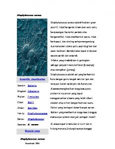
plex DNA. Since penicillinase plasmids are associated with the presence of circular duplex DNA (8, 12), we attempted to demonstrate such DNA in cleared lysates of transformants selected for plasmid markers. To control for possible artifacts of the preparative procedure, we prepared lysates from mixtures of separately labeled cultures. In each case, the transformant was labeled with 3H-thymidine, and the plasmid-positive donor strain, which served as a control, was labeled with "4C-thymidine (Fig. 2). Five of the transformants that received only the ero marker were tested, and none had detectable circular duplex DNA (Fig. 2A). The analytical technique is sufficiently sensitive to have detected circular duplex DNA amounting to 0.5% of that present in the plasmid-positive control (in other words, 200,000 atomic mass units/cell), and so it seems safe to conclude from these experiments that the erythromycin marker does not exist in these transformants as an autonomous circular duplex plasmid. Of the CadR transformants characterized in Table 3, however, all five showed typical circular duplex DNA bands in dye-buoyant density gradients as exemplified in Fig. 2B. Transformation with purified plasmid DNA. We have shown that transformation and transduction of plasmid markers are generally accompanied by the appearance of a character-
istic circular duplex DNA species in transformed clones. To establish definitively that the plasmid markers belong to this circular DNA, it is necessary to demonstrate specific transformation of these markers and no others upon purification of the DNA. In the experiments reported here, two different methods were used to obtain relatively pure circular duplex DNA from plasmid-positive S. aureus. One of the techniques is based on the reversible denaturation of circular duplex DNA at alkaline pH and irreversible denaturation of other DNA species at the same pH. After return to neutrality, the preparation was centrifuged to remove precipitated debris, desalted by chromatography on Sephadex G-100, and finally fractionated on nitrocellulose which separates native from denatured DNA. We used this technique to isolate circular duplex plasmid DNA from two strains of S. aureus harboring, respectively, X1 lde and T,l9. lde (mol wt 24 x 106) (12) is a high-frequency transducing element in which a large segment of the plasmid genome PI2.8 has been replaced by a section of the genome of the transducing phage 41l (7). The plasmid moiety includes the ero marker and a segment which is responsible for plasmid compatibility and maintenance and is required for plasmid replication. T1,,, is a plasmid of mol wt 2.66 x 10⁶ (7), whose only known marker is for tetracycline resistance. TABLE 2. Transformation of the plasmid markers Table 4 shows the results of transformaerythromycin (ero) and cadmium (cad) resistance by tion experiments with plasmid DNA isolated DNA from strain 8325 (PI258)a and purified by this method. The wild-type EroR transExpt Recipient CadR trans8325 and the nuc- mutant of 8325 were no. formants/ml formants/ml used as recipients in these experiments with erythromycin and tetracycline resistance as 1 8325 1.1 X 10³ 10 selective markers. Two Ero- and two Tet2 8325 nuc7 x 102 17 resistant transformants were isolated and la3 8325 nuc 1.4 x 101 7 4 8325 nuc8.7 x 102 13 beled with 3H-thymidine. Control cultures of 5 8325 nuc 2.2 x 10³ 23 the donor strains were labeled with "4C-thymidine, and lysates were prepared from mixtures aEach transformation mixture contained 10 ,ug of of donor cultures and respective transformants. donor DNA in a volume of 1 ml. TABLE 3. Co-transformation of resistance markers from plasmid PI2.8 among selected cadmium resistant transformants isolated from different experimentsa Resistance pattem Isolate no.5
1 2 3 4 5
Recipient
8325 8325 nuc 8325 nuc8325 nuc8325 nuc-
ero
asi
asa
pen
lea
cad
mer
R R R R S
S S S S R
S S S S
R R R R R
R R R R R
R R R R R
R R R R R
R
a Donor strain 8325(PI 6.) has the following resistance pattern: erythromycin (ero), arsenite (asi), arsenate (asa), penicillin (pen), lead (lea), cadmium (cad), and mercury (mer) resistant. R, Resistant; S, sensitive. bIsolate number corresponds to experiment number in Table 2.
142
LINDBERG AND NOVICK a
tr
35 _
,,
30
Ln
25
J . BACTrERIOL.
PA
B
3 20 F 15 4
cz
10
z w
S Gj
5
10
1S
20
25
30
10
15
20
30
25
FRACTION NUMBER
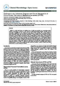
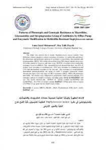
FIG. 1. Map of PI258. This map has been constructed from deletion (6) and recombination data (Novick and Brodsky, unpublished data). In addition to resistance markers, asa (arsenate), asi (arsenite), ant (antimony), ero (erythromycin), mer (mercury), cad (cadmium), bis (bismuth), lea (lead), and pen (penicillin, penicillinase), there are two control loci for penicillinase, penI and penB (11) as well as the structural locus, penZ, and mcr/seg, a locus involved in plasmid maintenance, compatibility, and replication. Markers in parentheses have not been ordered with respect to their near neighbors. Outside the main map are two plasmid deletions generated during transformation (see Table 3).
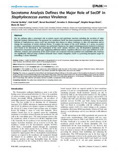
The mixed lysates were centrifuged to equilibrium in cesium chloride density gradients with ethidium bromide (Fig. 3). In all four tested transformants circular duplex DNA was detected as in the donor strains. The mixed cleared lysates were also subjected to band sedimentation in neutral sucrose gradients (10-30%). A major peak corresponding to the circular duplex plasmid DNA peak of the respective donor strain was obtained from all tested transformants (Fig. 4), showing that the transformed plasmid was essentially the same size as the donor plasmid from which it was derived. The minor peaks observed in these gradients are of uncertain origin (see Fig. 4 legend). The recipients in the transformation experiments are lysogenic for phages sll, 012, and 413. Phage 41l functions as a helper phage in the production of transducing particles from cells in which llde is established (7). This means that, in a culture medium from cells lysogenic for phage 441 and harboring 4llde, there is always production of transducing parti-
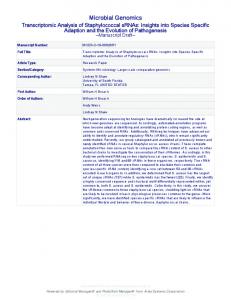
FIG. 2. A, Elution profiles of preparative cesium chloride density gradients of DNA from mixed cleared Iysates of the plasmid harboring strain RN492 (0, " C-labeled) and a .transformant of 8325 (0, 3Hlabeled) obtained after transformation with bulk DNA from strain 8325 (PI258) and initial selection for erythromycin resistance. Centrifugation was carried out at 20 C for 36 h at 42,000 rpm in a Spinco Ti 50 rotor in the presence of ethidium bromide (200 ,g/ml). Density increases from right to left in this and the succeeding diagrams. In each case, the denser peak contains the circular duplex DNA, which usually amounts to about 1% of the total in plasmid-carrying strains. The lighter peak contains plasmid and chromosome fragments and any linear or open circular plasmid molecules present. The data are plotted as percents of total counts recovered from the gradients. B, Same as (A) except that the tested strain (0, 3H-labeled) was a transformant of 8325 obtained after initial selection for cadmium resistance (isolate no. 1 in Table 2). TABLE 4. Transformation with circular duplex DNA isolated by selective renaturation from strains RN455 (carrying 011de) and RN1304 (carrying T,69), respectively Transformants/ml Recipient
DNA
EroR
8325 8325 nuc 8325 8325 nuc-
411dea /llde
T169b T.6,
TetR
127 217 30 17
aFinal concentration of DNA in transformation mixture = 5 ,g/ml. "Final concentration of DNA in transformation mixture = 4 ,g/ml.
cles due to the spontaneous release of 011. Five 44 lde transformants from each of the two recipients were, therefore, tested for their production of transducing particles. Culture supernatants sterilized by membrane filtration (Millipore Corp were titrated for plaque-forming activity and for ero-transducing activity. In all
Ccw
030
u0w
25 !5
w
m
143
S. AUREUS PLASMID-SPECIFIC TRANSFORMATION
VOL. 115, 1973 A
8
.2 Z) 0
20 T
-
-C
15 15
-
0
10 10
u I
w
11 z
s5
w u m w
IL
5
.. 10
1..5
1. 20
.. 1. 30 25 5 FRACTION NUMBER
10
IS
20
1.
1. 25
30
1.
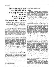
FIG. 3. A, Elution profiles of preparative cesium chloride gradients of DNA from mixed cleared lysates of strain RN455 (0, "4C-labeled) harboring plasmid '11de and a transformant of 8325 nuc (0, 'Hlabeled) obtained after transformation by prepared circular DNA of plasmid 'lide and selection for erythromycin resistance. Other details as under Fig. 1A. B, Same as (A) except that the two strains were RN1304 (0, "4C-labeled) harboring plasmid T,., and a transformant of 8325 nucr (0, 3H-labeled) obtained after transformation with prepared T5,6-DNA and selection for tetracycline resistance. 5
transducing titer was between 2 and 60% of plaque-forming titer, which is typical for 4llde strains. In addition, 441 lysates were prepared from two of the EroR transformants by UV irradiation. These lysates had Hft activity typical of llde (Table 5); therefore, the transformed plasmids were functionally as well as structurally indistinguishable from the donor 4llde plasmid. In two preliminary experiments, we also tested the transforming activity of DNA isolated from 4llde- and T169-harboring strains after centrifugation in cesium chloride gradients in the presence of ethidium bromide. After removal of ethidium bromide followed by dialysis, we found that plasmid characters could be transformed with DNA from both the heavy peak containing closed circular plasmid molecules and from the light peak which contained chromosomal fragments and any linear or nicked plasmid molecules present in the preparations (Table 6). The thymine-requiring mutant, however, could be transformed to thymine independence only with DNA from the light peak. For some reason tetracycline resistance was transformed at very low frequency with T1l. DNA prepared by this method as well as by the alkali denaturation method (Table 5). However, in controls without DNA no spontaneous TetR mutants were obtained; therefore we are confident that the TetR colonies obtained represent true transformants. The transformation frequencies obtained with these preparations were low because very little DNA was recovered. The cases,
15 20 10 FRACTION NUMBER
25
30
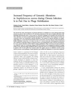
FIG. 4. Band sedimentation of cleared lysates in neutral sucrose. A 0.3-mi sample was sedimented through 5-ml 10 to 30%o neutral sucrose gradients containing 1 M NaCl and 50 mM EDTA at pH 7.5. Centrifugation was at 20 C and 45,000 rpm in a Spinco SW 65 rotor. Sedimentation was from right to left. A, Sedimentation profile for the lysate used in the experiment illustrated in Fig. 3A. Centrifugation was for 130 min. The main peak is the 54S circular duplex form of 'lide with a mol wt of 2.4 x 10⁷ (12). The nature of the material in the smaller peak (sedimenting at 100S) is unknown. B, Sedimentation profile for the lysate used in the experiment illustrated in Fig. 3B. Centrifugation was for 300 min. The main peak in this case is the 20S circular duplex form of T1*, with a mol wt of 2.7 x 10⁶ (8). The smaller peak (sedimenting at 13S) could be the open circular form of this plasmid. TABLE 5. Transducing ability of UV-induced lysates from transformants isolated after transformation with 011de DNA and selection for erythromycin resistance Strain PFU"/ml FUII/ml Strain
8325 8325 nuca PFU,
7 x 10⁶ 2 x 10⁶
EroR trans-
Trans-
ductants/mi
ductants/PFU
7.1 x 10⁶ 1-2 3.5 x 10⁶ 1.75 x 10⁻²
Plaque-forming units.
precise amount in this case is unknown because contaminating UV-absorbing material prevented accurate estimate by UV absorption.
144
LINDBERG AND NOVICK
TABLE 6. Transformation with DNA from strains RN455 and RN1304 isolated by centrifugation in cesium chloride gradients in presence of ethidium bromide Transformants/ml
Recipient
8325 nuc-
Donor
RN455a RN4555 RN13045 8325(PI258)thy- RN455b 8325(PI258)thy- RN1304`
8325-4(011) 8325-4(011)
Selective DNA marker from heavy peak
ero ero tet
thy+ thy+
43 60 13 20c 23c
DNA from light peak
103 87 10 173 143
aEthidium bromide removed by passage through Dowex 50 column. 'Ethidium bromide removed by extraction with n-butanol. c Equal to frequency of spontaneous reversion.
J. BACTERIOL.
possible reason for the differences seems instructive. The penicillinase plasmid, PI258, is a circular DNA molecule with a molecular weight of approximately 18 x 106 atomic mass units (12). It has been mapped by a combination of genetic and physical techniques (8), and we should like to emphasize two features of the map (Fig. 1) which seem significant in connection with these results. These are: (i) that there is only one region of the plasmid (mcr-seg, located between the markers ero and mer) that is essential for its survival as an autonomous replicon; and (ii) that the cad locus is relatively far from this region (perhaps 1800). With the above-mentioned bulk DNA, we obtained approximately 100 times as many transformants when initial selection was for erythromycin resistance as when selection was for cadmium resistance (Table 2). Moreover, in the EroR transformants, ero was the only detectable plasmid marker, and circular duplex plasmid DNA could not be demonstrated. We suggest, therefore, that the ero marker was carried on plasmid fragments rather than on intact plasmid molecules and that it has in these transformants become integrated into the host chromosome. The high frequency of this integration indicates a considerable degree of homology between the ero region of the plasmid and a special region(s) of the staphylococcal chromosome. In contrast, selection for cadmium resistance always resulted in the cotransformation of other plasmid markers and in the establishment of the incoming plasmid in its normal autonomous state (Table 3 and Fig. 2B). All of the CadR transformants examined, however, apparently carried small deletions involving unselected plasmid markers. Thus, it appears that transformation of the cad locus may also have involved broken, presumably linear plasmid molecules. The transformation results with these two markers are strongly reminiscent of a series of earlier transduction experiments in which plasmid deletions were generated by UV irradiation of the transducing phage (6). Whereas selection for cadmium resistance always resulted in the transduction of autonomous plasmids, with or without deletions of unselected markers, selection for erythromycin resistance gave rise, in addition to transductants carrying autonomous plasmids, to a class in which the ero locus was the only plasmid marker transduced. The properties of these transductants suggested that the ero locus was integrated into the host chromo-
DISCUSSION Transformation results with circular duplex DNA prepared from plasmid-carrying strains by alkali denaturation and selective renaturation or by dye-buoyant density centrifugation were clear and unequivocal. Plasmid markers, but not chromosomal markers, could be transformed by this material, and in all of the transformants tested the transformed markers became established as autonomous plasmids that were biologically and physically indistinguishable from those carried by the donor strains. As DNA from the less dense peak in dye-buoyant density gradients prepared from cleared lysates of plasmid-carrying strains was also active in the transformation of plasmid markers, we conclude that open circular DNA molecules (and possibly linear ones as well) are acceptable. Because preparations of circular duplex DNA always contain some open circular molecules, we are unable to draw any conclusion as to whether circular duplex molecules are active in transformation as such. As Cohen et al. (2) found a somewhat higher efficiency of transformation for closed than for open circular plasmid DNA, it seems likely that closed circular molecules are active as such in E. coli. Further studies on the relative transforming efficiencies of the various DNA allomorphs should settle this question for S. aureus. Transformation with bulk DNA prepared by phenol extraction from strain RN453 (carrying the penicillinase plasmid, P1258) gave results rather different from those obtained with purified plasmid DNA, and a consideration of the some.
VOL. 115, 1973
S. AUREUS PLASMID-SPECIFIC TRANSFORMATION
145
Since integration of fragments carrying only grants GM-14372 and K04-GM-12252 (to R.N.) from the the cad locus has not been observed, either in National Institute of General Medical Sciences. transformation or in transduction experiments, LITERATURE CITED we conclude that integration of this locus must be quite rare. Therefore, it appears that only 1. Clewell, D. B., and D. R. Helinski. 1969. Supercoiled circular' DNA-protein complex in Escherichia coli: intact plasmids or fragments that could be purification and induced conversion to an open circular converted to functional autonomous plasmids DNA form. Proc. Nat. Acad. Sci. U.S.A. 62:1159-1166. in the recipient gave rise to CadR transfor- 2. Cohen, S. N., A. C. Y. Chang, and L. Hsu. 1972. Nonchromosomal antibiotic resistance in bacteria: gemants. If so, such fragments must have fulfilled netic transformation of Escherichia coli by R-factor two conditions: the mcr-seg region must have DNA. Proc. Nat. Acad. Sci. U.S.A. 69:2110-2114. been included (Fig. 1) and the molecule must 3. Hudson, B., D. A. Clayton, and J. Vinograd. 1968. have been able to undergo circularization in the Complex mitochondrial DNA. Cold Spring Harbor Symp. Quant. Biol. 33:435-442. recipient. Presumably the difference in fre4. Lindberg, M., J. E. Sj6strom, and T. Johansson. 1972. quency of transformation of the two markers Transformation of chromosomal and plasmid characreflects the relative rarity in phenol-extracted ters in Staphylococcus aureus. J. Bacteriol. bulk DNA of molecules carrying cad that were 109:844-847. able to fulfill these two conditions and the 5. Novick, R. P. 1963. Analysis by transduction of mutations affecting penicillinase formation in Staphylococrelative ease of establishment by integration of cus aureus. J. Gen. Microbiol. 33:121-136. fragments carrying ero. 6. Novick, R. P. 1967. Penicillinase plasmids of StaphSince we have not yet completed a series of ylococcus aureus. Fed. Proc. 26:29-38. transformations involving purified PI258 DNA, 7. Novick, R. P. 1967. Properties of a cryptic high-frequency transducing phage in Staphylococcus aureus. Virology the above conclusions must remain tentative. 33:155-166. However, the results with 4llde suggest that, 8. Novick, R. P., and D. Bouanchaud. 1971. Extrachromowhere incoming plasmid molecules have a somal nature of drug resistance in Staphylococcus aureus. Ann. N.Y. Acad. Sci. 182:279-294. choice of recombining with the resident chromo9. Novick, R. P., and C. Roth. 1968. Plasmid-linked resistsome or becoming established as autonomous ance to inorganic salts in Staphylococcus aureus. J. plasmids, they choose overwhelmingly the latBacteriol. 95:1335-1342. ter course. Since the recipients are 41l lysogens 10. Radloff, R., W. Bauer, and J. Vinograd. 1967. A dyebuoyant density method for the detection and isolation and 4llde is probably composed of mostly of closed circular duplex DNA: the closed circular DNA phage genome, the probability of integration, in HeLa cells. Proc. Nat. Acad. Sci. U.S.A. based purely on considerations of homology, 57: 1514-1521. would seem to be quite high. Yet all of the five 11. Richmond, M. H. 1967. A second regulatory region involved in penicillinase synthesis in Staphylococcus 4llde transformants tested had autonomous aureus. J. Mol. Biol. 26:357-360. lde plasmids indistinguishable physically M. G., C. N. Gordon, R. P. Novick, and R. C. and biologically from the donor 1l1de plasmid. 12. Rush, Wamer. 1969. Penicillinase plasmid DNA from StaphACKNOWLEDGMENTS This study was supported by research grants from the Swedish Medical Research Council and from the Swedish Cancer Society (to M.L.), and by Public Health Service
ylococcus aureus. Proc. Nat. Acad. Sci. U.S.A. 63: 1304-1310. 13. Sjostrom, J. E., M. Lindberg, and L. Philipson. 1973. Competence for transfection in Staphylococcus aureus. J. Bacteriol. 113:576-585.