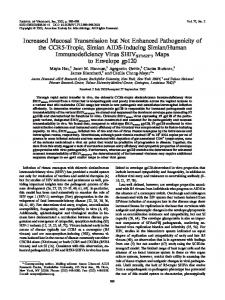
JOURNAL OF VIROLOGY, Apr. 1997, p. 3263–3267 0022-538X/97/$04.0010 Copyright q 1997, American Society for Microbiology
Vol. 71, No. 4
Polyclonal Bovine Sera but Not Virus-Neutralizing Monoclonal Antibodies Block Bovine Leukemia Virus (BLV) gp51 Binding to Recombinant BLV Receptor BLVRcp1 OTO ORLIK,1 JOZEF BAN,2 JURAJ HLAVATY,2 CESTMIR ALTANER,2 RICHARD KETTMANN,3 DANIEL PORTETELLE,3 AND GARY A. SPLITTER1* Department of Animal Health and Biomedical Sciences, University of Wisconsin—Madison, Madison, Wisconsin 537061; Department of Molecular Virology, Cancer Research Institute, Slovak Academy of Sciences, 812 32 Bratislava, Slovak Republic2; and Departments of Molecular Biology 2 and Microbiology 3, Faculty of Agronomy, B-5030 Gembloux, Belgium3 Received 19 August 1996/Accepted 26 December 1996
Bovine leukemia virus (BLV), a transactivating lymphotropic retrovirus, is the etiologic agent of enzootic lymphosarcoma or leukemia in cattle. Sera from BLV-infected animals possess high BLV-neutralizing antibody titres. The availability of the recombinant BLV receptor candidate, BLVRcp1, allowed us to determine a mechanism of virus neutralization by polyclonal sera and monoclonal antibodies (MAbs). Bovine sera from animals naturally infected with BLV blocked gp51 binding to recombinant BLVRcp1. In contrast, virusneutralizing MAbs specific for gp51 F, G, and H epitopes did not prevent gp51-receptor attachment. Furthermore, gp51 neutralization epitopes F, G, and H were accessible to antibodies following gp51 attachment to BLVRcp1. This finding implies that virus neutralization by MAbs to defined BLV gp51 epitopes can occur subsequent to virus engagement of the receptor while polyclonal sera can specifically block virus attachment to the receptor. In conclusion, these data suggest that cell infection by BLV is a multistep process requiring receptor binding (inhibited by polyclonal sera) followed by a second, postbinding event(s) at the cell membrane (inhibited by anti-gp51 MAbs). cuted by antibodies can be a key host factor, inducing virus latency, absence of viremia, and a long incubation period prior to tumor occurrence. Immunological analysis using monoclonal antibodies (MAbs) revealed two immunodominant B-cell epitope regions on the main structural protein, p24, of BLV (29). Similar analysis of envelope glycoprotein gp51 characterized eight distinct epitopes (11), and two additional epitopes were detected later (28). Linear epitopes A, B-B9, D, and E are not neutralizing and are localized near the carboxy-terminal end of gp51 (6, 14). Epitopes F, G, and H are conformational and virus neutralizing and localized at the amino-terminal end of gp51 (32, 33). Epitopes C, C1, and C2 are conformational, without virus-neutralizing activity (11, 28). Recently, a cDNA encoding the putative BLV receptor, BLVRcp1, was isolated and characterized (7, 8). Transfection of the BLVRcp1 cDNA into nonpermissive NIH 3T3 cells conferred sensitivity to BLV infection, and infection of transfected cells was prevented by anti-gp51 neutralizing MAbs to epitopes F, G, and H. BLVRcp1 cDNA encodes a 94-kDa protein in MDBK cells, and its chromosome localization in several species was determined (30); however, the physiological function of this protein has not been determined. The availability of putative BLV receptor BLVRcp1 provided the opportunity to study the engagement of viral gp51 with its receptor and investigate the mechanism of virus neutralization by polyclonal sera and MAbs. Recombinant BLVRcp1 preparation and characterization. To prepare recombinant receptor, the BLVRcp1 EcoRI-XbaI fragment (7) was inserted into the plasmid pMAL-c2 (New England BioLabs, Beverly, Mass.). Transformation of Escherichia coli DH5a resulted in expression of a fusion product containing maltose binding protein (MBP). Bacterial lysates of isopropylthiogalactoside-induced bacterial cells from the clone MBP-BLVRcp1, which is MBP containing BLVRcp1 cDNA,
Bovine leukemia virus (BLV) is a lymphotropic retrovirus of cattle that is closely related to human T-cell lymphotropic viruses 1 and 2 and causes enzootic lymphosarcoma/leukemia in a small percentage (0.6 to 5%) of infected animals. Up to 30% of infected animals develop persistent lymphocytosis, a benign and permanent increase in circulating B lymphocytes; however, these animals remain asymptomatic (21, 36). Seroconversion and lifelong presence of antibodies are the only indications of infection in the vast majority of infected animals (9, 18). Antibody response to BLV infection can be detected as soon as 2 weeks after infection, and antibody titer increases can be detected with time (24). BLV structural proteins, predominantly p24 and gp51, are targets for antibody response, and detection of antibodies is routinely used for diagnosis of BLV infection (5, 34). Antibodies to the transactivating protein Rex can be detected intermittently, and their occurrence likely correlates with episodes of Rex release from dead (or killed) infected cells (35). Antibodies from infected animals are virus neutralizing in vesicular stomatitis virus-BLV pseudotype neutralization and syncytium inhibition assays (4, 41) and mediate lysis of infected cells by complement fixation (31). Humoral immunity likely plays an important role in protection from progression of BLV infection. Passive immunization with antiBLV antibodies conferred protection from BLV infection (22). Resistance to BLV challenge after vaccination of sheep with gp51 antigen (27) and cattle with cells producing BLV envencoded glycoproteins and p24 (2) correlated with the presence of virus-specific antibodies. Despite the fact that tumors develop in the presence of antibodies, selection pressure exe* Corresponding author. Mailing address: Department of Animal Health and Biomedical Sciences, University of Wisconsin—Madison, 1655 Linden Dr., Madison, WI 53706. Phone: (608) 262-1837. Fax: (608) 262-7420. E-mail:
[email protected]. 3263
3264
NOTES
FIG. 1. Characterization of recombinant fusion product of BLVRcp1. (A) Bacterial proteins were separated by SDS-PAGE on a 10% gel and visualized by staining with Coomassie blue. MBP-BLVRcp1, bacterial lysate containing MBPBLVRcp1 fusion protein; Mal-MBP, control lysate. The arrow indicates the overexpressed, 120-kDa MBP-BLVRcp1 recombinant protein. (B) Western blot of bacterial lysates detected with rabbit antiserum R70 raised against amino acids 25 to 36 of BLVRcp1. Arrows indicate specific binding to the full-length, 120kDa MBP-BLVRcp1 and its 75-kDa degradation product. (C) Receptor binding assay on MBP-BLVRcp1 lysate detected by a pool of MAbs to gp51 epitopes A to H. BLV, tissue culture fluid from virus-producing FLK cell line containing disrupted BLV; gp51, BLV gp51 purified by affinity chromatography. Arrows indicate gp51 binding to the full-length, 120-kDa MBP-BLVRcp1 and its 75-kDa degradation product.
and the mock-transfected clone Mal-MBP were analyzed for expression of recombinant protein. Comparison of electrophoretic profiles in Coomassie-stained sodium dodecyl sulfatepolyacrylamide gel electrophoresis (SDS-PAGE) gels revealed the presence of a recombinant product with the predicted size 120 kDa in the lysate MBP-BLVRcp1 but not in the lysate Mal-MBP (Fig. 1A). To determine the antigenic authenticity of the recombinant MBP-BLVRcp1 fusion product, a rabbit antiserum, R70, raised against amino acids 25 to 36 of BLVRcp1, was used (7). In Western blot analysis, R70 diluted 20,0003 followed by incubation with secondary alkaline phosphatase (AP) conjugate and chromogenic substrate nitroblue tetrazolium chloride (NBT)–5-bromo-4-chloro-3-indolylphosphate p-toluidine salt (BCIP) specifically recognized a 120-kDa fusion product and its 75-kDa degradation product in the MBP-BLVRcp1 lysate. Bacterial lysate Mal-MBP did not react with the peptide antiserum R70 (Fig. 1B). Preimmune rabbit serum R70 did not bind to MBP-BLVRcp1 (data not shown). Furthermore, MBP-BLVRcp1 protein was tested in a receptor binding assay for binding of affinity-purified gp51 (25) or gp51 from FLK-VP-1 (ovine) (1), R(BLV) (rat), (3), BK(BLV) (bovine), or B/BLV (bat) cell line supernatants containing virions disrupted by 0.1% N-octylglucoside. Nitrocellulose membranes blotted with MBP-BLVRcp1 lysate were blocked with 5% nonfat milk in phosphate-buffered saline and incubated with purified gp51 or disrupted virions in TBS (10 mM Tris-HCl [pH 7.4], 150 mM NaCl, 0.05% Tween 20) for 16 h at 48C. Glycoprotein gp51 binding to the recombinant receptor was detected with a pool of MAbs to gp51 epitopes A to H (11, 28) followed by anti-mouse AP-labelled secondary antibody and chromogenic substrate NBT-BCIP. As shown in Fig. 1C, full-length, 120-kDa fusion recombinant MBP-BLVRcp1 as well as its 75-kDa degradation product bound gp51. Recombinant MBP-BLVRcp1 degradation was difficult to control, and various degradation patterns were observed with different lysate preparations. The bacterial lysates used for experiments in
J. VIROL.
FIG. 2. Electrophoretic analysis of purified MBP-BLVRcp1 subjected to SDS-PAGE (10% polyacrylamide) and stained with Coomassie blue. MBPBLVRcp1, full-length, purified fusion MBP-BLVRcp1 protein; Xa digested, MBP-BLVRcp1 protein after digestion with factor Xa. The arrow indicates recombinant BLVRcp1 protein separated from MBP.
Fig. 1, 3, and 5 were different. Bacterial lysate with the least degradation was observed in Fig. 5, in which only full-length, 120-kDa MBP-BLVRcp1 bound gp51. Two additional degradation products, 25 and 75 kDa, bound gp51 in the lysates shown in Fig. 3 or Fig. 1, respectively. BLV gp51 from all sources tested bound recombinant fusion BLVRcp1. Binding of gp51 to nitrocellulose strips with Mal-MBP lysate was not detected (data not shown). Next, we purified the recombinant fusion product from bacterial lysate by affinity chromatography on amylose resin columns (New England BioLabs). However, binding to the column was poor and several degradation products were copurified (data not shown). Therefore, nondegraded, fulllength MBP-BLVRcp1 protein was recovered after excision and electroelution of the 120-kDa band from SDS-PAGE gels (19). Specific cleavage of the fusion protein with factor Xa resulted in the appearance of two expected major protein bands, 75 and 42 kDa, corresponding to BLVRcp1 protein and MBP, respectively (Fig. 2). However, digestion with factor Xa was incomplete, with a minor, 120-kDa-band residue and nonspecific cleavage resulted in production of several minor degradation bands of 50, 60, and 66 kDa, while the 35-kDa band corresponded to factor Xa. Because purified and digested BLVRcp1 yielded small amounts and degraded protein, MBPBLVRcp1 bacterial lysate was used for BLVRcp1-gp51 blocking experiments. Inhibition of BLVRcp1-gp51 binding by MAbs. Previously, the amino-terminal portion of BLVRcp1 protein, encoded by the SmaI-SacI DNA fragment, was demonstrated to be crucial for gp51 binding and comprises the receptor binding site (7). However, the receptor binding site on gp51 has not been clearly identified. Therefore, we tested whether some of the previously characterized gp51 epitopes A to H might comprise the gp51 receptor binding domain (11, 28). Eleven individual gp51-specific MAbs or smaller pools of MAbs were tested to detect gp51 bound to BLVRcp1. Surprisingly, binding of gp51 to the 120-kDa, full-length MBP-BLVRcp1, as well as the 25-kDa degradation product was detected by pools of MAbs as well as all individual anti-gp51 MAbs (Fig. 3). Binding to the receptor was detected by antibodies only with addition of gp51,
VOL. 71, 1997
NOTES
3265
FIG. 4. Neutralization of gp51-BLVRcp1 binding by neutralizing MAbs. Medium containing gp51 was preincubated with individual MAbs prior to incubation with the BLVRcp1 blot. gp51-MAb immune complexes were detected by secondary AP-conjugated anti-mouse secondary antibody and chromogenic substrate. Letters F to H designate the epitope specificities of the gp51 neutralizing MAbs. MAb BLVp24-X48 was used as a control antibody.
FIG. 3. Virus binding assay using MBP-BLVRcp1 lysate detected by individual anti-gp51 MAbs and pools of MAbs. Nitrocellulose strips were incubated in tissue culture fluid containing (1) or lacking (2) gp51 from disrupted BLV. Bound MAbs were visualized by reaction with AP-conjugated anti-mouse secondary antibody and NBT-BCIP chromogenic substrate. Individual letters A to H indicate gp51-epitope specific MAbs used for detection. A,B and C,FG,H are pools of MAbs. MAb BLVp24-X48 possessing BLV anti-p24 specificity was used as a negative control (Neg). Arrows indicate gp51 binding to the full-length, 120-kDa MBP-BLVRcp1 and a 25-kDa degradation product.
confirming the specificity of the reactions. Protein bands detected on Western blots incubated without BLV represent nonspecific binding of relevant MAbs to bacterial lysate, particularly evident with MAb to epitope E (Fig. 3). Control MAb to BLV p24 (29) did not react with bound gp51. These data indicated that none of the gp51 epitopes A to H are directly involved in the receptor binding site and that virus-neutralizing epitopes F, G, and H were always exposed after gp51 receptor engagement. This finding was unexpected, because MAbs to these epitopes are virus neutralizing and inhibition of virusreceptor interaction was originally proposed as the mechanism of virus neutralization by MAbs (11). However, our data indicate that this explanation may not be correct. Furthermore, a pool of MAbs to conformational epitopes was superior for detecting BLVRcp1-bound gp51 (Fig. 3). The finding that virus-neutralizing MAbs can bind to receptor-bound gp51 is directly applicable in understanding the action of virus-neutralizing MAbs against gp51. Maintaining accessibility of neutralization epitopes F, G, and H after gp51 binding to the receptor indicates that neutralizing MAbs elicit their function following virus attachment to the receptor. To dissect the mechanism(s) of virus neutralization by MAbs, MAbs recognizing gp51 F, G, and H epitopes were tested for blocking gp51 binding to BLVRcp1 in the receptor binding assay. Medium containing gp51 was preincubated overnight with individual MAbs followed by incubation with the BLVRcp1 blots and detection using a pool of anti-gp51 MAbs. As predicted, based on our data shown in Fig. 3, none of the neutralizing MAbs prevented gp51 binding to the receptor BLVRcp1. Furthermore, the gp51-MAb complex itself bound to BLVRcp1 (Fig. 4). In these experiments, receptor bound-gp51 was directly detected with secondary anti-mouse AP-conjugate omitting the incubation with the anti-gp51 MAb pool. Immune complexes formed by gp51 and MAbs to neutralizing epitopes F, G, and H bound to 120-kDa MBPBLVRcp1 were detected by the secondary antibody. The control antibody to p24 did not produce immune complex binding to recombinant receptor (Fig. 4). However, when an anti-gp51 MAb pool was used prior to the secondary antibody the 120kDa band was observed, confirming the specificity of these
experiments (data not shown). Thus, we conclude that virusneutralizing MAbs were not blocking antibodies and that their neutralizing activity likely involves postbinding events in the process of cell infection. This finding is in agreement with a modeling study suggesting that the three loops in the head of gp51, proposed as the potential interaction site with its cellular receptor, did not contain F, G, and H epitopes (15). Additional postbinding events important for cell infection may include gp51 interaction with other functionally important segments of the receptor, a coreceptor molecule, proteolytic cleavage of the viral glycoprotein (10, 16), or proper exposure of the transmembrane protein gp30, which is required for fusion of virus and cell membrane (39). Similar to those neutralizing BLV, human immunodeficiency virus type 1 (HIV-1)-neutralizing MAbs specific to the gp120 V3 loop effectively blocked cell fusion and virus infectivity independent of gp120-CD4 binding (20). Also, neutralizing MAbs against Epstein-Barr virus prevented virus infectivity independent of receptor engagement (38). Blocking of BLVRcp1-gp51 binding by polyclonal sera. Bovine sera from BLV-infected or -immunized cattle possess virus-neutralizing activity (2, 4). To determine the mechanism of virus neutralization, bovine sera from seropositive animals naturally infected with BLV were tested for blocking gp51BLVRcp1 attachment. The anti-gp51 antibody titers of tested sera, determined by enzyme-linked immunosorbent assay (ELISA) (5), are shown in Table 1. Nonspecific binding of bovine sera to MBP-BLVRcp1 lysate was removed by adsorption on Mal-MBP lysate and nondisrupted E. coli. Medium containing gp51 was preincubated overnight at 48C with individual sera followed by incubation with the BLVRcp1 blots and detection of receptor-bound gp51 with a pool of gp51 MAbs and a secondary AP conjugate. Interestingly, in contrast to neutralizing MAbs, preincubation of gp51 with BLV-positive bovine sera prevented gp51 binding to BLVRcp1, as deTABLE 1. Antibody titers of bovine sera Animal no.
10 AB1 75 350 750c
Antibody titer a
ELISA
Blockingb
3,200 6,400 1,600 3,200 0
.800 200 50 200 0
a The ELISA titer is defined as the inverse value of the last serum dilution at which the optical density in the well with captured gp51 antigen is double that in the well with capturing MAb only. b The blocking titer, determined by receptor binding assay, is defined as the inverse value of the last serum dilution which completely blocks gp51 binding to BLVRcp1. c BLV-noninfected animal.
3266
NOTES
J. VIROL.
alternative BLV receptor as an explanation for failure of neutralizing MAbs to block gp51 binding to BLVRcp1. Unfortunately, four MAbs prepared to BLVRcp1 did not block gp51 binding to BLVRcp1 (data not shown); however, development of blocking anti-BLVRcp1 MAbs could dissect the possibility that other molecules can participate in BLV attachment in different cell types. This research was supported by the College of Agricultural and Life Sciences, by grants from the National Institutes of Health (RO1.CA 59127), BARD (95-34339-2556), and the Slovak Academy of Sciences, by the Fogarty International Center, (NCI, Bethesda, Md.), by the Belgian Services Federaux des Affaires scientifiques, techniques et cultures (SSTC), and by the Fonds Cancerologique de la C.G.E.R. REFERENCES
FIG. 5. Bovine serum blocking of gp51 binding to recombinant BLVRcp1. BLV gp51 was preincubated with bovine sera before addition to BLVRcp1 nitrocellulose strips. Detection of gp51 bound to MBP-BLVRcp1 was performed with a pool of anti-gp51 MAbs followed by an AP-conjugated anti-mouse secondary antibody and chromogenic substrate. Serum ID numbers are the same as animal numbers; serum dilutions are given as fold values. The arrow indicates gp51 bound to the 120-kDa MBP-BLVRcp1 protein.
duced by an absence of the 120-kDa band in some samples (Fig. 5). Bovine sera differed remarkably in blocking activity, and the blocking titers ranged from 50 in animal 75 to more than 800 in animal 10 (Table 1). To test the possibility that BLVRcp1-bound gp51 cannot be detected by MAbs (Fig. 5) because bovine antibodies occupied available gp51 epitopes, blots with missing bands were stained with AP-labelled antibovine secondary antibody. No bovine antibodies were detected by Western blot, confirming the specificity of the previous assays and the ability of bovine sera to block gp51 binding to receptor (data not shown). BLV-negative sera from animals 833 and 750 did not prevent binding of gp51 to BLVRcp1, and a strong 120-kDa band was detected (Fig. 5, animal 750). Blocking of gp51 binding to BLVRcp1 indicates that BLVpositive sera besides those with antibodies to F, G, and H epitopes (12) contain antibodies recognizing a receptor binding site (hypothesized to be just below the large globular head of gp51, as described in reference 15). Alternatively, these sera may induce conformational changes on gp51 that prevent receptor attachment. Similarly, serum samples from HIV-1-infected humans possess high titers of neutralizing antibodies that block gp120-CD4 binding (17). Since the blocking titers but not the ELISA titers of bovine sera differed greatly among animals, the gp51 binding site most likely is not a prominent antigenic region of gp51, and low-titer sera may not possess blocking activity. In fact, polyclonal sera developed in animals immunized with HIV-1 gp160 peptide displayed potent virusneutralizing activity but failed to block gp120-CD4 binding (37). Our preliminary experiments indicate that soluble BLVRcp1 protein blocks infection of OVK, an ovine kidney cell line, and MDBK cells by recombinant BLV (data not shown). Similarly, several soluble viral receptors, such as CD4 for HIV, soluble intercellular adhesion molecule 1 for rhinoviruses, and CD21 for Epstein-Barr virus, have been shown to bind virus and prevent viral infection (13, 23, 26, 40). Further blocking experiments with soluble BLVRcp1 performed with bovine lymphoid cells will address more closely the possibility of an
1. Altaner, C., J. Ban, V. Zajac, H. Rossler, S. Rosenthal, R. Kettmann, and A. Burny. 1985. Isolation and characterization of cell clones producing various amounts of bovine leukemia virus. Folia Biol. (Prague) 31:107–114. 2. Altaner, C., J. Ban, V. Altanerova, and V. Janik. 1991. Protective vaccination against bovine leukemia virus infection by means of cell-derived vaccine. Vaccine 9:889–895. 3. Altanerova, V., D. Portetelle, R. Kettmann, and C. Altaner. 1989. Infection of rats with bovine leukaemia virus: establishment of a virus-producing rat cell line. J. Gen. Virol. 70:1929–1932. 4. Astier, T., R. Mamoun, B. Guillemain, A. L. Parodi, and J. F. Duplan. 1978. Characterization of antibodies responsible for the inhibition of BLV induced early polycaryocytosis. Ann. Rech. Vet. 9:699–708. 5. Ban, J., E. Gieciova, O. Orlik, and C. Altaner. 1990. Use of monoclonal antibodies in an ELISA for the diagnosis of bovine leukemia virus infection. J. Virol. Methods 30:79–88. 6. Ban, J., S. Czene, C. Altaner, I. Callebaut, V. Krchnak, M. Merza, A. Burny, R. Kettmann, and D. Portetelle. 1992. Mapping of sequential epitopes recognized by monoclonal antibodies on the bovine leukemia virus external glycoproteins expressed in Escherichia coli by means of anti-peptide antibodies. J. Gen. Virol. 73:2457–2461. 7. Ban, J., D. Portetelle, C. Altaner, B. Horion, D. Milan, V. Krchnak, A. Burny, and R. Kettmann. 1993. Isolation and characterization of a 2.3-kilobase-pair cDNA fragment encoding the binding domain of the bovine leukemia virus cell receptor. J. Virol. 67:1050–1057. 8. Ban, J., A. T. Truong, B. Horion, C. Altaner, A. Burny, D. Portetelle, and R. Kettmann. 1994. Isolation of the missing 59-end of the encoding region of the bovine leukemia virus cell receptor gene. Arch. Virol. 138:379–383. 9. Bex, F., C. Bruck, M. Mammerickx, D. Portetelle, J. Ghysdael, Y. Cleuter, M. Leclercq, D. Dekegel, and A. Burny. 1979. Humoral antibody response to bovine leukemia virus infection in cattle and sheep. Cancer Res. 39:1118– 1123. 10. Broder, C. C., O. Nussbaum, W. G. Gutheil, W. W. Bachovchin, and E. A. Berger. 1994. CD26 antigen and HIV fusion? Science 264:1156–1159. 11. Bruck, C., S. Mathot, D. Portetelle, C. Berte, J. D. Franssen, P. Herion, and A. Burny. 1982. Monoclonal antibodies define eight independent antigenic regions on the bovine leukemia virus (BLV) envelope glycoprotein gp51. Virology 122:342–352. 12. Bruck, C., D. Portetelle, M. Mammerickx, S. Mathot, and A. Burny. 1984. Epitopes of bovine leukemia virus glycoprotein gp51 recognized by sera of infected cattle and sheep. Leuk. Res. 8:315–321. 13. Byrn, R. A., I. Sekigawa, S. M. Chamow, J. S. Johnson, T. J. Gregory, D. J. Capon, and J. E. Groopman. 1989. Characterization of in vitro inhibition of human immunodeficiency virus by purified recombinant CD4. J. Virol. 63: 4370–4375. 14. Callebaut, I., A. Burny, V. Krchnak, H. Gras-Masse, B. Wathelet, and D. Portetelle. 1991. Use of synthetic peptides to map sequential epitopes recognized by monoclonal antibodies on the bovine leukemia virus external glycoprotein. Virology 185:48–55. 15. Callebaut, I., D. Portetelle, A. Burny, and J. P. Mornon. 1994. Identification of functional sites on bovine leukemia virus envelope glycoproteins using structural and immunological data. Eur. J. Biochem. 222:405–414. 16. Callebaut, C., B. Krust, E. Jacotot, and A. G. Hovanessian. 1993. T cell activation antigen, CD26 as a cofactor for entry of HIV in CD41 cells. Science 262:2045–2050. 17. Chamat, S., P. Nara, L. Berquist, A. Whalley, W. J. Morrow, H. Kohler, and C. Y. Kang. 1992. Two major groups of neutralizing anti-gp120 antibodies exist in HIV-infected individuals. Evidence for epitope diversity around the CD4 attachment site. J. Immunol. 149:649–654. 18. Chander, S., B. S. Samagh, and A. S. Greig. 1978. BLV-antibodies in serial sampling over five years in a bovine leukosis herd. Ann. Rech. Vet. 9:797– 802. 19. Hager, D. A., and R. R. Burgess. 1980. Elution of proteins from sodium dodecyl sulfate-polyacrylamide gels, removal of sodium dodecyl sulfate, and
VOL. 71, 1997
20.
21.
22. 23. 24. 25.
26.
27. 28. 29. 30.
renaturation of enzymatic activity: results with sigma subunit of Escherichia coli RNA polymerase, wheat germ DNA topoisomerase, and other enzymes. Anal. Biochem. 109:76–86. Ivanoff, L. A., J. W. Dubay, J. F. Morris, S. J. Roberts, L. Gutshall, E. J. Sternberg, E. Hunter, T. J. Matthews, and S. R. Petteway, Jr. 1992. V3 loop region of HIV-1 gp120 envelope protein is essential for virus infectivity. Virology 187:423–432. Kettmann, R., A. Burny, I. Callebaut, L. Droogmans, M. Mammerickx, L. Willems, and D. Portetelle. 1994. Bovine leukemia virus, p. 39–81. In J. Levy, H. Fraenkel-Conrat, and R. Wanger (ed.), The viruses, vol. 3. The Retroviridae. Plenum Publishing Co., New York, N.Y. Kono, Y., K. Arai, H. Sentsui, S. Matsukawa, and S. Itohara. 1986. Protection against bovine leukemia virus infection in sheep by active and passive immunization. Jpn. J. Vet. Sci. 48:117–125. Marlin, S. D., D. E. Staunton, T. A. Springer, C. Stratowa, W. Sommergruber, and V. J. Merluzzi. 1990. A soluble form of intercellular adhesion molecule-1 inhibits rhinovirus infection. Nature 344:70–72. Meiron, R., J. Brenner, A. Gluckman, R. Avraham, and Z. Trainin. 1985. Humoral and cellular responses in calves experimentally infected with bovine leukemia virus (BLV). Vet. Immunol. Immunopathol. 9:105–114. Merza, M., J. Sober, B. Sundquist, I. Toots, and B. Morein. 1991. Characterization of purified gp51 from BLV integrated into iscom: physiochemical properties and serum antibody response to the integrated gp51. Arch. Virol. 120:219–232. Moore, M. D., M. J. Cannon, A. Sewall, M. Finlayson, M. Okimoto, and G. R. Nemerow. 1991. Inhibition of Epstein-Barr virus infection in vitro and in vivo by soluble CR2 (CD21) containing two short consensus repeats. J. Virol. 65:3559–3565. Onuma, M., T. Hodatsu, S. Yamamoto, M. Higashihara, S. Masu, T. Mikami, and H. Izawa. 1984. Protection by vaccination against bovine leukemia virus infection in sheep. Am. J. Vet. Res. 45:1212–1215. Orlik, O., J. Ban, E. Gieciova, V. Altanerova, and C. Altaner. 1993. Preparation and characterization of monoclonal antibodies directed against glycoproteins of bovine leukemia virus. Acta Virol. 37:377–387. Orlik, O., J. Ban, E. Gieciova, V. Altanerova, and C. Altaner. 1993. Two immunodominant regions revealed by monoclonal antibodies on the main structural protein p24 of bovine leukemia virus. Viral Immunol. 6:245–254. Popescu, C. P., J. Bosher, H. C. Hayes, J. Ban, and R. Kettmann. 1995. Chromosomal localization of the BLV receptor candidate gene in cattle, sheep, and goat. Cytogenet. Cell Genet. 69:50–52.
NOTES
3267
31. Portetelle, D., C. Bruck, A. Burny, D. Dekegel, M. Mammerickx, and J. Urbain. 1978. Detection of complement-dependent lytic antibodies in sera from bovine leukemia virus-infected animals. Ann. Rech. Vet. 9:667–674. 32. Portetelle, D., D. Couez, C. Bruck, R. Kettmann, M. Mammerickx, M. Van Der Maaten, R. Brasseur, and A. Burny. 1989. Antigenic variants of bovine leukemia virus (BLV) are defined by amino acid substitutions in the NH2 part of the envelope glycoprotein gp51. Virology 169:27–33. 33. Portetelle, D., C. Dandoy, A. Burny, J. Zavada, H. Siakkou, H. Gras-Masse, H. Drobecq, and A. Tartar. 1989. Synthetic peptides approach to identification of epitopes on bovine leukemia virus envelope glycoprotein gp51. Virology 169:34–41. 34. Portetelle, D., M. Mammerickx, and A. Burny. 1989. Use of two monoclonal antibodies in an ELISA test for the detection of antibodies to bovine leukemia virus envelope protein gp51. J. Virol. Methods 23:211–222. 35. Powers, M. A., D. Grossman, L. C. Kidd, and K. Radke. 1991. Episodic occurrence of antibodies against bovine leukemia virus Rex protein during the course of infection of sheep. J. Virol. 65:4959–4965. 36. Schwartz, I., and D. Levy. 1994. Pathobiology of the bovine leukemia virus. Vet. Res. 25:521–536. 37. Skinner, M. A., A. J. Langlois, C. B. McDanal, J. S. McDougal, D. P. Bolognesi, and T. J. Matthews. 1988. Neutralizing antibodies to an immunodominant envelope sequence do not prevent gp120 binding to CD4. J. Virol. 62:4195–4200. 38. Stocco, R., G. Sauvageau, I. Stefanescu, and J. Menezes. 1990. Effect of monoclonal antibodies to Epstein-Barr virus envelope glycoprotein on Epstein-Barr virus neutralization and binding to target cell receptors. Intervirology 31:295–300. 39. Voneche, V., D. Portetelle, R. Kettmann, L. Willems, K. Limbach, E. Paoletti, J. M. Ruysschaert, A. Burny, and R. Brasseur. 1992. Fusogenic segment of bovine leukemia virus and simian immunodeficiency virus are interchangeable and mediate fusion by means of oblique insertion in the lipid bilayer of their target cells. Proc. Natl. Acad. Sci. USA 89:3810–3814. 40. Willey, R. L., M. A. Martin, and K. W. C. Peden. 1994. Increase in soluble CD4 binding to and CD4-induced dissociation of gp120 from virions correlates with infectivity of human immunodeficiency virus type 1. J. Virol. 68:1029–1039. 41. Zavada, J., L. Cerny, A. D. Alstein, and Z. Zavadova. 1978. Pseudotype particles of vesicular stomatitis virus with surface antigens of bovine leukemia virus-VSV (BLV) as a sensitive probe for detecting antibodies in the sera of spontaneously infected cattle. Acta Virol. 22:91–96.