Black brackets indicate samples taken from .... Packer and Moses Lagog, for assistance with the collection of field ... Mary R. Galinski and John W. Barnwell.
Am. J. Trop. Med. Hyg., 61(4), 1999, pp. 518–525 Copyright q 1999 by The American Society of Tropical Medicine and Hygiene
POLYMORPHISM AT THE MEROZOITE SURFACE PROTEIN-3a LOCUS OF PLASMODIUM VIVAX: GLOBAL AND LOCAL DIVERSITY MARIAN C. BRUCE, MARY R. GALINSKI, JOHN W. BARNWELL, GEORGES SNOUNOU, AND KAREN P. DAY Wellcome Trust Centre for the Epidemiology of Infectious Disease, University of Oxford, Oxford, United Kingdom; Division of Infectious Diseases, Department of Medicine, Emory University, School of Medicine, Atlanta, Georgia; Biology and Diagnostic Branch, Division of Parasitic Diseases, Centers for Disease Control and Prevention, Atlanta, Georgia; Department of Infection and Tropical Medicine, Wellcome Centre for Clinical Tropical Medicine, Imperial College School of Medicine, University of London, London, United Kingdom
Abstract. Allelic diversity at the Plasmodium vivax merozoite surface protein-3a (PvMsp-3a) locus was investigated using a combined polymerase chain reaction/restriction fragment length polymorphism (PCR/RFLP) protocol. Symptomatic patient isolates from global geographic origins showed a high level of polymorphism at the nucleotide level. These samples were used to validate the sensitivity, specificity, and reproducibility of the PCR/RFLP method. It was then used to investigate PvMsp3a diversity in field samples from children living in a single village in a malariaendemic region of Papua New Guinea, with the aim of assessing the usefulness of this locus as an epidemiologic marker of P. vivax infections. Eleven PvMsp-3a alleles were distinguishable in 16 samples with single infections, revealing extensive parasite polymorphism within this restricted area. Multiple infections were easily detected and accounted for 5 (23%) of 22 positive samples. Pairs of samples from individual children provided preliminary evidence for high turnover of P. vivax populations. Due to the necessity of having to analyze a large number of samples before drawing conclusions about the genetic make-up of parasite populations, molecular epidemiologic markers must have a high degree of polymorphism that is quickly and easily detectable. Plasmodium vivax markers reported to date have either insufficient polymorphism,15 require complex methods or sequencing for detection of the polymorphism,16–18 or require large volumes of blood for serologic19 and biochemical methods.20,21 Thus, we sought a molecular epidemiologic marker that could be used for analysis of P. vivax populations. Here we present a PCR/RFLP protocol based on PvMsp-3a genes and demonstrate its utility in analyses of P. vivax parasites from geographically diverse symptomatic cases and asymptomatic infections from children living in a single village in Papua New Guinea.
Epidemiologic analyses of the population structure of Plasmodium parasites within and between endemic areas is essential for understanding the role of parasite diversity in the transmission of malaria as well as for designing and evaluating malaria vaccines.1 Several large-scale studies have been conducted for P. falciparum, where the presence and dynamics of either single or multiple polymorphic antigenencoding genes have been investigated.2–6 Comparable studies using highly polymorphic markers have yet to be reported for P. vivax. Here we present a P. vivax polymerase chain reaction/restriction fragment length polymorphism (PCR/RFLP) protocol that will facilitate such analyses. Using this protocol, we demonstrate that multiple genotypes of P. vivax are present in an endemic area of Papua New Guinea and provide preliminary evidence for a rapid turnover of P. vivax genotypes within individuals. The analysis is based on the evaluation of the presence and number of P. vivax merozoite surface protein-3a (PvMsp-3a) alleles.7 Proteins on the surface of Plasmodium merozoites are targets of naturally acquired8–10 and vaccine-induced immunity against malaria.11,12 The PvMsp-3a7 is a member of an Msp3 gene family that is genetically related to P. falciparum merozoite surface protein-3 (PfMsp-3).9 In P. falciparum, this molecule is known to induce antibody-dependent cellularmediated inhibition, a form of immunity naturally acquired through repeated malaria infection.13,14 Thus, there is an added interest in the P. vivax Msp-3a antigen family as immunogens and vaccine candidates. Amplification and sequence analysis of a series of PvMsp3a genes from laboratory maintained and patient isolates of P. vivax from diverse geographic regions (Central and South America, India, and southern Asia) indicate a high degree of polymorphism in these genes and encoded proteins. The considerable sequence diversity tends to be largely restricted to the central domain of the molecule. Nevertheless, conserved flanking areas exist that enable PCR-based amplification of this gene almost in its entirety from most P. vivax samples tested to date (Galinski MR, Barnwell JW, unpublished data). The polymorphic nature of these molecules suggests that they may be under immune selection pressure.
MATERIALS AND METHODS
Parasites. Samples of parasites were of 4 types. 1) Symptomatic patient samples. Venous blood was collected in EDTA Vacutainerst (Becton Dickinson UK, Ltd., Oxford, United Kingdom) from symptomatic residents in the United Kingdom attending the Northwick Park Hospital during 1997, who had contracted malaria while traveling abroad. The geographic origin of the isolates is as follows: 14/97, 16/97, 23/97, and 24/97—India; 26/97—Sudan; 47/ 97—Sri Lanka. Five samples had microscopy counts of less than 200 parasites/ml of blood, while one, 47/97, had a count of 1,500 parasites/ml. Samples were pretested for the presence of other species of Plasmodium using species-specific PCR amplification22 and were found to contain only P. vivax. 2) Asymptomatic field samples. Fingerprick blood samples were obtained from asymptomatic children between 4 and 14 years of age residing in 11 houses in the village of Gonoa, Madang, Papua New Guinea. Gonoa is situated in an area of intense malaria transmission where all 4 species of human Plasmodium (P. falciparum, P. vivax, P. malariae, and P. ovale) are transmitted.23 The mean number of P. vivax infectious bites in this region is 133 bites/person/year.24 Chil-
518
519
P. VIVAX GLOBAL AND LOCAL DIVERSITY
dren in the 4–14-year-old age range tolerate infections for long periods without clinical symptoms, having only between 0.5 and 1 clinical episodes every 6 months.25 Children were enrolled into the study following informed consent from parents or guardians. Samples were collected from children every 3 days for 60 days. Ethical clearance was granted for longitudinal surveillance in the absence of treatment by the Medical Research Advisory Committee of Papua New Guinea. A total of 39 P. vivax-positive samples, mostly pairs of samples from each of 20 children (but for one child 3 samples and for 2 others 1 sample) were selected from the series of samples taken at 3-day intervals. Species identity and parasite density was determined by microscopy of Giemsa-stained thick and thin blood smears. Enumeration of parasites was carried out per 200 leukocytes and counts were converted to parasites per microliter of blood, assuming a standard leukocyte count of 8000/ml. Blood was stored frozen in EDTA tubes (Sarstedt, Ltd., Leicester, United Kingdom) at 2708C until use. Samples were selected to represent a range of P. vivax densities (40–7,600 parasites/ml of blood) and time intervals between samples (3–57 days). In addition to P. vivax parasites, 9 samples contained other Plasmodium species (P. falciparum or P. malariae) at various densities (160–2,240 parasites/ml). 3) Laboratory strains. Plasmodium vivax laboratory strains Belem originating from Brazil26 and Sal I from El Salvador27 were obtained by venipuncture from infections maintained in Saimiri monkeys.28 Plasmodium falciparum isolates from Africa (3D729) and Papua New Guinea (Muz37.2, Muz12.2, Muw51,25 and 177630) were grown in culture31 and used in control samples as described below. 4) Control samples. Symptomatic patient sample 47/97, which contained predominantly ring-stage parasites, was diluted in whole human blood (O1, hematocrit 5 40%) to create control samples with P. vivax parasite densities ranging from 1,000 to 0.02 parasites/ml of blood. Other control samples were prepared containing 1,000, 250, 100, and 25 parasites/ml of 47/97 mixed with 1,000 or 500 P. falciparum ring-stage parasites/ml (isolate 3D7). Additional samples were also prepared containing the equivalent density of greater than 10,000 parasites/ml of the remaining four P. falciparum cultured isolates. Extraction of DNA. Plasmodium vivax genomic DNA was extracted from 100 ml of infected venous blood, using standard phenol/chloroform methods, following washing of cells in phosphate-buffered saline (PBS), saponin lysis of red blood cells with 0.1% saponin in PBS, and digestion with proteinase K (adapted from Robson and others32). The DNA samples were resuspended in 100 ml of sterile, deionized water, and stored at 2708C. Asymptomatic fingerprick and control samples were treated as follows. Twenty-microliter volumes of blood were transferred to filter paper (no. 3; Whatman International, Ltd., Maidstone, United Kingdom), and the DNA was extracted using a Chelext boiling method33 (Chelext 100 Resin; Bio-Rad Laboratories, Hercules, CA) with extracts having a final volume of 200 ml. Polymerase chain reaction amplification of the PvMsp3a gene. Nested PCR amplification of PvMsp3a genes was carried out in reaction volumes of 20 ml using 1–2 ml of DNA extracts in the primary round and 1–0.01 ml of the primary reaction in the nested round. Primary (P) and nested
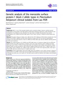
FIGURE 1. Diagrammatic representation of the Plasmodium vivax merozoite surface protein-3a (PvMsp-3a) gene from the Belem laboratory strain showing the position of the primary (P1 and P2) and nested (N1 and N2) oligonucleotide primers. Numbers refer to nucleotide positions within the Belem sequence. Primers were designed within conserved and semi-conserved regions that flank the central a-helical region of the molecule.7 bp 5 basepairs.
(N) oligonucleotide PCR primers were designed from regions of the molecule known to be conserved within 6 strains of P. vivax (Galinski MR, Barnwell JW, unpublished data) (Figure 1): P1—59 CAGCAGACACCATTTAAGG 39; P2—59 CCGTTTGTTGATTAGTTGC 39; N1—59 GACCAGTGTGATACCATTAACC 39; N2—59 ATACTGGTTCTTCGTCTTCAGG 39. Computer programs, Amplify (Engels W, University of Wisconsin. Madison, WI) and Primers 1.2 (Andersen R, Bristol, G, University of California, Los Angeles, CA) were used to check for the absence of significant primer-dimer and hairpin features in the primer sequences. One unit of Taq polymerase (Promega, Madison, WI) was used per reaction, with oligonucleotide primers at a final concentration of 0.1 mM and each of the 4 deoxynucleotide triphosphates at 0.15 mM, in reaction buffer (supplied with Taq) containing 2.5 mM MgCl2. Cycling conditions were as follows: primary reaction, 948C for 3 min, 1 cycle, then 948C for 30 sec, 568C for 30 sec, 688C for 2.5 min, 35 cycles; nested reaction, 948C for 30 sec, 578C for 30 sec, 688C for 2.5 min, 30 cycles. Products were visualized under UV illumination after electrophoresis on 0.8% agarose gels containing 0.25 mg/ml of ethidium bromide. Sizing of products was carried using a standard curve drawn from DNA markers run adjacently (0.1 mg/lane, 1-kb ladder; Gibco-BRL, Gaithersburg, MD). Restriction fragment length polymorphism analysis of PvMsp-3a amplification products. Approximately 4 ml of each PCR product was digested individually with the restriction enzymes Hha I and Alu I in 20-ml reaction volumes (5 units of enzyme/reaction; Promega) in buffer supplied with the enzymes at 378C for 4–5 hr. The DNA fragments were visualized under UV illumination after electrophoresis on 1.8% agarose gels containing 0.25 mg/ml of ethidium bromide. Sizing of products was carried using a standard curve drawn from DNA markers run adjacently (0.1 mg/lane, marker XIV; Roche Diagnostics, Ltd., Lewes, United Kingdom). RESULTS
Polymorphism in both size and sequence of PvMsp3a alleles has previously been observed and evaluated in the P. vivax laboratory strains Belem and Sal I and in 4 clinical
520
BRUCE AND OTHERS
isolates of diverse geographic origin (Galinski MR, Barnwell JW, unpublished data). Here we investigate an additional 6 geographically diverse isolates from symptomatic patients. The samples from the symptomatic individuals were used to evaluate the sensitivity, reproducibility, and potential allelic amplification bias in the PCR/RFLP method. Symptomatic infections were acquired by patients while traveling and were likely to be the result of infection from a single infected mosquito bite. These samples were therefore more likely to contain single P. vivax genotypes than those from superinfected individuals living under endemic conditions. The PCR/RFLP method was then used to determine the level of PvMsp-3a polymorphism within asymptomatic infections from the very small geographic area of a single malaria endemic village in Papua New Guinea. By analyzing multiple samples taken from the same individuals at different time points we were able to assess turnover of P. vivax parasite populations within semi-immune children. Laboratory and symptomatic patient samples. Nested amplification products generated from the Belem and Sal I laboratory strains were of the approximate size (1,900 basepairs [bp]) expected from the known sequences (1,896 bp and 1,908 bp, respectively7 (Galinski MR, Barnwell JW, unpublished data) (Figure 2a, lanes 1 and 2). The PvMsp-3a alleles from the 6 symptomatic patient samples appeared to be identical in size to alleles from the laboratory strains when analyzed on agarose gels (Figure 2a, lanes 3–8). Despite the lack of detectable size variation in the amplification products of the 6 symptomatic samples, RFLP analysis revealed substantial diversity at the nucleotide level (Figure 2b and c). The sizes of the RFLP fragments of the Belem and Sal I strains were as expected from their previous sequence characterization (Figure 2 lanes 1 and 2; see legend); fragments , 100 bp could not be resolved. The RFLP patterns of all 6 symptomatic isolates showed size conservation of the largest fragments in Hha I (Figure 2b, approximately 1,000 bp) and Alu I digests (Figure 2c, approximately 550 bp) while smaller fragments showed variation in size. The sum of the fragment sizes did not always equal the size of the intact PCR products, indicating non-resolvable variation in the size of the uncut amplification products. Two pairs of samples (16/97 and 23/97, Figure 2b, lanes 6 and 7; 47/97 and 24/97, Figure 2b, lanes 4 and 8) have identical RFLP patterns when cut with Hha I. When Alu I was used, identity was detected only in samples 47/97 and 24/97 (Figure 2c, lanes 4 and 8). Using the combination of both RFLP patterns, 7 different alleles of PvMsp-3a could be distinguished in the 8 different samples. The 2 identical alleles were from samples 47/97 and 24/97, which originated from different geographic regions, Sri Lanka and India, respectively. Validation of the PCR/RFLP technique. Triplicate PvMsp-3a amplification and RFLP analysis from the laboratory and symptomatic patient samples showed no variation in the size of the PCR product or RFLP pattern, demonstrating the reproducibility of these procedures using samples containing parasites from a single infection. The sensitivity of the amplification conditions reached its limit at around 100 ring-stage parasites/ml of blood, as determined from PCR analysis of the dilution series containing strain 47/97. Products were inconsistently amplified from samples containing fewer than 100 parasites/ml, indicating randomness
in the amplification of alleles present at low density. No interference in the amplification sensitivity or change in the RFLP pattern was observed in samples containing up to an additional 1,000 P. falciparum parasites/ml. Amplification products were not obtained from samples containing only P. falciparum parasites, demonstrating the species specificity of the primers. Triplicate analysis of samples containing equal volumes of DNA extracts from the 6 symptomatic patient isolates revealed consistent results, which showed composite RFLP patterns consisting of fragments from both alleles (Figure 2, lanes 9 and 10) even when the ratio of target DNA from each sample differed by up to 7:1. However, in a minority of cases the intensity of fragments of each pattern was not equal, indicating that PCR bias may occur with particular combinations of alleles tested, as has been found with some P. falciparum PCR typing systems (Molecular Epidemiology in Malaria Collaborative Research Network,34 unpublished data). Asymptomatic field samples. The PvMsp-3a amplification products were obtained from 22 of 39 asymptomatic samples tested in triplicate. Positive samples were obtained from 16 children. Although the number of positive replicates obtained per sample was correlated with P. vivax density (correlation coefficient 5 0.453, degrees of freedom 5 37, P 5 0.003), sensitivity was reduced compared with symptomatic samples. Other Plasmodium species (P. falciparum or P. malariae) were also present in 9 samples but were not inhibitory to specific amplification of the P. vivax alleles, in line with results from control samples. Amplification products showed a major size polymorphism (Figure 3a). Products were predominantly of 2 sizes (approximately 1,900 and 1,100 bp). The size of the largest product is in agreement with that observed for laboratory and symptomatic samples. Digestion with either Hha I or Alu I yielded fragment sizes that were highly polymorphic between samples (Figure 3b and c). The sum of the RFLP fragment sizes was significantly greater than the size of the uncut product in some samples (Figure 3, lanes 4, 20, and 21), indicating the presence of more than 1 PvMsp-3a allele in these, as well as in samples where multiple uncut fragments were present (Figure 3, lanes 23–26). Faint bands observed in samples with multiple products may represent novel alleles but could also be recombinant alleles formed during PCR amplification.35 Eighteen of twenty-two samples gave multiple positive results from the triplicate analysis and 13 (72%) of these had identical RFLP patterns. The remaining sample replicates showed either different uncut amplification products or different RFLP patterns (Figure 3, white brackets). Non-identical results probably represent stochastic variation in amplification of multiple parasite genotypes present within a sample. This phenomenon, called allelic dropout,36 has also been observed in P. falciparum PCR amplification techniques (Molecular Epidemiology in Malaria Collaborative Research Network,34 unpublished data). From the 16 samples in which single infections were detected (Figure 3, lanes 1–3 and 6–16, and 2 samples not shown, see figure legend) 9 different Hha I and 9 different Alu I RFLP patterns were detected. When data from both analyses are combined, a total of 11 distinct PvMsp-3a alleles can be differentiated, indicating a greater sensitivity
P. VIVAX GLOBAL AND LOCAL DIVERSITY
521
FIGURE 2. Uncut Plasmodium vivax merozoite surface protein-3a (PvMsp-3a) polymerase chain reaction amplification products (a) and restriction fragment length polymorphism (RFLP) patterns after digestion with the restriction enzymes Hha I (b) and Alu I (c) from the laboratory strains Belem (lane 1) and Sal I (lane 2) and symptomatic patient samples (lanes 3–8: samples 26, 47, 14, 16, 23 and 24/97 respectively). Products amplified from mixtures of target DNA from symptomatic patient samples 14 and 16 (lane 9) and 23 and 24 (lane 10) are also shown. DNA size markers are shown in lanes labeled m with sizes shown in basepairs (bp). From nucleotide sequences the predicted RFLP fragment sizes after digestion of nested amplification products from Belem and Sal I are Belem, Hha I (963, 330, 254, 211, 75, and 63 bp); Alu I (492, 258, 201, 198, 172, 156, 146, 114, 60, 39, 33, and 27 bp); Sal I, Hha I (975, 465, 405, and 63 bp); Alu I (537, 258, 189, 172, 156, 153, 150, 146, 69, 51, and 27 bp)7 (Galinski MR, Barnwell, JW unpublished data).
from the use of 2 restriction enzymes. Three genotypes were detected in more than 1 sample and 2 genotypes were found in more than 1 child. Six pairs of samples from individual children with time intervals of 3–45 days yielded PCR products. Samples taken from 2 children at 3-day intervals showed identity (Fig-
ure 3, lanes 7 and 8, and 15 and 16). Turnover of parasite populations was demonstrated in samples taken at time intervals of 15 and 42 days (Figure 3, lanes 9 and 10, and 13 and 14). In the remaining 2 children, mixed parasite populations were detected in 1 or both samples. In each case, however, RFLP patterns indicated persistence of at
522
BRUCE AND OTHERS
FIGURE 3. Uncut Plasmodium vivax merozoite surface protein-3a (PvMsp-3a) polymerase chain reaction amplification products (a) and restriction fragment length polymorphism (RFLP) patterns after digestion with the restriction enzymes Hha I (b) and Alu I (c) from asymptomatic field samples taken from semi-immune children resident in a single village in Papua New Guinea. Black brackets indicate samples taken from same child at different time points. The time interval between these samples is shown in days above the brackets. White brackets indicate non-identical, duplicate amplifications from a single sample. Single results are indicated with an asterisk. All other samples yielded identical replicate results. Data from 2 samples from 2 children are not shown. These had identical amplification products and RFLPs as the sample in lane 14. One was a single amplification and the other had an identical replicate. DNA size markers are shown in lanes labeled m with sizes shown in basepairs.
least one genotype over the 15- and 45-day intervals between samples. DISCUSSION
We have investigated size and sequence polymorphism in the recently described merozoite surface molecule of P. vivax (PvMsp-3a ). While initial studies showed at least 4 alleles based on size differences between PCR products from 20 isolates (Galinski MR, Barnwell JW, unpublished data), we demonstrate here that the inclusion of an RFLP analysis of PCR products increases the number of distinguishable alleles. Polymorphism is demonstrated here in isolates from geographically diverse origins as well as within samples from a very restricted area, suggesting that diversity is not only linked to geographic origin. An allele with identical
RFLP patterns was detected in 2 symptomatic patient samples from different geographic regions. The sharing of alleles between different regions suggests that there may be functional constraints on the number of potential alleles at this locus. Since this molecule may be a potential P. vivax vaccine candidate, further analysis of the polymorphism within this molecule is desirable to determine the association between the observed allelic variation and the naturally acquired or induced immunologic response. The polymorphic nature of PvMsp-3a makes it an ideal marker for distinguishing different infections in epidemiologic studies. The PvMsp-3a alleles were shown to be present in 2 major size forms in field samples from Papua New Guinea. The smaller form is indicative of size differences in the major central a-helical repeat region of the molecule.7 The size differences observed when multiple PvMsp-3a al-
523
P. VIVAX GLOBAL AND LOCAL DIVERSITY
leles were amplified is only a first indication of polymorphism. Although sequencing would provide fine detail of the diversity, the alternative use of the PCR/RFLP protocol to detect sequence diversity is more suited to epidemiologic studies in which large numbers of samples are analyzed. This protocol can facilitate the process of addressing epidemiologic questions even when applied to field samples from asymptomatic carriers. Furthermore, the use of more than 1 enzyme to produce the RFLPs is more discriminatory in analyses of asymptomatic field samples. A total of 11 different PvMsp-3a alleles were detected in 16 samples from 12 children in which single infections were identified. This level of allelic diversity is as great as that found in epidemiologic markers of infection used for P. falciparum.4,5,37,38 Detection of multiple infections in 5 of 22 of the positive field samples was possible from size determination of products and RFLP analyses. One limitation of the PCR/RFLP protocol is that it is often unable to determine the actual number of genotypes present in a single sample when more than one genotype is present. This is due to the inability to define the association of the multiple RFLP fragments from each genotype in mixed infections. Our analysis of the diversity at the PvMsp-3a locus of P. vivax is consistent with the presence of multiple genotypes within parasite populations in endemic regions and coinfection of genotypes within individuals. The proportion of mixed P. vivax genotype infections, where multiple PvMsp3a genes are present, is 23%. This is less than that seen in 2 recent studies. In Papua New Guinea, 65% of P. vivax samples were mixed, based on sequence data from multiple P. vivax loci17 and in India 43% were mixed, based on multiple alloenzyme analysis.21 Our data are closest to the results of other investigators,18,39 who showed approximately 10% mixed infections using serological techniques and sequence analysis, respectively, in Sri Lanka. Small sample sizes and differences in sample collection and detection techniques make it impossible to draw meaningful epidemiologic conclusions from these comparisons. A slightly lower sensitivity of detection was observed in field samples (approximately 400 parasites/ml of blood) compared to those from symptomatic patients (100 parasites/ml of blood). This could be due to a number of factors: 1) inhibitors of the PCR may be present in the crude DNA extracts obtained from field samples; 2) the stages of the P. vivax erythrocytic cycle present in the peripheral circulation contain varying numbers of nuclei (ring stages contain 1, schizonts contain up to 24) and therefore density, as counted only as the number of cells, will not necessarily be related to the number of target DNA molecules present within a sample; 3) alleles from parasites present at low density will be amplified in a random fashion as observed with control samples. The PCR/RFLP protocol has been used here to assess the intra-host dynamics of P. vivax populations in pairs of samples with varying time intervals from individual children. Maintenance of the same infection was apparent in 2 pairs of samples taken from individual children at 3-day intervals, although there was also evidence for the maintenance of infections up to 45 days. Turnover of the P. vivax population was demonstrated for two pairs of samples collected over 15- and 42-day intervals. This interval is relatively short
compared with the duration of P. vivax infections, which can persist for many months in non-immune individuals. In cases where samples from the same child showed a consistent RFLP pattern, reinfection with the same genotype of P. vivax, rather than persistence of an infection, could be inferred. This is unlikely to be the case at 3-day intervals but could be a possible explanation of apparent persistence at longer intervals. In the absence of population frequency data of each PvMsp-3a allele, the likelihood of reinfection with an identical genotype cannot be calculated. Given the large number of alleles distinguishable within the small number of samples analyzed here, reinfection with an identical genotype would appear unlikely. The small number of paired samples allows us only to conclude that the rate of turnover can vary between individuals and for different infections. A large-scale longitudinal analysis is underway using all of the samples acquired from these children, of which those described here are only a small subset. Using the new PCR/RFLP protocol described, this study will provide a detailed description of the dynamics of P. vivax infections in endemic regions. Plasmodium falciparum population dynamics indicate that in highly endemic regions parasite turnover can also occur over a short time scale.40,41 In a region such as Papua New Guinea where P. falciparum and P. vivax often coexist within human hosts, interaction between species may be an important factor affecting the rate of turnover. Such questions can now be addressed and should provide a greater understanding of the epidemiology and transmission dynamics of malaria. Acknowledgments: We thank the people of Gonoa village for their longstanding cooperation, and the staff of the Papua New Guinea Institute of Medical Research in Madang, especially Dr. Michael Packer and Moses Lagog, for assistance with the collection of field samples. Thanks are also given to David Walliker and Michael Alpers for support. Financial support: Collection of field samples was funded by a joint grant to Karen P. Day, David Walliker and Michael Alpers from The European Commission. Karen P. Day is funded by a Program Grant from The Wellcome Trust. Marian C. Bruce was funded by a studentship from the Medical Research Council of the United Kingdom and by the Wellcome Trust. Mary R. Galinski and John W. Barnwell are funded by grants from the National Institutes of Health (AI24710-12 and U01-AI37545) and WHO/TDR (950440 and 910495). Authors’ addresses: Marian C. Bruce and Karen P. Day, Wellcome Trust Centre for the Epidemiology of Infectious Disease, University of Oxford, South Parks Road, Oxford OX1 3FY, United Kingdom. Mary R. Galinski, Division of Infectious Diseases, Department of Medicine, Emory University, School of Medicine, Emory Vaccine Center, Atlanta, GA 30329. John W. Barnwell, Biology and Diagnostic Branch, Division of Parasitic Diseases, Centers for Disease Control and Prevention, Mailstop F-13, Building 22B, 4770 Buford Highway, Atlanta, GA 30341. Georges Snounou, Department of Infection and Tropical Medicine (Lister Unit), Wellcome Centre for Clinical Tropical Medicine, Imperial College School of Medicine, Northwick Park Hospital, Harrow, Middlesex, HA1 3UJ, United Kingdom. Reprint requests: Marian C. Bruce and Karen P. Day, Wellcome Trust Centre for the Epidemiology of Infectious Disease, University of Oxford, South Parks Road, Oxford OX1 3FY, United Kingdom. REFERENCES
1. Read A, Day KP, 1992. The genetic structure of malaria parasite populations. Parasitol Today 8: 239–242.
524
BRUCE AND OTHERS
2. Conway DJ, Greenwood BM, McBride JS, 1992. Longitudinal study of Plasmodium falciparum polymorphic antigens in a malaria-endemic population. Infect Immun 60: 1122–1127. 3. Babiker HA, Satti G, Walliker D, 1995. Genetic changes in the population of Plasmodium falciparum in a Sudanese village over a three-year period. Am J Trop Med Hyg 53: 7–15. 4. Contamin H, Fandeur T, Bonnefoy S, Skouri F, Ntoumi F, Mercereau-Puijalon, 1995. PCR typing of field isolates of Plasmodium falciparum. J Clin Microbiol 33: 944–951. 5. Paul REL, Packer MJ, Walmsley M, Lagog M, Ranford-Cartwright LC, Paru R, Day KP, 1995. Mating patterns in malaria parasite populations of Papua New Guinea. Science 269: 1709–1711. 6. Roper C, Elhassan IA, Hviid L, Giha H, Richardson W, Babiker H, Satti GMH, Theander TG, Amot DE, 1996. Detection of very low levels of Plasmodium falciparum infections using the nested polymerase chain reaction and reassessment of the epidemiology of unstable malaria in Sudan. Am J Trop Med Hyg 54: 325–331. 7. Galinski MR, Corredor-Medina C, Povoa M, Crosby J, Ingravallo P, Barnwell JW, 1999. Plasmodium vivax merozoite surface protein-3 contains coiled-coil motifs in an alanine-rich central domain. Mol Biochem Parasitol 101: 131–147. 8. Riley EM, Morris-Jones S, Blackman MJ, Greenwood BM, Holder AA, 1993. Longitudinal study of naturally acquired cellular and humoral immune responses to a merozoite surface protein (MSP1) of Plasmodium falciparum in area of seasonal malaria transmission. Parasite Immunol 15: 513–524. 9. Oeuvray C, Bouharoun-Tayoun H, Gras-Masse H, Bottius E, Kaidoh T, Aikawa M, Filgueira M-C, Tartar A, Druilhe P, 1994. Merozoite surface protein-3: a malaria protein inducing antibodies that promote Plasmodium falciparum killing by cooperation with blood monocytes. Blood 84: 1594–1602. 10. Taylor RR, Allen SJ, Greenwood BM, Riley EM, 1998. IgG3 antibodies to Plasmodium falciparum merozoite surface protein 2 (MSP2): increasing prevalence with age and association with clinical immunity to malaria. Am J Trop Med Hyg 58: 406–413. 11. Pasloske BL, Howard RL, 1994. The promise of asexual malaria vaccine development. Am J Trop Med Hyg 50 (suppl 4): 3–10. 12. Perera KLRL, Handunnetti SM, Holm I, Longacre S, Mendis K, 1998. Baculovirus merozoite surface protein 1 C-terminal recombinant antigens are highly protective in a natural primate model for human Plasmodium vivax malaria. Infect Immun 66: 1500–1506. 13. Bouharoun-Tayoun H, Attanath P, Sabchareon A, Chonsuphajaisiddhi T, Druilhe P, 1990. Antibodies that protect humans against Plasmodium falciparum blood stages do not on their own inhibit parasite growth and invasion in vitro, but act in cooperation with monocytes. J Exp Med 172: 1633–1641. 14. Sabchareon A, Burnouf T, Ouattara D, Attanath P, BouharounTayoun H, Chantavanich P, Foucault C, Chongsuphajaisiddhi T, Druilhe P, 1991. Parasitologic and clinical response to immunoglobulin administration in falciparum malaria. Am J Trop Med Hyg 45: 297–308. 15. Sattabongkot J, Suwanabun N, Rongnoparut P, Wirtz RA, Kain KC, Rosenberg R, 1994. Comparative test of DNA probes for detection of Plasmodium vivax circumsporozoite protein polymorphs VK 247 and VK 210. J Infect Dis 169: 464–466. 16. Mann VH, Good MF, Saul A, 1995. Diversity in the circumsporozoite protein of Plasmodium vivax: does it matter? Parasitol Today 11: 33–36. 17. Kolakovich KA, Ssengoba A, Wojcik K, Tsuboi T, Al-Yaman F, Alpers MP, Adams JH, 1996. Plasmodium vivax: favored gene frequencies of the merozoite surface protein-1 and the multiplicity of infection in a malaria endemic region. Exp Parasitol 83: 11–18. 18. Premawansa S, Snewin VA, Khouri E, Mendis KN, David PH, 1993. Plasmodium vivax: recombination between potential allelic types in the merozoite surface protein Msp 1 in parasites isolated from patients. Exp Parasitol 76: 192–199. 19. Udagama PV, David PH, Peiris JSM, Ariyarantne YG, Perera KLRL, Mendis KN, 1987. Demonstration of antigenic poly-
20. 21. 22.
23.
24.
25.
26.
27.
28.
29.
30.
31.
32.
33.
34.
35. 36.
37.
38.
39.
morphism in Plasmodium vivax malaria with a panel of 30 monoclonal antibodies. Infect Immun 55: 2604–2611. Joshi H, Subbarao SK, Raghavendra K, Sharma VP, 1989. Plasmodium vivax: enzyme polymorphism in isolates of Indian origin. Trans R Soc Trop Med Hyg 83: 179–181. Joshi H, Subbarao SK, Nanda N, Ghosh SK, Carter R, Sharma VP, 1997. Genetic structure of Plasmodium vivax isolates in India. Trans R Soc Trop Med Hyg 91: 231–235. Snounou G, Viriyakosol S, Zhu XP, Jarra W, Pinheiro L, Rosario VE, Thaithong S, Brown KN, 1993. High sensitivity of detection of human malaria parasites by the use of nested polymerase chain reaction. Mol Biochem Parasitol 61: 315–320. Cattani JA, Tulloch JI, Vrbova H, Jolley D, Gibson FD, Moir JS, Heywood PF, Alpers MP, Stevenson A, Clancy R, 1986. The epidemiology of malaria in a population surrounding Madang, Papua New Guinea. Am J Trop Med Hyg 35: 3–15. Burkot TR, Graves PM, Cattani JA, Wirtz RA, Gibson FD, 1987. The efficiency of sporozoite transmission in the human malarias, Plasmodium falciparum and P. vivax. Bull World Health Organ 65: 375–380. Cox MJ, Kum D, Tavul L, Narara A, Raiko A, Alpers M, Medley G, Day KP, 1994. Dynamics of malaria parasitaemia associated with febrile illness in children from a rural area of Madang, Papua New Guinea. Trans R Soc Trop Med Hyg 88: 191–197. Arnot DE, Barnwell JW, Tam JP, Nussenzweig V, Nussenzweig RS, Enea V, 1985. Circumsporozoite protein of Plasmodium vivax: gene cloning and characterization of the immunodominant epitope. Science 230: 815–818. Gibson HL, Tucker JE, Kaslow DC, Krettli AU, Collins WE, Kiefer MC, Bathurst IC, Barr PJ, 1992. Structure and expression of the gene for Pv200, a major blood-stage surface antigen of Plasmodium vivax. Mol Biochem Parasitol 50: 325– 334. Barnwell JW, Nichols ME, Rubinstein P, 1989. In vitro evaluation of the role of the Duffy blood group in erythrocyte invasion by Plasmodium vivax. J Exp Med 169: 1795–1802. Walliker D, Quakyi IA, Wellems TE, McCutchan TF, Szarfman A, London WT, Corcoran LM, Burkot TR, Carter R, 1987. Genetic analysis of the human malaria parasite Plasmodium falciparum. Science 236: 1661–1666. Forsyth KP, Philip G, Smith T, Kum E, Southwell B, Brown GV, 1989. Diversity of antigens expressed on the surface of erythrocytes infected with mature Plasmodium falciparum parasites in Papua New Guinea. Am J Trop Med Hyg 41: 259– 265. Piper KP, Roberts DJ, Day KP, 1998. Plasmodium falciparum: analysis of the antibody specificity to the surface of the trophozoite-infected erythrocyte. Exp Parasitol 91: 161–169. Robson KJH, Hall JRS, Davies LC, Crisanti A, Hill AVS, Wellems TE, 1990. Polymorphism of the TRAP gene of Plasmodium falciparum. Proc R Soc Lond Biol Sci 242: 205–216. Kyes S, Craig AG, Marsh K, Newbold CI, 1993. Plasmodium falciparum: a method for the amplification of S antigens and its application to laboratory and field samples. Exp Parasitol 77: 473–483. Bjorkman A, do Rosario VE, Snounou G, Walliker D, 1998. Standardizing PCR for molecular epidemiology studies of malaria. Parasitol Today 14: 85. Meyerhans A, Vartanian J-P, Wain-Hobson S, 1990. DNA recombination during PCR. Nucleic Acids Res 18: 1687–1691. Gagneux P, Boesch C, Woodruff DS, 1997. Microsatellite scoring errors associated with noninvasive genotyping based on nuclear DNA amplified from shed hair. Mol Ecol 6: 861–868. Babiker HA, Ranford-Cartwright LC, Currie D, Charlwood JD, Billingsley P, Teuscher T, Walliker D, 1994. Random mating in a natural population of the malaria parasite Plasmodium falciparum. Parasitology 109: 413–421. Felger I, Tavul L, Kabintik S, Marshall V, Genton B, Alpers M, Beck H-P, 1994. Plasmodium falciparum: extensive polymorphism in merozoite surface antigen 2 alleles in an area with endemic malaria in Papua New Guinea. Exp Parasitol 79: 106–116. Udagama PV, Gamage-Mendis AC, David PH, Peiris JSM, Per-
P. VIVAX GLOBAL AND LOCAL DIVERSITY
era KLRL, Mendis KN, Carter R, 1990. Genetic complexity of Plasmodium vivax parasites in individual human infections analyzed with monoclonal antibodies against variant epitopes on a single parasite protein. Am J Trop Med Hyg 42: 104– 110. 40. Daubersies P, Sallenave Sales S, Magne S, Trape JF, Contamin H, Fandeur T, Rogier C, Mercereau-Puijalon O, Druilhe P,
525
1996. Rapid turnover of Plasmodium falciparum populations in asymptomatic individuals living in a high transmission area. Am J Trop Med Hyg 54: 18–26. 41. Farnet A, Snounou G, Rooth I, Bjorkman A, 1997. Daily dynamics of Plasmodium falciparum subpopulations in asymptomatic children in a holoendemic area. Am J Trop Med Hyg 56: 538–547.