of peptide termini with their respective pockets in the class I molecule, and imply a role for the minor ..... as an illustration of the dramatic effect seen with some of.
Polymorphisms in Pockets of Major Histocompatibility Complex Class I Molecules Influence Peptide Preference By Eric M. Rohren,* Larry R . Pease,* H i d d e L. Ploegh,~ and Ton N. M. Schumacher~ From the *Department of Immunology, Mayo Clinic, Rochester, Minnesota 55905; and the
r 02139
Institute of Technology Center for Cancer Research, Cambridge, Massachusetts
Summary The set of peptides that is bound by a given major histocompatibility complex class I product can be described by one or two properly spaced anchor residues, and two properly spaced peptide termini, approximately 8-10 residues apart. Using radiolabeled peptide libraries, we examined whether mutations in those "pockets" in class I K b molecules that do not seem critically involved in the interaction with the peptide anchor residues, do exert an effect on the set of preferred peptides. We find that mutations in all the pockets found in the structure of K s have a significant effect on the peptide preference of the molecule, and their recognition by cytotoxic T cells. Alterations in substrate specificity are also observed for mutations involving residues that interact with main chain atoms in both peptide termini. These findings challenge a static view of the interaction of peptide termini with their respective pockets in the class I molecule, and imply a role for the minor pockets in peptide selectivity. he isolation of naturally processed peptides as bound by different class I products has revealed that for each allele T these peptides adhere to a "motif" (1, 2), characterized by the (almost) exclusive occurrence of one or a few amino acids at one or two positions within the sequence. For K b molecules, the prime anchor residue seems to be Tyr/Phe-5 (1) (for nonamer peptides Tyr/Phe-6 [3]). At other positions in these peptide motifs, a much greater variety of amino acids is observed, suggesting that either the side chains at these positions contact solvent, or that a much greater variety of side chain residues is tolerated by the regions in the class I molecule that combine with them. Resolution of the crystal structure of K b molecules, complexed with either the Sendai-N- or vesicular stomatitis virus (VSV)l-N-derived Kb-restricted peptides (4-6) showed that the Tyr-5 side chain docks in one of the pockets found in class I molecules that has been implicated in peptide binding (7, 8). Apart from the interaction of the Tyr residue with pocket C, and the peptide termini with their respectivepockets, other side- and mostly main chain atoms interact to some extent with amino acids forming the minor pocket D and other residues lining the peptide binding groove (4-6). (We use the prefix 'major' for pocket C in K b, since it seems the prime determinator of peptide specifidty for this allele. Pockets 1Abbreviation used in this paper: VSV, vesicular stomatitis virus.
1713
B, D, and E, which have a less defined role in peptide preference of K b molecules are called 'minor pockets'. Pockets A and F are considered separately, since they interact (at least in part) with invariable parts of the peptide chain, and were therefore thought to be of limited importance to class I specificity [4-7].) However, it is hard to assess the relative importance of the observed interactions based on structural data only. Naturally occurring class I mutants (such as Kbml), in which only amino acids in supposedly minor pockets, or the pockets that combine with the peptide termini, are different from those in K b, appear in some cases unable to present any of the known Kb-restricted epitopes (9). A critical role for these pockets in the determination of peptide binding specificity is therefore likely, but direct proof is lacking. To investigate more directly the role of these minor pockets, and those regions in the molecule that combine with the peptide termini in the determination of substrate specificity, we have confronted a series of K b mutants with a radiolabeled peptide library (10), and monitored peptide selection by twodimensional display of class I captured peptides. We find that alterations throughout the class I structure have a major effect on the set of peptides bound, not only in those regions of the molecule that combine with variable parts of the peptide, but also in those parts that interact with main chain atoms of the peptide termini.
J. Exp. Med. 9 The Rockefeller University Press 9 0022-1007/93/06/1713/09 $2.00 Volume 177 June 1993 1713-1721
Materials and Methods Generation of H-2 K b Mutants and Cell Culture. Class I variant genes were prepared by site-directed mutagenesis of the K b gene (11-16). The wild-type Kb molecule, and Kb163A are encoded by exons 1-8 of K b. All other variants are composed of exons 1-3 of K b and exons 4-8 of the Ld gene, and can be immunoprecipitated using a mAb against the c~3 domain of Ld (17, 18). Sequences of exons that were mutagenized were confirmed by sequencing. Class I genes were cotransfected with the herpes simplex thymidine kinase gene in L cells. Cells surviving HAT selection were screened for expression of the transferred genes by FACS| (Becton Dickinson & Co., Mountain View, CA) using 28-14-8S or B8-24-3. All mutants are named after the residue involved, followed by the single letter code of this amino acid in the mutant. LPS blasts were obtained from spleen cells by a 4-d culture of cells in the presence of LPS (30/xg/ml LPS-B; Difco Laboratories, Inc., Detroit, MI). Peptide Labeling and Binding to Class I Molecules. The peptide mixture "poly8" was synthesized by standard t-boc chemistry, and has been described previously (10). Peptides were labeled by chloramine-T catalyzed iodination (19), and used within 24 h after iodination. Before peptide binding experiments, ceUswere cultured at 26~ for 2 d in Hepes-huffered DMEM/10% FCS. Cells were incubated with 1/zM of the radiolabded poly8 mixture at 22~ for 2.5 h in Hepes-buffered DMEM without serum (total vol 5 ml). Subsequently, cells were washed, and lysed in Triton X-100 lysis buffer (10). After removal of call debris, and preclearing of lysates, class I molecules were immunoprecipitated, using antiexon-8 (20) for proteins containing the H-2 K b re3 domain, and 28-14-8S (17, 18), for proteins containing the H-2 Ld re3 domain. Immunoprecipitates were washed three times. Class I-associated peptides were liberated by two cycles of TFA extraction (200/A of 1% TFA for 20 min at room temperature). Extracts were faltered over a 0.22-~tm filter and lyophilized. Peptides were dissolved in 0.05% TFA and analyzed by reverse phase HPLC on a column (model C18; Millipore Corp. Waters Chromatography, Milford, MA). Buffers used were: A, H20/ 0.05% TFA; B, acetonitrile/0.05% TFA. Elution profiles were performed for: 0-3 min, 95-95% A; 3-5 min, 95-85% A; 5-23 rain, 85-60% A; 23-25 min, 60--35% A; 25-27 rain, 35-35% A; and 27-33 rain, 35-95% A, at a flow rate of 1.7 ml/min. 0.3-rain fractions were collected, and fractions 41-65 were lyophilized. Fractions were dissolved in 6/A H20 and 3/~1 of each sample was applied to silica gel 60 plates (EM Science, Gibbstown, NJ). TLC was performed in freshly made N-butanol/H20/pyridine/acetic acid 2:1:0.75:0.25. Plates were exposed to Kodak X-AR5 films at -70~ Note that TLC were exposed until the central part of the autoradiogram was of approximately equal intensity (with the exception of Kb97R, for which there are no peptides to be found in this region), therefore only alterations in the relative intensity of spots are relevant. The set of peptides selected by all mutants was displayed by twodimensional HPLC/TLC at least once and by one-dimensional HPLC in an independent experiment for confirmation. CTL Assays. The CTL clone 33(21) was provided by Jim Sheil (University of West Virginia, Morgantown, WV) and was maintained in RPMI 1640 (Sigma Chemical Co., St. Louis, MO)/10% FCS/5% Con A supernatant. The CTL clone bm8 anti-B10-4.23 was a kind gift of Jeffrey A. Bluestone (University of Chicago, Chicago, IL) and was maintained in DMEM (Sigma Chemical Co.)/10% FCS. The C3H anti-C3H.SW T cell line was prepared by mixing 5 x 10⁶ irradiated C3H.SW splenocytes with 5-7 x 1714
106 C3H splenocytes. Responder cells were tested on day 5 for activity, then restimulated with irradiated splenocytes on day 7. Thereafter, cells were maintained in IMDM (Sigma Chemical Co.)/10% FCS containing 10-20 U/m111-2, and restimulated weekly. L cells expressing transfected class I molecules were used in a standard cytotoxicity assay. Briefly, target cells were labeled with 1.0 mCi NaSICR per 107 cells for 45 min at 37~ 5 x 10³ target cells were subsequently incubated with CTL, in a total vol of 200/A (in the presence of 5 #M VSV-8 for clone 33). Plates were incubated at 37~ for 8 h, centrifuged, and 150/zL samples were removed for 3,-spectrometry. Maximal release was determined by the addition of 10% Triton X-100 (Sigma Chemical Co.). Spontaneous release was determined by incubating target cells for 8 h at 37~ in the absence of CTL. Specificlysis was calculated as: (Experimental release - spontaneous release)/(maximum release - spontaneous release).
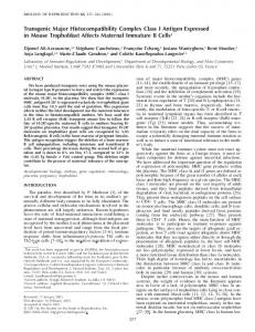
Results and Discussion A synthetic peptide library comprising 432 different peptides, based largely on the sequences of the Kb-restricted Sendai-N and VSV-N epitopes, was produced by the simultaneous coupling of different amino acids and has been described ([10], Fig. 1). For peptide selection experiments, peptides in this library were labeled by iodination of Tyr residues. Although iodination of the Tyr anchor residue in these peptides could conceivably alter their binding properties, such an effect would likely be similar for all mutant class I products, and should not hamper a comparison of their peptide preference. To examine the effect of naturally occurring mutations in the different pockets, we generated a series of mutants of the K b molecule (Table 1, [11, 22]). L cells were transfected with genes encoding K b molecules and the various mutants, and expression of the transfected products was determined by FACS| (see legends to Fig. 3). To ascertain that the binding properties of K b molecules were independent of the cell type in which they were expressed, we first established that the sets of peptides captured by K ~ molecules as present in B6 LPS blasts, and when expressed in L cells are indeed identical (Fig. 2). The reproducibility of this assay has been shown previously by a comparison of the set of peptides captured by K b molecules on LPS blasts of three different mouse strains (10). Most of the class I molecules we examined are hybrids between the oq and Cez domains of K b and the c~3, transmembrahe and cytoplasmic domains of L a. However, comparison 1
2
3
4
S
NH 2- A
P
R
G
G
H
Y
Y
V
D
F
6
7
8
L -COOH
P
A
Q
G
K
5 T
amino acid nO
Figure I. Sequence of the poly8 peptide library. Sequences are given in single letter code. Note that both the octamer version of the Sendai-N epitope (for which a nonamer with a 1-amino acid e~tension at the NH2 terminus is optimal for binding to Kb), and the VSV-N epitope are represented once in this mixture.
Polymorphismsin MHC I Molecule Pockets Influence Peptide Preference
Table 1.
Amino Acid Substitutions in the Various K ~ Mutants
Mutant
Position and nature of substitution
5A.44 Kb163A KSSM Kb45V Kb24S Kb73W Kb74S Kb155Y Kb152A Kb97R Kb77S
wt K b plus c~3 tm* and ct of L a 163: T-A 5: L-M 45: Y-V 24: E-S 73: S-W 74: F-S 155: R-Y 152: E-A 97: V-R 77: D-S
Natural occurrence NA K bin1~ K k, Dr, D f, and others K r, D b, Dq, L d, and others D r and D' K bins, K k, D b, and many others K d, D b, D q, L d, and others K r, K v, and Kq K bml, K ~, and La K bin1, K f, D d, D b, and others K a, K k, K r, K', and others K bin3, K d, K k, D b, and others
Pocket NA A A B B C C D E C and E F
Amino acid substitutions in Kb mutants. Alleles in which substitutions at the positions indicated occur are only given for mouse class I. Assignment of the different amino acids to the various pockets in Kb was based on references 4, 5, and 24. * tm, transmembrane domain; ct, cytoplasmic tail; NA, not applicable. of K b and Kb/L d molecules (which have identical peptide binding regions and differ only in the membrane proximal regions) both with respect to CTL recognition (data not shown) and peptide preference, demonstrated that these molecules behave identically (compare Fig. 2 bottom with Fig. 3 A). Fig. 3 shows a comparison of the set of peptides captured by wild-type K b molecules (A) and variants thereof. To facilitate a comparison of the effect of the various mutations on substrate preference, we have tabulated the occurrence of all peptide spots observed in the two-dimensional separations in Fig. 4. The transfectant (Kb97R) was included as an illustration of the dramatic effect seen with some of the substitutions. Residue 97 contributes to both pockets C and E, and the amino acid substitution is major: V to R. This mutation (Kb97R, Fig. 3 B) leads to a sharp reduction in the number of peptides from our peptide library with which the molecule can combine. A similar mutation in the K d molecule also results in a class I molecule with a greatly impaired peptide binding repertoire (Abastado, J.-P. et al., unpublished observations). A comparison of the more complex mutants bm8 45V, and bm8 45V-24A shows that not all mutations in the peptide binding groove exert an effect on peptide preference, and underscores the reproducibility of the assay (see Fig. 6). The Minor Pockets. Single amino acid substitutions in pockets D (155 R to Y, Fig. 3 C) and E (152 E to A, Fig. 3 D) respectively, both give a slight but distinct alteration in the peptide preference of the class I molecule. These two substitutions are both present in the natural variant K bin1,
Figure 2. Kb moleculesexpressedin different cell types have the same peptide binding specificity.LPS blasts of B6 mice, and L cells transfected with Kb were cultured at 26~ for 48 h, and resuspended in Hepes1715
Rohren et al.
buffered DMEM containing 1 #M of radiolabded polyS. Cells were incubated with peptidesfor 2.5 h at 22~ and washedthree times in medium. MHC class I moleculeswere isolated and Kb-associatedpeptides were extracted with 1% TFA, and separated by two-dimensional HPLC/TLC as described (10). Separationalong the x-axis is HPLC, and along the y-axis is TLC.
Figure 3. Naturally occurring polymorphismsin MHC class I pockets affectpeptide preference. L cells transfectedwith Kb moleculesand variants thereofwere cultured at 260C for 48 h and incubatedwith radiolabeledpolyS. ClassI-associatedpeptideswere displayedby two dimensionalHPLC/TLC as for Fig. 2. Expression of transfected genes was examinedby FACS| using B8-24-3 (obtained from American Type Culture Collection cell lines and hybridomas)for Kb163A, and 28-14-8s (17, 18) for all other cell lines, and is given as peak channel value. 5A44: 178; Kb163A 151; Kb24S: 153; Kb73W: 145; Kb74S: 178; Kb155Y: 178; and Kb152A: 182; Kb77S: 168. Background staining was less than 55. Note that close examination of the set of peptides captured by Kb molecules and Kb45V does reveal slight differences.However, differencesof a similar magnitude can be observed between different experiments for the set of peptides captured by Kb molecules. Therefore, only differencesclearly beyond experimental variation are considered significant. and are the main determinants of the K bml phenotype with respect to CTL recognition (Van Bleek, G. M., and S. G. Nathenson, manuscript in preparation). Molecular modelling, and more recently, the elucidation of the crystal structure of K b, predict that in K b the 45 side chain is hardly accessible to peptide because of steric hindrance by the large side chain of E24, and so is not predicted to play a role in peptide selection (4). Indeed, modification of residue 45 in pocket B (Y45V, Fig. 3 E) does not lead to a dearcut alteration in the set of peptides selected (see legend to Fig. 3). However, a single change at position 24 in pocket 1716
B (Kb24S, Fig. 3 F) shifts peptide preference, and suffices to impose a K bms phenotype on the K b molecule (Kbins, Fig. 3 G). The importance of this amino acid substitution with regard to allo-CTL recognition has been described earlier (22). The parallel change in peptide binding capacities observed here underscores the importance of peptide recognition in class I-restricted allogeneic T cell responses. Thus, naturally occurring amino acid substitutions in all the three minor pockets exert an effect on the set of peptides with which the class I molecule combines, indicating that the structure of pockets that do not interact with the pro-
Polymorphismsin MHC I Molecule Pockets Influence Peptide Preference
1.2 I 2.1 2.2 2.3 3.1 0 3.2 4.1 4.2 4.3
oil
I
1.1
0 0 0 0
eoe
,0 0
'OJ
'0
' O i O O: OiO: O:O[
~
0i00
o:
o'o1
5.1 0 s.2,JO 5.3 .O 6.1 0
]0 01 9
1
t
7.1!10 7.2! 0
I
13.4'.' 9 I i3.5. i 14.1., ~)' 14.2,. D, 14,3..0.
0
0[0
14"4..0.0
9
14.8 0
7.4!.
*
7.3 i9 7.51~0
9149149149
:0 00: Q ; 01. 9 O f 9 O
0 0:0 O: 0i , 0 00: , 0: 010 "Oi : O: !00:
7.8.0 8.1 , 0 8.2 8.3:0 8.4 8.5 ',0 8.8 9 9.1 b 9.2 9.3 '9 9.4 9.s ',0 9.6 0
'0 00
00: 9
'OOO ,
O
,
*
15,1
0
O
15.4"0'
9 0:0
0
00 O:O ;0 O:O Oi 9 9 1 4 9 O:OIO
O:O O:O 00
,
O!O
,
15.6., 18.1,, 16.2,, 16.3 '18.4)
_ 16.5]1 17.1!
'
0
9
9 01 9 '00 010 9149 0 1 9 9 1 4 9 1 4 09 1 0 9 1 4 9
'00]O
"
0 O: : 0 01
',C) 0 : 0
'
)o
10
: *
11.5
11.B![ I
,
O:
)o'.
!0
!0
I
17.3i 17.4![
:OO
0:0
!OO0O
O[CJ 0:
'
L
[
O;O
O:O!OO
O:
010
O'
0:0 O:O
0[ @ oc OiO
OO
O' 0
,
_
21,1
21.2" [ 21.3" ~J 2 1 , 4 : .
L 22.1.
010
[
iO [0
L
.9
Oi
J
9
:0
:O O : g
i010
0
0 9 0
'1
20.3,
9
010
'0
19.2" 19.3::
,202
9
'
9
2o.I,,
0i0
!0 9 0
'O 0
18.2L~ ~1B.3~
~ 1~.1
9
0
0
Oi :0 9 0 0 Oi 9 O:q)O o 0 9 o o 0:0 0(1 0 Oq-) O 0 9 :0 :0 0:0 0:0 :0 0:0
'
O = :1o:o o ~176176176 ! 17.8 O : O . O (-l: 0 1B.I;,
0:0
1o.1 0 10.2 ' 8 10.3 10.4 , 0 10.5 , 0 11.1 11.2:0 11.3 , 11.4 ! 9
17.2/i
0 0 O: 0010 00:O 0 i0 ] 0 'l ]0 o OiO 0 0 0 !0 0100' 9149 010 010
0
15.5, 0
_
O 0 0 0 0 0 0 0 1
10 I0 !0
15.2"0' 15.3
9 1 4 9e 9
00I OIO;0!O
'O,;
0:0
O0O
9
i4.5,,0,
0
OO O
9 6;:0 e i e ' 9
! i
0:0 0:0
]C'
IV O:O
6.31 9
OIO
010 [0 OiO 9149
J 9 o : o o:ql o r 9 1 4 9 9
6.2J .O
12.5: 9
13.2"0', [13.3
0:0 9 9149 9149
010.i 9
!
0'
0
12.6 12.7',i" 12.8 13.1
ID:O O : O ~:o 01 0 : 0 O:
:0 o}o 0 :0 ] 9149 ]0 0:0 9
4.4 0
12.1 12,2
O
O'
12.3:0 12.4 ]
010
0
'Oi
9 0 00Oi 09 0
0
0 O Oi 9149
0
'0 010
0
8 i0 :0
,0
I
:e
'0 [ [0
0
)0 0] 9
9169 :9 [ Oi 010 0
Figure 4. Naturally occurring polymorphisms in class I pockets affect peptide preference. For each class I molecule assayed, 22 individual HPLC fractions were resolved by TLC. The grid in Fig. 3 K identifies and numbers each of these 22 fractions. For a given TIC track we numbered the resolved peptides, and tabulated the occurrence of that particular spot for each of the mutants analyzed (e.g., 12.6 refers to HPIC fraction 12, and the index to the number of the spot, counting from the origin (bottom). Peptides were thus scored as being abundantly present (0), absent (box left open), or of intermediate intensity (O). While this evaluation cannot be considered strictly quantitative, it does facilitate the comparison of individual mutants. The total number of spots scored for all mutants combined is 96. Note that some peptides (e.g., 5.2, 7.5, 13.4) are bound by all, or almost all mutants tested.
posed anchor residues does influence the spectrum of peptides presented. Such an effect could either be due to steric hindrance or to an interaction between peptide and class I molecule in the minor pockets. In some cases, a reduction
1717
Rohren et al.
in size of the side chain (e.g., 152 E to A) leads to a reduced capacity to combine with certain peptides. At least in these cases, amino acid residues in the minor pockets may actually contribute to the affinity of the class I molecule for the peptide.
Pocket
A
B
C
C
D
E
F
'~
1
40 Clone 33 + V S V %SpeGlClcLySkl=atE:T 7,5:~ 30 2O 10 0
5A.44
Kb163A
Igb248
1~73W
I