ANTICANCER RESEARCH 25: 2503-2508 (2005)
Possible Mechanism Connected with the Progression of Submucosal Invasion in Colorectal Neoplasias C.A. RUBIO
Gastrointestinal and Liver Pathology Research Laboratory, Department of Pathology, Karolinska Institute and Hospital, Stockholm, Sweden
Abstract. Previous studies in human and in experimentallyinduced colorectal carcinomas showed that the material retained in dilated glands at the growing tumour edge was directly released into the peritumoral extracellular matrix (ECM) through glandular faults called glandular pores. In this work, the characteristics of the glands at the growing edge in adenomas with submucosal invasion were investigated in 56 patients. Sections (stained with hematoxylin-eosin, and MNF 116) from 56 colonic adenomas with submucosal invasion were reviewed. Pore formation was defined as a glandular defect characterised by lack of a group of epithelial cells in the deeper portion of the glands. Numerous dilated submucosal glands with pores were found in 46 (82%) of the 56 neoplasias. Through those pores retained mucin, neutophils and/or necrotic material, containing proteolytic enzymes, were siphoned off into the surrounding tissues, causing disruption of the paratumoral ECM and facilitating host penetration by neoplastic cells. Glandular continuity seems to be restored by the proliferation of malignant cells from the tip of the free borders of the pores into the disrupted ECM. The sealing of the glandular flaws would permit the re-accumulation of intraglandular proteolytic material, a mechanism that would replicate a new wave of host invasion, thus ensuring a stepwise, but continual, tumour progression at the growing edge in untreated patients. In order to study some of the determinants for the acquisition of an invasive phenotype, several parameters pertinent to neoplastic cells have been investigated, e.g. histological configuration and immunohistochemical expression (7), cellular proliferation (26), p53 expression
Correspondence to: C.A. Rubio, MD, PhD, Gastrointestinal and Liver Pathology Research Laboratory, Department of Pathology, Building P1/02, Karolinska Institute and University Hospital, 17176 Stockholm, Sweden. Fax: 46 8 51774524, e-mail:
[email protected] Key Words: Colon, rectum, adenomas, progression, invasive carcinoma.
0250-7005/2005 $2.00+.40
(26), K-ras mutations (26), cyclin E and CDK (10), bcl (26), K(+) ion channels from the HERG1 protein family (9) and carbonic anhydrase-related protein VIII (CA-RP VIII) (14). On the other hand, in order to assess the role played by the extracellular matrix (ECM) in tumour penetration, other parameters have been explored, e.g. angiogenesis (28), telomerase activation (6), cathepsin B (4), CD10 expression (15), increased membrane type 1 matrix metalloproteinase (12), TGF-‚ signalling in fibroblasts (2) and trimeric laminin 5 expression (11). Notwithstanding, despite the burgeoning literature, the mechanism whereby the neoplastic glands invade the juxtaposed normal matrix of the host remains poorly understood. In a preliminary investigation (17), we found dilated neoplastic glands with pores at the growing edge of sporadic colorectal adenocarcinomas. It was observed that, through the glandular pores, the retained glandular material, namely mucin, inflammatory cells and/or necrotic material, was being siphoned off directly into the juxtaposed extracellular matrix (ECM). These proteolytic enzyme-containing materials (5, 8, 16, 18, 29, 30) disrupt the paratumoral anatomy of the ECM (19). It was inferred that this mechanism would facilitate tumour penetration. Subsequent studies on large sections from 112 colectomies and rectal amputates (20) revealed that those histological parameters were equally frequent in colonic and in rectal adenocarcinomas. As preoperative irradiation was administered only to patients with rectal tumours, it was inferred that those glandular pores were neither evoked nor abrogated by irradiation. Studies of tumour stage indicated that glandular pore formation was unrelated to the ability of colorectal tumours to metastasise to regional lymph nodes (20). Further studies on the growing tumour edge in sporadic colorectal carcinomas in patients with inflammatory bowel disease (19), in carcinomas from patients with hereditary nonpolyposis colorecral cancer (HNPCC) (21), and in chemically-induced colonic carcinomas in rats (22), as well
2503
ANTICANCER RESEARCH 25: 2503-2508 (2005)
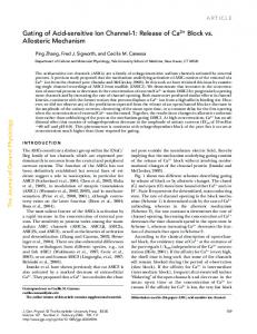
Figure 1. Overview showing a colonic adenoma with submucosal invasion. Note dilated submucosal neoplastic glands with pore formation at the invading tumour edge (H&E 4x).
as in sporadic oesophageal adenocarcinomas in patients with Barrett’s oesophagus (23), showed similar dilated glands, pore formation and release of glandular products into the paratumoral ECM. In a more recent study of colorectal adenomas without submucosal invasion (24), we found a similar pore formation in some of the adenomatous glands in the lamina propria facing the muscularis mucosa. Through those pores, the retained mucin and/or granulocytes were released into the lamina propria. It was speculated that the release of proteolytic material through glandular pores would disrupt the surrounding lamina propria mucosa, a mechanism that would facilitate the intramucosal penetration by neoplastic cells, setting aflame a committed process of host invasion. The aim of the present work was to examine colorectal adenomas with neoplastic glands invading the submucosal layer in order to explore whether invading submucosal neoplastic glands also displayed pore formation.
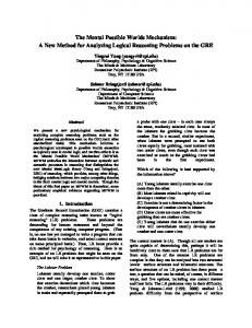
Figure 2. Detail from a submucosal neoplastic gland at the invading tumour edge. Note pore formation at arrows and release of the glandular mucins with inflammatory cells directly into the peritumoral ECM (H&E 10x).
2504
Rubio: Invasion in Adenomas
Figure 3. Detail from a submucosal neoplastic gland at the invading tumour edge. Note pore formation at arrows and direct contact between the glandular material and the peritumoral ECM (MNF 116 immunostain, 40x).
Materials and Methods Histological sections from 56 colonic adenomas presenting submucosal invasion were studied. Sections were stained with hematoxylin and eosin (H&E) and MNF 116 immunostain (Dako, Denmark). Adenomas were histologically classified into tubular, villous, mixed (tubulo-villous), serrated and microtubular, as reported elsewhere (15). Pore formation was defined as a glandular defect characterised by the lack of a group of epithelial cells in the deeper portion of the glands. A semi-quantitative estimation of the number of submucosal neoplastic glands with pores was done as follows: none to occasional, and ≥5.
Results Of the 56 adenomas showing submucosal invasion, 3 were tubular, 4 tubulo-villous, 38 villous, 9 serrated and 2 microtubular. Thus, the majority (68%) of the adenomas with submucosal invasion in this study were villous adenomas.
Figure 4. Another example of pore formation in a submucosal neoplastic gland at the invading tumour edge. Note broad pore formation at arrows, the release of the glandular contents and destruction of the periglandular ECM (MNF 116 immunostain, 40x).
2505
ANTICANCER RESEARCH 25: 2503-2508 (2005) Numerous dilated submucosal glands with pores were found in 46 (82%) of the 56 neoplasias: in 1 (33%) of the 3 tubular adenomas, in all 4 (100%) tubulo-villous adenomas, in 31 (82%) of the 38 villous adenomas, in 8 (89%) of 9 serrated adenomas, and in the 2 (100%) microtubular adenomas. In the remaining 10 (18%) of the 56 neoplasias, from none to occasional dilated submucosal glands with pores were found: in 2 (67%) of 3 the tubular adenomas, in none of the 4 tubulovillous adenomas, in 7 (18%) of the 38 villous adenomas, and in 1 (11%) of the 9 serrated adenomas. The retained materials in the dilated glands with pores were mucin, granulocytes and/or necrotic material (Figures 1 and 2). The occurrence of glandular pores was clearly visualized in sections stained with MNF 116 immunostain (Figures 3 and 4).
Discussion The mechanism of tumour invasion has been studied extensively (3, 32). These studies have focused on the kinetic ability of cancer cells to migrate into the surrounding matrix. The authors maintain that tumour cell locomotion is the single most important parameter accountable for the local progression of the tumour (3, 32). Proteolytic enzymes native to the ECM, e.g. metalloproteinases (MMPs), cathepsins and serine proteases (1, 13), cause the disintegration of the paratumoral ECM, thus encouraging tumour cell progression. Masaki et al. (13) maintain that the proteolytic degradation of the ECM by matrix metalloproteinases (MMPs) is one of the essential events in tumour invasion. At present, the 21 members of the human MMP gene family are classified into subgroups of proteolytic enzymes: collagenases, stromelysins, matrilysins, gelatinases, membrane-type MMPs (MT-MMPs) and other MMPs (13). Friedl and Wolf (3) postulated that the peritumoral breakdown of the ECM would generate localised matrix defects and remodelling along migration tracts. On the other hand, increased collagen degradation by MMPs in the bowel wall can also be achieved in the absence of a growing tumour, in experimentally-induced colonic obstruction (27). Moreover, the failure of broadspectrum MMP inhibitors in clinical trials has opened the door for other proteases to be considered as relevant drug targets in anticancer therapies (30). Recently, Joyce et al. (5) demonstrated that, although ECM degradation has largely been attributed to MMPs, it is now clear that different classes of cancer cell proteases contribute to tumour penetration, with cathepsins being involved directly in the degradation of ECM. Degradation of the ECM (including laminin, fibronectin, and collagen) may also come about through the modulation of protease-sensitive regulatory networks, involving other proteases and non-
2506
proteases such as anexin II (found at the cellular surface of cancer cells) (5, 30). Other enzymes, produced by cancer cells and whose activity is enhanced in tumours, include heparanase (8) and serine/threonine kinase AKT (29). The neo-production of lysozymes by colorectal tumour cells has been demonstrated in materials discharged through tumoral glandular pores into the peritumoral ECM (18). Furthermore, neutrophils are known to release lysosomal enzymes into the ECM (31). It may be argued that pore formation in invading tumour glands (described here and elsewhere) (17-24) is a haphazard event. But if that is the case, why are glandular pores more often found at the growing edge of submucosal or overt invasive carcinomas (17-22) than in adenomas without invasion (24)? And why does this phenomenon occur at the growing edge and not within the tumour mass? In the present investigation, we found that, at the invading front, 98% or 55 of the 56 adenomas with submucosal invasion had one or more submucosal dilated glands with pores. In 82% of the tumours submucosal glands with pores recorded at the invading tumour front were numerous. As in the case of overt colorectal carcinomas (1722), the accumulated intra-glandular material was discharged through glandular pores into the peritumoral ECM. It is assumed that the proteolytic enzymes released through the pores would lead to the breakdown of the peritumoral ECM at the growing tumour edge, thus facilitating host penetration by neoplastic cells. To remodel the defective glands, malignant cells proliferating from the tip of the free borders of the pores would invade the enzymaticallydisrupted matrix, with the goal of achieving glandular continuity. The sealing of the glandular flaws would permit the re-accumulation of new intra-glandular proteolytic material, a mechanism that would replicate a new wave of host invasion at the growing edge, thus ensuring a stepwise, but everlasting, tumour progression in untreated patients.
References 1 Ahokas K, Lohi J, Illman SA, Llano E, Elomaa O, Impola U, Karjalainen-Lindsberg ML and Saarialho-Kere U: Related matrix metalloproteinase-21 is expressed epithelially during development and in cancer and is up-regulated by transforming growth factorbeta1 in keratinocytes. Lab Invest 83:1887-1899, 2003. 2 Bhowmick NA, Chytil A, Plieth D, Gorska AE, Dumont N, Shappell S, Washington MK, Neilson EG and Moses HL: TGFbeta signaling in fibroblasts modulates the oncogenic potential of adjacent epithelia. Science 303: 848-851, 2004. 3 Friedl P and Wolf K: Tumour-cell invasion and migration: diversity and escape mechanisms. Nat Rev Cancer 3: 362-374, 2003. 4 Hazen LG, Bleeker FE, Lauritzen B, Bahns S, Song J, Jonker A, Van Driel BE, Lyon H, Hansen U, Kohler A and Van Noorden CJ: Comparative localization of cathepsin B protein and activity in colorectal cancer. J Histochem Cytochem 48: 1421-1430, 2000.
Rubio: Invasion in Adenomas
5 Joyce JA, Baruch A, Chehade K, Meyer-Morse N, Giraudo E, Tsai FY, Greenbaum DC, Hager JH, Bogyo M and Hanahan D: Cathepsin cysteine proteases are effectors of invasive growth and angiogenesis during multistage tumourigenesis. Cancer Cell 5: 443-453, 2004. 6 Kanamaru T, Tanaka K, Kotani J, Ueno K, Yamamoto M, Idei Y, Hisatomi H and Takeyama Y: Telomerase activity and hTERT mRNA in development and progression of adenoma to colorectal cancer. Int J Mol Med 10: 205-210, 2002. 7 Kurisu Y, Shimoda T, Ochiai A, Nakanishi Y, Hirata I and Katsu KI: Histologic and immunohistochemical analysis of early submucosal invasive carcinoma of the colon and rectum. Pathol Int 49: 608-616, 1999. 8 Kurokawa H, Katsube K, Podyma KA, Ikuta M, Iseki H, Nakajima M, Akashi T, Omura K, Takagi M and Yanagishita M: Heparanase and tumour invasion patterns in human oral squamous cell carcinoma xenograft. Cancer Sci 94: 277-285, 2003. 9 Lastraioli E, Guasti L, Crociani O, Polvani S, Hofmann G, Witchel H, Bencini L, Calistri M, Messerini L, Scatizzi M, Moretti R, Wanke E, Olivotto M, Mugnai G and Arcangeli A: herg1 gene and HERG1 protein are overexpressed in colorectal cancers and regulate cell invasion of tumour cells. Cancer Res 64: 606-611, 2004. 10 Li JQ, Miki H, Ohmori M, Wu F and Funamoto Y: Expression of cyclin E and cyclin-dependent kinase 2 correlates with metastasis and prognosis in colorectal carcinoma. Hum Pathol 32: 945-953, 2001. 11 Lohi J, Oivula J, Kivilaakso E, Kiviluoto T, Frojdman K, Yamada Y, Burgeson RE, Leivo I and Virtanen I: Basement membrane laminin-5 is deposited in colorectal adenomas and carcinomas and serves as a ligand for alpha3 beta1 integrin. APMIS 108: 161-172, 2000. 12 Malhotra S, Newman E, Eisenberg D, Scholes J, Wieczorek R, Mignatti P and Shamamian P: Increased membrane type 1 matrix metalloproteinase expression from adenoma to colon cancer: a possible mechanism of neoplastic progression. Dis Colon Rectum 45: 537-543, 2002. 13 Masaki T, Sugiyama M, Matsuoka H, Abe N, Izumisato Y, Sakamoto A and Atomi Y: Matrix metalloproteinases may contribute compensationally to tumour invasion in T1 colorectal carcinomas. Anticancer Res 23: 4169-4174, 2003. 14 Miyaji E, Nishimori I, Taniuchi K, Takeuchi T, Ohtsuki Y and Onishi S: Overexpression of carbonic anhydrase-related protein VIII in human colorectal cancer. J Pathol 201: 37-45, 2003. 15 Ogawa H, Iwaya K, Izumi M, Kuroda M, Serizawa H, Koyanagi Y and Mukai K: Expression of CD10 by stromal cells during colorectal tumour development. Hum Pathol 33: 806-811, 2002. 16 Rao J: Molecular mechanisms of glioma invasiveness: the role of proteases. Nature Rev Cancer 3: 489-500, 2003. 17 Rubio CA: Colorectal carcinomas. Possible mechanism of local tumour progression. Anticancer Res 23: 347-350, 2003. 18 Rubio CA: Colorectal adenomatous cells produce lysozyme. Anticancer Res 23: 5165-5172, 2003.
19 Rubio CA: Adenocarcinoma in inflammatory colorectal disease. Histologic features pertinent to local tumour progression. Anticancer Res 23: 4313-4318, 2003. 20 Rubio CA: Further studies on the histological characteristics linked to local tumour invasion in colorectal carcinomas. Anticancer Res 23: 3555-3560, 2003. 21 Rubio CA and Lindhblom A: Histological changes pertinent to local tumour progression in Hereditary Nonpolyposis Colorectal Cancer (HNPCC). Anticancer Res 24: 1765-1768, 2004. 22 Rubio CA: Possible mechanism of local tumour progression in experimentally-induced colorectal carcinomas in rats. In Vivo 17: 97-100, 2003. 23 Rubio CA and Lagergren J: Histologic features pertinent to local tumour progression in Barrett’s adenocarcinoma. Anticancer Res 23: 3015-3018, 2003. 24 Rubio CA: Possible mechanism pertinent to intramucosal invasion in sporadic colonic adenomas. Anticancer Res 24: 2033-2036, 2004. 25 Sakashita M, Aoyama N, Minami R, Maekawa S, Kuroda K, Shirasaka D, Ichihara T, Kuroda Y, Maeda S and Kasuga M: Glut1 expression in T1 and T2 stage colorectal carcinomas: its relationship to clinicopathological features. Eur J Cancer Review 37: 204-209, 2001. 26 Saleh HA, Jackson H and Banerjee M: Immunohistochemical expression of bcl-2 and p53 oncoproteins: correlation with Ki67 proliferation index and prognostic histopathologic parameters in colorectal neoplasia. Appl Immunohistochem Mol Morphol 8: 175-182, 2000. 27 Syk I, Mirastschijski U, Jeppsson BW and Agren MS: Experimental colonic obstruction increases collagen degradation by matrix metalloproteinases in the bowel wall. Dis Colon Rectum 46: 1251-1259, 2003. 28 Takahashi Y, Ellis LM and Mai M: The angiogenic switch of human colon cancer occurs simultaneous to initiation of invasion. Oncol Rep 10: 9-13, 2003. 29 Tanno S, Yanagawa N, Habiro A, Koizumi K, Nakano Y, Osanai M, Mizukami Y, Okumura T, Testa JR and Kohgo Y: Serine/threonine kinase AKT is frequently activated in human bile duct cancer and is associated with increased radioresistance. Cancer Res 64: 3486-3490, 2004. 30 Turk V, Kos J and Turk B: Cysteine cathepsins (proteases). On the main stage of cancer? Cancer Cell 5: 409-410, 2004. 31 Voetman A, Weening R, Hamers M, Meerhof L, Bot A and Roos D: Phagocytosing human neutrophils inactive their own granular enzymes. J Clin Invest 67: 1541-1549, 1981. 32 Wells A: Tumor invasion: role of growth factor-induced cell motility. In: Advances in Cancer Research (Vande Woude G and Klein G, eds.), Academic Press 78: 31-90, 2000.
Received December 27, 2004 Accepted April 6, 2005
2507