toes syndrome.9 10. We had the ... Discussion. We report a post-traumatic movement disorder .... and moving toes associated with tarsal tunnel syndrome.
J Neurol Neurosurg Psychiatry 1998;64:673–675
673
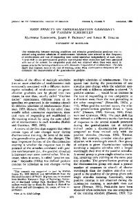
SHORT REPORT
Post-traumatic stimulus suppressible myoclonus of peripheral origin Frédéric Assal, Michel R Magistris, François J G Vingerhoets
Abstract A patient is described who presented with myoclonus of the first dorsal interosseus muscle of the right foot. This myoclonus occurred 18 months after trauma of the cutaneous branch of the deep peroneal nerve on the dorsal aspect of the foot. Tactile stimulation in the dermatome of this nerve, or an anaesthetic block of the deep peroneal nerve stopped the myoclonus. The diVerent innervation between the eVerent motor activity responsible for the movements and the sensory aVerence suppressing it points firmly towards involvement of central connections. However, abolition of the movement by anaesthesia suggests the presence of a peripheral ectopic generator. This finding confirms that focal myoclonus can have its origin in the peripheral nervous system and may be modulated by sensory inputs. (J Neurol Neurosurg Psychiatry 1998;64:673–675) Keywords: post-traumatic movement disorders; myoclonus; peripheral nerve; plasticity
Clinique et Policlinique de Neurologie, H U G, Geneva, Switzerland F Assal M R Magistris F J G Vingerhoets Correspondence to: Dr FJG Vingerhoets, Policlinique de Neurologie, HUG, 24 rue Micheli-du-Crest, 1211 Genève 14, Switzerland. Telephone 0041 22 372 8318; fax 0041 22 372 8332. Received 24 July 1997 and in revised form 30 October 1997 Accepted 12 November 1997
Post-traumatic movement disorders may follow injuries of the central or peripheral nervous system.1 2 Trauma resulting in a focal lesion may contribute to the understanding of the neural circuitry involved in the development of various movement disorders. It has been hypothesised that peripheral nerve lesions could alter sensory inputs and induce central reorganisation generating the movement disorder.3–6 This theory is based mainly on experimental evidence but clinical findings are scarce.3–8 Similar mechanisms have been proposed to explain the painful leg and moving toes syndrome.9 10 We had the unique opportunity to study a myoclonus of the first dorsal interosseous muscle of the right foot that followed a direct trauma to the cutaneous branch of the deep peroneal nerve. The very restricted localisations of both the nerve lesion and the myoclonus has allowed us to explore part of the physiopathology of post-traumatic focal myoclonus.
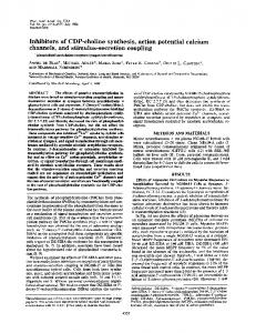
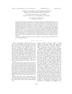
Case report A 24 year old carpenter dropped a heavy piece of wood on the dorsum of his right foot in August 1993. This caused local haematoma but no fracture. Eighteen months later he noticed a continuous painless involuntary movement of the second toe occurring at rest. This movement disappeared during voluntary movements of the toe or when walking, and was absent during sleep. Unchanged after six months, this symptom led to a neurological referral to our clinic. There was a continuous rhythmic movement of adduction of the second right toe. A hypoaesthesia and slight dysaesthesia involved a triangular area overlying the first intermetatarsal space and the base of the first and second toes (fig 1). This area corresponds to the sensory innervation of the cutaneous branch of the deep peroneal nerve. A Tinel’s sign was found near the proximal angle of the hypoaesthetic area on the dorsum of the foot, 4 cm proximal to the web space. Gentle rubbing of the hypoaesthetic skin surface consistently suppressed the rhythmic contractions, which resumed immediately after cessation of the stimulus. Neither similar stimulation of any other skin areas nor passive manoeuvres
1
3 2 Figure 1 Stimulus suppressive myoclonus. Hatched surface corresponds to the hypoaesthesic area. Arrow shows the myoclonus of the second toe. (1) Location of the Tinel’s sign. (2) Site of xylocaine injection that causes complete anaesthesia of the hatched zone without aVecting the myoclonus. (3) Site of xylocaine injection that suppresses the myoclonus.
674
Assal, Magistris, Vingerhoets
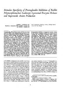
Figure 2 EMG recording of the first dorsal interosseus muscle shows bursts of spontaneous activity at 1.6 Hz lasting up to 400 ms, corresponding to the myoclonus. Black bar shows when the tactile stimulus was applied.
performed on the right foot aVected the myoclonus. Neurological examination was otherwise normal. A standard EEG showed no epileptiform activity. Back averaged EEG and back averaged potentials at the lumbar spinal level showed no activity correlated with the myoclonus. An MRI of the spinal cord and lumbosacral plexus was normal. An electroneuromyography (Vicking IVNicolet) was performed. The sensory-motor conduction studies of the peroneus superficialis, sural, deep peroneal, and tibial nerves were normal. Needle EMG of the extensor digitorum brevis was normal. No abnormal activity was noted in other muscles (flexor digitorum pedis brevis, flexor hallucis brevis, abductor digiti minimi pedis). The concentric needle EMG recordings of the first dorsal interosseus muscle showed bursts of spontaneous rhythmic activity occurring at 1.6 Hz and lasting between 200 and 400 ms. This activity was synchronous to the clinical myoclonus. Each burst included the activity of several motor unit action potentials (fig 2). Motor responses of the first dorsal interosseus muscle were elicited by stimulation of the tibial nerve and not by stimulation of the deep peroneal nerve, confirming a normal innervation. A pharmacological test was performed. Injection of 1 ml 2% xylocaine along the cutaneous branch of the deep peroneal nerve, distal to the site of Tinel’s sign, anaesthetised the skin territory of the deep peroneal nerve. This did not aVect the myoclonus but subsequent tactile stimulation of the triangular skin surface no longer stopped the abnormal movement. A second xylocaine injection 15 minutes later 2 cm proximal to the site of the Tinel’s sign suppressed the abnormal movement for five minutes. Discussion We report a post-traumatic movement disorder that was suppressed by a tactile stimulus. The movements were more rhythmic and sinusoidal than classic myoclonus, and the EMG bursts were longer in duration than what has been described in this condition. These characteristics were similar to those found in the painful
legs and moving toes syndrome9 10 but our patient had no pain, and one toe only was involved. Therefore we favoured the term of myoclonus that has already been applied to similar movements in previous reports.5 11 12 Myoclonus may arise from diVerent generators located in the CNS—namely, in the cortex, brainstem reticular formation, or spinal cord.11 12 Some cortical and reticular myoclonus have been termed reflex myoclonus because they may be triggered by sensory stimuli. Similarly, some spinal myoclonus may be stimulus sensitive.12 Our patient had no clinical, radiological, or electrophysiological evidence for such a CNS origin of the myoclonus. In addition, the myoclonus was suppressed rather than triggered by the sensory stimulus. A peripheral origin of this myoclonus is strongly supported by its suppression after an anaesthetic block of the cutaneous branch of the deep peroneal nerve proximal to the site of the Tinel’s sign. Such a peripheral generator to a myoclonus has been hypothesised in cervical and lumbosacral plexus3–6 and, rarely, in peripheral nerve lesions as in spasms of amputation stumps.13 However, these lesions were not so precisely localised. In our case, the peripheral ectopic activity could have been generated by a small neuroma at the site of the lesion. A central relay is suggested by the localisation of the myoclonus to the first dorsal interosseus muscle. In our patient, we verified that this muscle was normally innervated by the tibial nerve. The diVerence of innervation between the generator of the myoclonus, dependent on the deep peroneal nerve, and the eVector, innervated by the tibial nerve, imply a relay within the spinal cord, probably at the level L5-S1. The 18 month delay between injury and appearance of the myoclonus suggests a possible central reorganisation before the myoclonus started. However, in our case this reorganisation was neither permanently established nor independent as the myoclonus could be suppressed: (a) by tactile stimulation of the sensory territory of the injured deep peroneal nerve; (b) by the proximal anaesthetic block.
675
Myoclonus of peripheral origin
The modulation of an abnormal movement by sensory input may be related to various mechanisms. It has been proposed that lesion of a sensory nerve may lead to abnormal sensory spinal aVerences causing loss of local inhibitory spinal interneurons.7 8 This lack of inhibition could allow the activation of segmental á-motor neurons generating the abnormal movement. In our case, the sensory stimulation would stop the myoclonus by overcoming this very limited inhibitory deficit. Alternatively, sensory modulation may have a peripheral origin. Ectopic impulses generated at the level of a neuroma may be blocked through axonal reflex by the potentials caused by the tactile stimuli. In both central or peripheral hypotheses, the anaesthetic block distal to the peripheral generator would prevent the tactile suppression, as we found. To conclude, our finding confirms that posttraumatic myoclonus may have its origin in the peripheral nervous system. It strongly supports that such peripheral origin implies a generation of a continuous peripheral abnormal activity at the lesion site. In addition it shows that, even after months, the abnormal activity can still be modulated by sensory input.
This work was supported by a grant from the Swiss National Science Foundation (grant No 32–51 090.97).
1 Jankovic J. Post-traumatic movement disorders: central and peripheral mechanisms. Neurology 1994;44:2006–14. 2 Koller WC, Wong GF, Lang A. Posttraumatic movement disorders: a review. Mov Disord 1989;4:20–36. 3 Said G, Bathien N. Myoclonies rythmées du quadriceps en relation avec un envahissement sarcomateux du nerf crural. Rev Neurol 1977;133:191–8. 4 Sotaniemi KA. Paraspinal myoclonus due to spinal root lesion. J Neurol Neurosurg Psychiatry 1985;48:722–3. 5 Glocker FX, Deuschl G, Volk B, et al. Bilateral myoclonus of the trapezius muscles after distal lesion of an accessory nerve. Mov Disord 1996;11:571–5. 6 Banks G, Nielsen VK, Short MP, et al. Brachial plexus myoclonus. J Neurol Neurosurg Psychiatry 1985;48:582–4. 7 Wall PD, Gutnick M. Ongoing activity in peripheral nerves: the physiology and pharmacology of impulses originating from a neuroma. Exp Neurol 1974;43:580–93. 8 Gourin-Lippmann R, Devor M. Ongoing activity in severed nerves: source and variation in time. Brain Res 1978;159: 406–10. 9 Dressler D, Thompson PD, Gledhill RF, et al. The syndrome of painful legs and moving toes. Mov Disord 1994;9:13–21. 10 Pla MER, Dillingham RD, Spellman NT, et al. Painful legs and moving toes associated with tarsal tunnel syndrome and accessory soleus muscle. Mov Disord 1996;11:82–6. 11 Jankovic J, Pardo R. Segmental myoclonus: clinical and pharmacological study. Arch Neurol 1986;43:1025–31. 12 Davis SM, Murray NMF, Diengdoh JV, et al. Stimulussensitive spinal myoclonus. J Neurol Neurosurg Psychiatry 1981;44:884–8. 13 Marion MH, Gledhill RF, Thompson PD. Spasms of amputation stumps: a report of 2 cases. Mov Disord 1989;4:354– 8.