for visualizing local translation in dendrites. One technical challenge in the field was separating existing proteins from newly synthesized proteins. We were able ...
Posttranscriptional Regulation of Intrinsic Plasticity Kimberly F. Raab-Graham, PhD Center for Learning and Memory Section of Neurobiology University of Texas at Austin Austin, Texas
© 2010 Raab-Graham
Posttranscriptional Regulation of Intrinsic Plasticity
Introduction
In 1894, Santiago Ramon y Cajal proposed that learning results from changes in synaptic strength (Cajal, 1894). This idea is consistent with the basic premise for memory formation and suggests that modifying independent synapses provides a large capacity to store information (Martin et al., 2000). Nearly 80 years after Cajal’s initial proposition, a landmark study by Bliss and Lomo demonstrated that excitatory synapses in the hippocampus can undergo persistent changes in synaptic strength that can be sustained for hours or even several days. These persistent changes are the cellular basis for a model of learning and memory that is commonly referred to as “long-term potentiation” (LTP) (Bliss and Lomo, 1973). Today, we know that synaptic strengthening requires changes in the number and/or conductance of glutamate receptors (specifically AMPA and NMDA type) with the induction of LTP. (Malenka and Bear, 2004; Shepherd and Huganir, 2007; Kessels and Malinow, 2009). However, new questions emerge if we consider the fact that LTP is Hebbian in nature, i.e., that synapses can be modified by previous experience (Hebb, 1949). How do changes in glutamate receptor activity influence the propensity of a synapse and nearby synapses to respond to presynaptic release? What are the underlying mechanisms that change postsynaptic responsiveness or, in other words, set the plasticity threshold, a process that Abraham and Bear termed “metaplasticity” (Abraham and Bear, 1996)? Can these metaplastic changes be compartmentalized in such a way that they are restricted to a synapse, a dendritic branch, or a dendritic tree?
Anatomy of a Neuron
The nature of a neuron is polarized. It naturally divides into microdomains of specifically targeted proteins that facilitate the structural and functional differences between the dendrites and the axon. Dendrites themselves can be subdivided into individual compartments based on several factors: their proximity to the soma, their branching patterns from the apical trunk, and the synaptic inputs they receive (Spruston, 2008). The proteins that make up these specialized microdomains determine the synaptic efficacy of the individual compartment. In neurons, the relative density and dendritic localization of ion channels play important functional roles in synaptic integration, plasticity, and neuronal excitability (Frick and Johnston, 2005). Historically, dendrites were viewed as electrically passive, in contrast to the electrical excitability of the soma © 2010 Raab-Graham
(Johnston et al., 1996). However, this idea has been found to be inconsistent with the observation that action potential firing exceeds what would be predicted by the summation of LTP-induced EPSPs. Bliss and colleagues described this phenomena as a “nonsynaptic component of LTP” (Bliss and GardnerMedwin, 1973). We now know that neuronal dendrites are not passive and that, in fact, they have a rich and complex distribution of ion channels (Zhang and Linden, 2003; Frick and Johnston, 2005; Bloodgood and Sabatini, 2008). The first indication that voltage-gated ion channels are “plastic” during LTP came from a series of experiments that initially discovered that sodiumdependent action potentials can propagate back into the dendrites from the soma (i.e., back-propagating action potential, or bAP) (Johnston et al., 1996). To test whether LTP causes changes in ion channel properties that alter membrane excitability (often referred to as “intrinsic plasticity”), bAPs were paired with synaptic stimulation (Frick et al., 2004; Frick and Johnston, 2005). Using calcium imaging and dendritic recordings, Johnston and colleagues found that, at the site of stimulation, there was an increase in bAP amplitude and an enhanced calcium signal, suggesting an increase in local dendritic depolarization (Frick et al., 2004). To account for this unexpected heightened excitability, changes in channel-gating and endocytosis of potassium channels have been proposed (Frick et al., 2004; Kim et al., 2007). Another possible mechanism, as our work suggests, is that NMDA activity triggers the posttranscriptional repression of potassium channel mRNA translation (Raab-Graham et al., 2006). These critical experiments open the field to questioning how changes in synaptic efficacy are coupled to changes in dendritic excitability, both locally and globally (Zhang and Linden, 2003; Frick and Johnston, 2005; Kim and Linden, 2007). Thus, this chapter will focus first on the importance of local translation and repression during LTP. Second, it will discuss the identification and the regulation of local translation of ion channel mRNAs in neuronal dendrites. Third, it will describe how site-specific changes in dendritic excitability are important for synaptic plasticity and how, if left unchecked, they may be involved in neurodegeneration.
Significance of mRNA Translation and Repression in Long-term Potentiation
LTP consists of two phases: early and late. It is widely believed that late-phase LTP serves as a useful model
47
Notes
48
Notes
for the consolidation phase of memory formation (Pittenger and Kandel, 2003). For several years, we have known that the conversion of early LTP to late phase requires protein synthesis (Krug et al., 1984; Frey et al., 1993; Abel et al., 1997). In 1983, Bliss and colleagues set out to examine changes in protein synthesis during late LTP using two-dimensional protein electrophoresis of 35S-methionine–labeled proteins that were synthesized during LTP in vivo (Fazeli et al., 1993). Remarkably, this technique revealed not only an increase in protein synthesis but also a corresponding decrease in the relative abundance of certain proteins. Interestingly, in their discussion, the authors stated that LTP was accompanied by the “reduction in synthesis (or an increase in degradation).” Since then, proteosomemediated protein degradation during neuronal activity has been an active area of research (Hegde and DiAntonio, 2002). Nonetheless, the idea of suppression of protein synthesis has been largely ignored.
mTOR, Plasticity, and Memory
Consistent with the requirement for protein synthesis during late-phase LTP, signaling of the mammalian target of rapamycin (mTOR) pathway is required for the maintenance of late LTP. mTOR is a serine/threonine kinase whose primary function is to promote mRNA translation initiation (Hay and Sonenberg, 2004). In hippocampal slices, elegant electrophysiological experiments that block mTOR signaling via the mTOR-specific inhibitor rapamycin, prior to LTP induction, reduced the magnitude of LTP for more than 5 h. (Tang et al., 2002; Cammalleri et al., 2003; Vickers et al., 2005). Further, inventive genetic approaches that resulted in increased basal phosphorylation of mTOR in mice have shown enhanced late LTP (Hoeffer et al., 2008). In line with the findings of these reports, behavioral tasks designed to assess learning and memory in rodents have indicated that mTOR activity is required for memory formation, consolidation, and reconsolidation (Casadio et al., 1999; Parsons et al., 2006; Bekinschtein et al., 2007; Blundell et al., 2008; Hoeffer et al., 2008).
Local Translation and Neuronal Plasticity
Unexpectedly, mTOR has been found both in the cell body and in the dendrites of neurons, suggesting that protein translation also occurs in the dendrites (Tang et al., 2002). These data challenge the view that all proteins are synthesized in the cell body and transported to the dendrites. Further support for the dendritic protein translation hypothesis has been
found in the ability to detect polyribosomes, mRNAs, translation machinery, as well as components of the secretory pathway (endoplasmic reticulum and golgi membranes) in dendrites (Bramham and Wells, 2007). Moreover, numerous biochemical assays on synaptosomes (isolated presynaptic and postsynaptic nerve endings) and severed dendrites have demonstrated the incorporation of radioactive amino acids in proteins in the absence of cell bodies (Schuman, 1997; Steward, 1997). Finally, the use of molecular tools that we and others have developed to visualize local protein synthesis in dendrites have advanced our understanding as to how local translation contributes to neuronal plasticity (Aakalu et al., 2001; Raab-Graham et al., 2006). Thus, local translation provides the unique advantage over somatic translation and protein trafficking by making available a specific source of new proteins in response to site-specific changes in synaptic strength (Schuman et al., 2006).
Characterization of Dendritic Kv1.1 Local Synthesis
Based on the evidence that voltage-gated ion channels are important in dendritic signaling, Patrick Haddick and I performed our own screen, looking for synaptic mRNAs while I was a postdoctoral fellow in the laboratory of Dr. Lily Jan at the University of California, San Francisco. Using microarrays and quantitative real-time PCR, we compared mRNA isolated from synaptosomes with mRNA isolated from the hippocampus. We determined that 4% of all transcripts assayed, including 202 known genes, reside at the synapse. These mRNAs report a synaptosome “intensity value” greater than CaMKII (a message that has been previously reported to be localized to dendrites) and a synaptosome-to-hippocampus ratio of greater than 1.2. Interestingly, several transcripts encoding ion channels and neurotransmitter receptors known to be involved in synaptic plasticity were enriched in the synaptosomal fraction. The first transcript we characterized from our microarray data was the dendrotoxin-sensitive, voltage-gated potassium channel Kv1.1, which controls the frequency of the action potential (Tanouye and Ferrus, 1985). Although a dendrotoxinsensitive Kv current has been described in CA1 pyramidal neurons, the molecular identity of this current is unknown (Chen and Johnson, 2010) and the specific role for Kv1.1 in the dendrites of hippocampal neurons has not been established. We verified our microarray data by detecting endogenous Kv1.1 mRNA in dendrites of cultured © 2010 Raab-Graham
Posttranscriptional Regulation of Intrinsic Plasticity
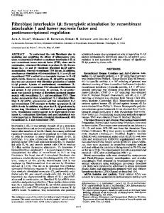
hippocampal neurons using in situ hybridization. The results were surprising because the prior thinking held that Kv1.1 was expressed exclusively in the axon in the hippocampus (Schechter, 1997; Southan and Owen, 1997; Geiger and Jonas, 2000; Monaghan et al., 2001; Raab-Graham et al., 2006). Thus, Kv1.1 mRNA trafficking provides a mechanism for the protein to escape the restrictive protein-encoded trafficking signals that direct the protein to the axon in order to be expressed in the dendrites in an activity-dependent manner. To confirm the dendritic localization of the channel, we demonstrated that the mTOR inhibitor rapamycin increased the total Kv1.1 protein in the CA1 region of the hippocampus and the surface expression in cultured neurons (Fig. 1A,B). These results were contrary to what we expected, because
mTOR activity was supposed to promote translation. However, what we found was that mTOR activity represses the translation of Kv1.1 mRNA. To verify this unusual finding, we decided to visualize local dendritic protein synthesis of Kv1.1. To do this, we developed an improved method for visualizing local translation in dendrites. One technical challenge in the field was separating existing proteins from newly synthesized proteins. We were able to solve this problem through the use of the photoconvertible protein Kaede (Mizuno et al., 2003) and multiphoton microscopy. Ultraviolet (UV) light induces a specific proteolytic cleavage of Kaede, converting its fluorescence from green into red. This property allowed us to develop a translational reporter by fusing Kaede to Kv1.1. Newly synthesized Kaede–Kv1.1 is distinguished
Figure 1. Inhibition of mTOR by rapamycin increases the local translation of Kv1.1 mRNA in central neuronal dendrites. A, Acute hippocampal slices treated with carrier or rapamycin (200 nM) for 75 min. Slices were fixed and stained with an antibody against Kv1.1. Note the significant increase in Kv1.1 expression in CA1 and dentate gyrus with mTOR inhibition by rapamycin. B, Surface staining of Kv1.1 protein of treated hippocampal neurons. Map2-positive dendrites show a significant increase in Kv1.1 protein in the dendrite greater than 50 µm from the soma. Scale bar, 20 µm. Raab-Graham et al. (2006), their Fig. 1A,D, reprinted with permission. © 2010 Raab-Graham
49
Notes
50
Notes
by its green chromophore and spectrally separated from the previously synthesized red protein (RaabGraham et al., 2006) (Fig. 2A). With tools in hand, we went on to demonstrate that Kaede–Kv1.1 is locally synthesized in dendrites upon inhibition of mTOR (Fig. 2B). These results led us to form a model proposing that the suppression of Kv1.1 mRNA translation is an imperative positive-feedback mechanism during
NMDA/mTOR–dependent neuronal activity that may be important for memory formation (Fig. 3). This model suggests that, at the site of synaptic stimulation, mTOR actively suppresses the translation of Kv1.1 mRNA, resulting in fewer channels on the surface of the dendrite, thereby creating a functional unit of the dendrite to be more excitable. Furthermore, at inactive synapses, Kv1.1 mRNA is translated and expressed on the membrane, possibly to increase the plasticity threshold.
Figure 2. Local translation assay using photoconvertible protein Kaede. A, Schematic of local translation assay using the photoconvertible protein Kaede. Left side shows neuron with boxed dendrites; right side shows enlargement. Green puncta indicate local translation “hot spots.” Kaede initially appears green, but with UV exposure, is converted to red. New protein synthesis is monitored by the appearance of new green protein over time (arrowheads). B, Live imaging of neurons expressing Kaede–Kv1.1 treated with carrier (DMSO) or rapamycin (200 nM) before, immediately after (0 min), and 60 min after the first UV exposure to photoconvert Kaede-Kv1.1 into red protein. Representative grayscale images show green fluorescence in neurons. Dendrite is outlined in orange within the black box, with arrows pointing to a single translational “hot spot” for Kaede-Kv1.1 (Raab-Graham et al., 2006). © 2010 Raab-Graham
Posttranscriptional Regulation of Intrinsic Plasticity
51
Notes
Figure 3. Model of positive feedback mechanism for the specific enhancement of dendritic excitability during NMDA/mTOR– mediated synaptic activity. Neuron on the left has an inactive synapse (gray) and mTOR is not active, thus permitting Kv1.1 local translation (green, RNA; yellow, channel subunit). Neuron on the right shows an active synapse (purple); NMDA-R (NMDA receptor) activity (white) turns on the mTOR signaling pathway via the PI3 kinase. mTOR suppresses the translation and insertion of Kv1.1 channels in dendrites. Activity-dependent changes in Kv1.1 expression may affect dendritic signaling via synaptic input and bAPs.
Misregulation of mTOR Leads to Seizure and Cognitive Decline
Patients with neurological diseases caused, at least in part, by overstimulation of the mTOR pathway experience seizures and cognitive defects. Overactive mTOR has been implicated in diseases such as epilepsy, tuberous sclerosis complex (TSC), Fragile X syndrome, and Alzheimer’s disease (AD) (Meikle et al., 2007, 2008; Pei and Hugon, 2008; Zeng et al., 2009; Sharma et al., 2010), all of © 2010 Raab-Graham
which have epileptic seizures associated with them. Interestingly, increased seizure susceptibility directly correlates with a decrease in kv1.1 gene expression (Smart et al., 1998; Rho et al., 1999). Recently, Mucke and colleagues provided evidence that the human amyloid precursor protein transgenic mouse, a model for AD, has spontaneous, nonconvulsant seizure activity in hippocampal and cortical circuits similar to what is observed
52
Notes
in temporal lobe epilepsy. They suggest that the aberrant excitatory neuronal activity may lead to the cognitive impairment observed in AD (Palop et al., 2007). Both the cognitive deficits and the neuronal hyperexcitability observed in AD models parallel the phenotype observed in other mTORrelated diseases such as TSC. Furthermore, treating rat and mouse models of temporal lobe epilepsy and TSC with rapamycin eliminated the observed spontaneous seizures (Meikle et al., 2007, 2008; Zeng et al., 2009) and memory deficits observed in these rats and mice (Meikle et al., 2007; Ehninger et al., 2008). It remains to be determined whether the hyperexcitability of these neurons results from mTOR-repression of Kv1.1 mRNA translation. Reducing seizure activity in the brain may reduce the cognitive deficits observed in mTOR-related diseases such as AD and TSC. Although Kv1.1 is one of many transcripts whose translation is regulated by mTOR activity, its physiological role in controlling neuronal excitability is not compensated by other voltagegated potassium channels (Smart et al., 1998; Rea et al., 2002; Tavazoie et al., 2005; Gong et al., 2006; Slipczuk et al., 2009). These neurological diseases are perfect examples of how positive feedback mechanisms, if unregulated, can lead to uncontrolled neuronal excitability, thus causing neuronal circuits to become unstable and rendering the storage of new information labile (Turrigiano and Nelson, 2000). Therefore, homeostatic mechanisms need to be identified that release translational repression of Kv1.1 mRNA translation in order to prevent instability and neurodegeneration.
Considerations for Future Research
Our findings contradict the dogma that mTOR signaling promotes global cap-dependent translation. Our data suggest that cap-dependent translation of select mRNAs is induced while the translation of other mRNAs is actively repressed. In light of these data, it is interesting to reconsider the implications of earlier studies that demonstrated the reduction in late-phase LTP by the mTOR inhibitor rapamycin. It is clear that mTOR promotes the translation of important transcripts that enhance synaptic plasticity (Gong et al., 2006; Kelly et al., 2007); however, whether the mRNAs repressed by mTOR activity are memory suppressor proteins is a key unanswered question. We recently addressed this question using a mouse model for memory impairment that overexpresses the RNA-binding protein HuD and discovered that HuD promotes the translation of Kv1.1 mRNA. CA-1 pyramidal neurons from these
mice have increased Kv1.1 expression, decreased firing rates, and an increased action potential threshold. Moreover, the most striking result of this study is that calcium signals provoked by bAPs in these mice are specifically reduced in oblique dendrites when compared with wild-type littermates (P. Huang, N. Sosanya, P.Y. Chang, K. Nguyen, N.I. Perrone-Bizzozero, and K.F. Raab-Graham, unpublished observations). These data suggest that translational regulation of Kv1.1 may result in both local and global changes in intrinsic excitability and may be important for memory storage. Determining what other mRNAs are suppressed by mTOR activity, addressing how mTOR activity represses mRNA translation, and establishing whether mTOR activity regulates small noncoding regulatory RNAs, such as microRNAs, are questions for future investigation.
Acknowledgments
I would like to thank Dr. Lisa Conti, Dr. Peggy Huang, and Natasha Sosanya for helpful discussion and critical reading of this manuscript. I apologize to my colleagues whose work was not included in this manuscript and regret that all relevant work could not be cited owing to space limitations. The work described within was supported in part by the National Science Foundation Grant IOS: 1026527 to K.R.G.
References
Aakalu G, Smith WB, Nguyen N, Jiang C, Schuman EM (2001) Dynamic visualization of local protein synthesis in hippocampal neurons. Neuron 30:489-502. Abel T, Nguyen PV, Barad M, Deuel TA, Kandel ER, Bourtchouladze R (1997) Genetic demonstration of a role for PKA in the late phase of LTP and in hippocampus-based long-term memory. Cell 88:615-626. Abraham WC, Bear MF (1996) Metaplasticity: the plasticity of synaptic plasticity. Trends Neurosci 19:126-130. Bekinschtein P, Katche C, Slipczuk LN, Igaz LM, Cammarota M, Izquierdo I, Medina JH (2007) mTOR signaling in the hippocampus is necessary for memory formation. Neurobiol Learn Mem 87:303-307. Bliss TV, Gardner-Medwin AR (1973) Longlasting potentiation of synaptic transmission in the dentate area of the unanaesthetized rabbit following stimulation of the perforant path. J Physiol 232:357-374. © 2010 Raab-Graham
Posttranscriptional Regulation of Intrinsic Plasticity
Bliss TV, Lomo T (1973) Long-lasting potentiation of synaptic transmission in the dentate area of the anaesthetized rabbit following stimulation of the perforant path. J Physiol 232:331-356.
Geiger JR, Jonas P (2000) Dynamic control of presynaptic Ca(2+) inflow by fast-inactivating K(+) channels in hippocampal mossy fiber boutons. Neuron 28:927-939.
Bloodgood BL, Sabatini BL (2008) Regulation of synaptic signalling by postsynaptic, non-glutamate receptor ion channels. J Physiol 586:1475-1480.
Gong R, Park CS, Abbassi NR, Tang SJ (2006) Roles of glutamate receptors and the mammalian target of rapamycin (mTOR) signaling pathway in activity-dependent dendritic protein synthesis in hippocampal neurons. J Biol Chem 281:1880218815.
Blundell J, Kouser M, Powell CM (2008) Systemic inhibition of mammalian target of rapamycin inhibits fear memory reconsolidation. Neurobiol Learn Mem 90:28-35. Bramham CR, Wells DG (2007) Dendritic mRNA: transport, translation and function. Nat Rev Neurosci 8:776-789.
Hay N, Sonenberg N (2004) Upstream and downstream of mTOR. Genes Dev 18:1926-1945. Hebb DO (1949) Organization of behavior. New York: Wiley.
Cajal SR (1894) La dine structure des centres nerveux. Proc R Soc Lond 55:444-468.
Hegde AN, DiAntonio A (2002) Ubiquitin and the synapse. Nat Rev Neurosci 3:854-861.
Cammalleri M, Lutjens R, Berton F, King AR, Simpson C, Francesconi W, Sanna PP (2003) Time-restricted role for dendritic activation of the mTOR-p70S6K pathway in the induction of latephase long-term potentiation in the CA1. Proc Natl Acad Sci USA 100:14368-14373.
Hoeffer CA, Tang W, Wong H, Santillan A, Patterson RJ, Martinez LA, Tejada-Simon MV, Paylor R, Hamilton SL, Klann E (2008) Removal of FKBP12 enhances mTOR-Raptor interactions, LTP, memory, and perseverative/repetitive behavior. Neuron 60:832-845.
Casadio A, Martin KC, Giustetto M, Zhu H, Chen M, Bartsch D, Bailey CH, Kandel ER (1999) A transient, neuron-wide form of CREBmediated long-term facilitation can be stabilized at specific synapses by local protein synthesis. Cell 99:221-237.
Johnston D, Magee JC, Colbert CM, Cristie BR (1996) Active properties of neuronal dendrites. Annu Rev Neurosci 19:165-186.
Chen X, Johnson D (2010) The elusive D-current: shaping action potentials in the dendrites? Hauppauge, NY: Nova Science Publishers. Ehninger D, Han S, Shilyansky C, Zhou Y, Li W, Kwiatkowski DJ, Ramesh V, Silva AJ (2008) Reversal of learning deficits in a Tsc2+/– mouse model of tuberous sclerosis. Nat Med 14:843-848. Fazeli MS, Corbet J, Dunn MJ, Dolphin AC, Bliss TV (1993) Changes in protein synthesis accompanying long-term potentiation in the dentate gyrus in vivo. J Neurosci 13:1346-1353. Frey U, Huang YY, Kandel ER (1993) Effects of cAMP simulate a late stage of LTP in hippocampal CA1 neurons. Science 260:1661-1664. Frick A, Johnston D (2005) Plasticity of dendritic excitability. J Neurobiol 64:100-115. Frick A, Magee J, Johnston D (2004) LTP is accompanied by an enhanced local excitability of pyramidal neuron dendrites. Nat Neurosci 7:126-135.
© 2010 Raab-Graham
Kelly MT, Crary JF, Sacktor TC (2007) Regulation of protein kinase Mzeta synthesis by multiple kinases in long-term potentiation. J Neurosci 27:3439-3444. Kessels HW, Malinow R (2009) Synaptic AMPA receptor plasticity and behavior. Neuron 61:340-350. Kim J, Jung SC, Clemens AM, Petralia RS, Hoffman DA (2007) Regulation of dendritic excitability by activity-dependent trafficking of the A-type K+ channel subunit Kv4.2 in hippocampal neurons. Neuron 54:933-947. Kim SJ, Linden DJ (2007) Ubiquitous plasticity and memory storage. Neuron 56:582-592. Krug M, Lossner B, Ott T (1984) Anisomycin blocks the late phase of long-term potentiation in the dentate gyrus of freely moving rats. Brain Res Bull 13:39-42. Malenka RC, Bear MF (2004) LTP and LTD: an embarrassment of riches. Neuron 44:5-21. Martin SJ, Grimwood PD, Morris RG (2000) Synaptic plasticity and memory: an evaluation of the hypothesis. Annu Rev Neurosci 23:649-711.
53
Notes
54
Notes
Meikle L, Talos DM, Onda H, Pollizzi K, Rotenberg A, Sahin M, Jensen FE, Kwiatkowski DJ (2007) A mouse model of tuberous sclerosis: neuronal loss of Tsc1 causes dysplastic and ectopic neurons, reduced myelination, seizure activity, and limited survival. J Neurosci 27:5546-5558. Meikle L, Pollizzi K, Egnor A, Kramvis I, Lane H, Sahin M, Kwiatkowski DJ (2008) Response of a neuronal model of tuberous sclerosis to mammalian target of rapamycin (mTOR) inhibitors: effects on mTORC1 and Akt signaling lead to improved survival and function. J Neurosci 28:5422-5432. Mizuno H, Mal TK, Tong KI, Ando R, Furuta T, Ikura M, Miyawaki A (2003) Photo-induced peptide cleavage in the green-to-red conversion of a fluorescent protein. Mol Cell 12:1051-1058. Monaghan MM, Trimmer JS, Rhodes KJ (2001) Experimental localization of Kv1 family voltagegated K+ channel alpha and beta subunits in rat hippocampal formation. J Neurosci 21:5973-5983. Palop JJ, Chin J, Roberson ED, Wang J, Thwin MT, Bien-Ly N, Yoo J, Ho KO, Yu GQ, Kreitzer A, Finkbeiner S, Noebels JL, Mucke L (2007) Aberrant excitatory neuronal activity and compensatory remodeling of inhibitory hippocampal circuits in mouse models of Alzheimer’s disease. Neuron 55:697-711.
Schechter LE (1997) The potassium channel blockers 4-aminopyridine and tetraethylammonium increase the spontaneous basal release of [3H]5hydroxytryptamine in rat hippocampal slices. J Pharmacol Exp Ther 282:262-270. Schuman EM (1997) Synapse specificity and longterm information storage. Neuron 18:339-342. Schuman EM, Dynes JL, Steward O (2006) Synaptic regulation of translation of dendritic mRNAs. J Neurosci 26:7143-7146. Sharma A, Hoeffer CA, Takayasu Y, Miyawaki T, McBride SM, Klann E, Zukin RS (2010) Dysregulation of mTOR signaling in Fragile X syndrome. J Neurosci 30:694-702. Shepherd JD, Huganir RL (2007) The cell biology of synaptic plasticity: AMPA receptor trafficking. Annu Rev Cell Dev Biol 23:613-643. Slipczuk L, Bekinschtein P, Katche C, Cammarota M, Izquierdo I, Medina JH (2009) BDNF activates mTOR to regulate GluR1 expression required for memory formation. PLoS One 4:e6007. Smart SL, Lopantsev V, Zhang CL, Robbins CA, Wang H, Chiu SY, Schwartzkroin PA, Messing A, Tempel BL (1998) Deletion of the K(V)1.1 potassium channel causes epilepsy in mice. Neuron 20:809-819.
Parsons RG, Gafford GM, Helmstetter FJ (2006) Translational control via the mammalian target of rapamycin pathway is critical for the formation and stability of long-term fear memory in amygdala neurons. J Neurosci 26:12977-12983.
Southan AP, Owen DG (1997) The contrasting effects of dendrotoxins and other potassium channel blockers in the CA1 and dentate gyrus regions of rat hippocampal slices. Br J Pharmacol 122:335-343.
Pei JJ, Hugon J (2008) mTOR-dependent signalling in Alzheimer’s disease. J Cell Mol Med 12:25252532.
Spruston N (2008) Pyramidal neurons: dendritic structure and synaptic integration. Nat Rev Neurosci 9:206-221.
Pittenger C, Kandel ER (2003) In search of general mechanisms for long-lasting plasticity: Aplysia and the hippocampus. Philos Trans R Soc Lond B Biol Sci 358:757-763.
Steward O (1997) mRNA localization in neurons: a multipurpose mechanism? Neuron 18:9-12.
Raab-Graham KF, Haddick PC, Jan YN, Jan LY (2006) Activity- and mTOR-dependent suppression of Kv1.1 channel mRNA translation in dendrites. Science 314:144-148. Rea R, Spauschus A, Eunson LH, Hanna MG, Kullmann DM (2002) Variable K+ channel subunit dysfunction in inherited mutations of KCNA1. J Physiol 538:5-23. Rho JM, Szot P, Tempel BL, Schwartzkroin PA (1999) Developmental seizure susceptibility of Kv1.1 potassium channel knockout mice. Dev Neurosci 21:320-327.
Tang SJ, Reis G, Kang H, Gingras AC, Sonenberg N, Schuman EM (2002) A rapamycinsensitive signaling pathway contributes to longterm synaptic plasticity in the hippocampus. Proc Natl Acad Sci USA 99:467-472. Tanouye MA, Ferrus A (1985) Action potentials in normal and Shaker mutant Drosophila. J Neurogenet 2:253-271. Tavazoie SF, Alvarez VA, Ridenour DA, Kwiatkowski DJ, Sabatini BL (2005) Regulation of neuronal morphology and function by the tumor suppressors Tsc1 and Tsc2. Nat Neurosci 8:1727-1734.
© 2010 Raab-Graham
Posttranscriptional Regulation of Intrinsic Plasticity
Turrigiano GG, Nelson SB (2000) Hebb and homeostasis in neuronal plasticity. Curr Opin Neurobiol 10:358-364. Vickers CA, Dickson KS, Wyllie DJ (2005) Induction and maintenance of late-phase longterm potentiation in isolated dendrites of rat hippocampal CA1 pyramidal neurones. J Physiol 568:803-813. Zeng LH, Rensing NR, Wong M (2009) The mammalian target of rapamycin signaling pathway mediates epileptogenesis in a model of temporal lobe epilepsy. J Neurosci 29:6964-6972. Zhang W, Linden DJ (2003) The other side of the engram: experience-driven changes in neuronal intrinsic excitability. Nat Rev Neurosci 4:885-900.
© 2010 Raab-Graham
55
Notes