World J Orthop 2015 December 18; 6(11): 983-990 ISSN 2218-5836 (online)
Submit a Manuscript: http://www.wjgnet.com/esps/ Help Desk: http://www.wjgnet.com/esps/helpdesk.aspx DOI: 10.5312/wjo.v6.i11.983
© 2015 Baishideng Publishing Group Inc. All rights reserved.
ORIGINAL ARTICLE Observational Study
Postural spinal balance defined by net intersegmental moments: Results of a biomechanical approach and experimental errors measurement Benjamin Blondel, Elke Viehweger, Bertrand Moal, Patrick Tropiano, Jean-Luc Jouve, Virginie Lafage, Raphael Dumas, Stéphane Fuentes, Gérard Bollini, Vincent Pomero Open-Access: This article is an open-access article which was selected by an in-house editor and fully peer-reviewed by external reviewers. It is distributed in accordance with the Creative Commons Attribution Non Commercial (CC BY-NC 4.0) license, which permits others to distribute, remix, adapt, build upon this work non-commercially, and license their derivative works on different terms, provided the original work is properly cited and the use is non-commercial. See: http://creativecommons.org/ licenses/by-nc/4.0/
Benjamin Blondel, Elke Viehweger, Patrick Tropiano, JeanLuc Jouve, Stéphane Fuentes, Gérard Bollini, Laboratoire d’analyse du mouvement, Spine Unit, Université Aix-Marseille, CHU Timone, 13005 Marseille, France Benjamin Blondel, Elke Viehweger, Jean-Luc Jouve, Vincent Pomero, Ecole Doctorale 463, Sciences du Mouvement Humain, Université Aix-Marseille, 13005 Marseille, France Benjamin Blondel, Bertrand Moal, Virginie Lafage, Depart ment of Spine Surgery, Hospital for Joint Diseases, New-York University, New York, NY 10002, United States
Correspondence to: Benjamin Blondel, MD, PhD, Laboratoire d’analyse du mouvement, Spine Unit, Université Aix-Marseille, avenue Jean-Moulin, CHU Timone, 13005 Marseille, France.
[email protected] Telephone: +33-4-91384430 Fax: +33-4-91384247
Raphael Dumas, Université de Lyon, F-69622 Lyon, France Raphael Dumas, IFSTTAR, LBMC, F-69675 Bron, France
Received: May 29, 2015 Peer-review started: May 30, 2015 First decision: July 30, 2015 Revised: August 4, 2015 Accepted: September 29, 2015 Article in press: September 30, 2015 Published online: December 18, 2015
Raphael Dumas, Université Lyon 1, LBMC, F-69373 Lyon, France Author contributions: Viehweger E, Jouve JL and Bollini G designed research; Blondel B, Viehweger E, Moal B, Tropiano P and Fuentes S performed research; Moal B and Lafage V contributed new reagents or analytic tools; Moal B, Lafage V and Pomero V analyzed data; Blondel B wrote the paper. Supported by The Association pour le Développement des Recherches biologiques et Médicales and the French Society of Orthopaedics and trauma surgery (Blondel B).
Abstract AIM: To describe initial results and experimental error measurement of a protocol analyzing Human posture through sagittal intersegmental moments.
Institutional review board statement: The study was reviewed and approved by the Spine Unit Institutional Review Board. Informed consent statement: All study participants, or their legal guardian, provided informed written consent prior to study enrollment.
METHODS: Postural analysis has been recently imp roved by development of three-dimensional radiographic imaging systems. However, in various situations such as global sagittal anterior malalignment interpretation of radiographs may not represent the real alignment of the subject. The aim of this study was to present initial results of a 3D biomechanical protocol. This protocol is
Conflict-of-interest statement: There are no conflicts of interest to report. Data sharing statement: No additional data are available.
WJO|www.wjgnet.com
983
December 18, 2015|Volume 6|Issue 11|
Blondel B et al . Sagittal articular moments and posture
obtained in a free standing position and characterizes postural balance by measurement of sagittal inter segmental net moments. After elaboration of a specific marker-set, 4 successive recordings were done on two volunteers by three different operators during three sessions in order to evaluate the experimental error measurement. A supplementary acquisition in a “radiographic” posture was also obtained. Once the data acquired, joint center, length, anatomical frame and the center of mass of each body segment was calculated and a mass affected. Sagittal net intersegmental moments were computed in an ascending manner from ground reaction forces at the ankles, knees, hips and the lumbosacral and thoraco-lumbar spinal junctions. Cervicothoracic net intersegmental moment was calculated in a descending manner.
http://www.wjgnet.com/2218-5836/full/v6/i11/983.htm DOI: http://dx.doi.org/10.5312/wjo.v6.i11.983
INTRODUCTION The clinical relevance of sagittal plane spino-pelvic parameters has continuously been demonstrated since [1] their description by Duval-Beaupère et al and Legaye [2] et al and multiple outcomes-related studies have reported correlations between sagittal radiographic parameters and Health Related Quality of Life scores [3-5] pre or postoperatively . These reports demonstrated that sagittal vertical axis (SVA, defined by the offset between the C7 plumb-line and the postero-superior corner of S1), Pelvic Tilt (PT, defined as the angle between a line drawn from the center of the femoral heads to the midpoint of the sacrum and the vertical), and more recently Pelvic Incidence minus Lumbar Lordosis (reflecting spino-pelvic harmony) are the 3 most important parameters due to their correlation with [6,7] clinical outcomes for patients with spinal deformities . With the exception of the pelvic incidence, those para meters are not fixed and are affected by the aging process of the spine where an increase of the thoracic kyphosis is usually associated with a pelvic retroversion (increased PT) and a loss of lumbar lordosis acting as compensatory mechanisms to maintain the head over [7] the pelvis; then finally leading to a positive SVA . This chain of correlation is also visible in patients with spinal deformities leading to a progressive anterior [6] malalignment associated with an increased disability . Most of the work accomplished so far in this do main is based on plain radiograph. Despite recent advancements, there are limitations to biplanar radio graphic acquisitions and significant differences have been reported between bi-dimensional and three[8,9] dimensional postural analyses . Furthermore, while posture is a dynamic condition with constant reciprocal interactions in an effort to maintain the head over the pelvis, radiograph only represents a snapshot of posture and therefore can lead to misinterpretation [10,11] of sagittal alignment . This last point is especially important for patients with an anterior malalignment who will adopt an “artificial” posture during radiographic exam in order to “fit” into the cassette footprint. In the context, it appears evident that there is a need for new evaluation tools for postural analysis independent from radiographs. The aim of this study is to report the experimental error measurement and the impact of the radiographic posture from a protocol characterizing the net intersegmental moments which occurs at the center of musculo-skeletal joints during a free standing posture.
RESULTS: Based on average recordings, clinical inter pretation of net intersegmental moments (in N.m) showed a dorsal flexion on the ankles (8.6 N.m), a flexion on the knees (7.5 N.m) and an extension on the hips (8.5 N.m). On the spinal junctions, it was flexion moments: 0.34 N.m on the cervico-thoracic; 6.7 N.m on the thoraco-lumbar and 0.65 N.m on the lumbo-sacral. Evaluation of experimental error measurement showed a small inter-trial error (intrinsic variability), with higher inter-session and inter-therapist errors but without important variation between them. For one volunteer the “radiographic” posture was associated to significant changes compared to the free standing position. CONCLUSION: These initial results confirm the technical feasibility of the protocol. The low intrinsic error and the small differences between inter-session and intertherapist errors seem to traduce postural variability over time, more than a failure of the protocol. Characterization of sagittal intersegmental net moments can have clinical applications such as evaluation of an unfused segment after a spinal arthrodesis. Key words: Posture; Sagittal alignement; Biomechanical evaluation; Intersegmental net moments © The Author(s) 2015. Published by Baishideng Publishing Group Inc. All rights reserved.
Core tip: Postural evaluation is commonly performed using full-spine radiographs. However, a biomechanical approach using a dedicated protocol is possible in order to evaluate sagittal intersegmental net moments. Results from this study confirmed the technical feasibility of the protocol. Furthermore, these results revealed postural variability over time. Such evaluation may have various clinical applications such as evaluation of an unfused segment after a spinal arthrodesis. Blondel B, Viehweger E, Moal B, Tropiano P, Jouve JL, Lafage V, Dumas R, Fuentes S, Bollini G, Pomero V. Postural spinal balance defined by net intersegmental moments: Results of a biomechanical approach and experimental errors measurement. World J Orthop 2015; 6(11): 983-990 Available from: URL:
WJO|www.wjgnet.com
MATERIALS AND METHODS Study sample
After informed consent, two healthy adult volunteers
984
December 18, 2015|Volume 6|Issue 11|
Blondel B et al . Sagittal articular moments and posture were recruited for this pilot study. Both volunteers had no previous history of back pain, any known spinal disorder or previous back surgery.
several seconds, then “radiographic” posture was recorded one time. Between each therapist evaluation, markers were all removed and the volunteer was equipped again. Finally, changes in spinal net intersegmental mo ments were calculated during a trunk flexion/extension for one volunteer. Analysis of the acquired data was conducted on a one-second record sample, with the less body sway, in order to calculate mean and maximum net reaction moments in the sagittal plane at each joint center, with an evaluation of the intra-subject, the intra-observer (inter-session) and inter-observer (inter-therapist) [16] variability according to Schwartz’s methodology . Ana lysis between free standing and “radiographic” posture was done by comparison of mean spinal net moments for each volunteer and a significant difference was defined as a difference superior to the experimental error measure. Results of estimated moments were interpreted using the convention as follows: Positives values were associated to extension moments on spinal junctions and knees, to dorsal flexion of the ankles and pelvic retroversion. Negatives values were associated to flexion moments on spinal junctions and knees, to plantar flexion of the ankles and pelvic anteversion.
Marker set
The protocol used for this study was based on a set of markers that could be used to calculate the center of [12] mass of pre-defined segments, previously published and summarized hereafter. Based on published re [13-15] ports , a set of 36 markers was used to divide the body in 10 segments (head, thorax, abdomen, pelvis, thighs, legs and feet): 4 markers were placed on the head, 8 on the thorax, 1 on the abdomen, 3 on the pelvis, 4 on each thigh, 4 on each leg and 3 on each foot. Among these 36 markers, the spine was described using 6 markers allowing a description of thoracic and lumbar curvatures (C7, T6, T8, T12, L3 and S1). This set of markers was defined in order to be able to localize the centers of mass of each body segment using easily recognizable anatomical landmarks, then using anthropometric tables and the height/weight of the subject, a mass was attributed to each segment [13] according to Dumas et al .
Data acquisitions
Once equipped, each volunteer was asked to adopt a free standing position (with horizontal gaze) without external constraint or support, with shoulders flexed at 30° and fingers tips on zygomatic processes (i.e., the “radiographic” posture). The location of the markers over time were recorded using a Vicon® (Vicon, Oxford, United Kingdom) optoelectronic system with 6 high resolution infrared cameras and a 100Hz sampling fre quency. Each foot was positioned over a forceplate (AMTI, United States) in order to collect the ground reaction force of the subject. The sagittal net intersegmental moments (ankles, knees, hips, hip-pelvis complex, lumbosacral junction, thoraco-lumbar junction and cervico-thoracic junction) were calculated in an asc ending manner (i.e., going upward from the ground reaction forces) between each body segment previously identified except for the cervico-thoracic junction where the sagittal net intersegmental moment was calculated using a descending manner from the center of mass and mass of the head and neck.
RESULTS Study sample
The first volunteer was a 30-year-old male, 180 cm in height and 80 kg in weight. The second volunteer was a 26-year-old female, 158 cm in height and 52 kg in weight.
Results of mean and maximum net intersegmental moments
Results from analysis on the 2 volunteers revealed similar orientation of net sagittal moments on each joint (Table 1). Based on all the recordings acquired from the two volunteers, it was possible to estimate mean net intersegmental moments as follows: (1) A mean dorsal flexion moment of -10.4 N.m on the right ankle and -6.8 N.m on the left; (2) A mean moment of -7.15 N.m on the right knee and -7.95 N.m on the left; (3) A mean moment of 7.26 N.m on the right hip and 9.72 N.m on the left; and (4) On the spinal junctions: a mean moment of -0.65 N.m at the lumbosacral junction, -6.72 N.m at the thoracolumbar junction and -0.34 N.m at the cervicothoracic junction. Detailed results for each volunteer are summarized in Table 1. According to the convention described in this protocol, the clinical interpretation of the net interseg mental moments applied on the different joints was: ankle dorsal flexion, knee flexion, hip extension (pelvic retroversion) and a flexion at the different spinal junctions. In other words, in order to maintain a free standing posture muscular system will have to generate
Experimental error measurement
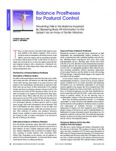
In order to estimate experimental errors of this pro tocol, measurements were repeated according to the [16] experimental design described by Schwartz et al . Postural data from the two volunteers were acquired by three trained therapists, during three different sessions with one week interval (Figure 1). During each session, the volunteer was asked to stand 4 times in a free standing position with each foot on a force platform (with a free walk between each trial), without modifications of the markers, and postural data were recorded during
WJO|www.wjgnet.com
985
December 18, 2015|Volume 6|Issue 11|
Blondel B et al . Sagittal articular moments and posture
2 volunteers
3 therapists
3 sessions
4 trials
Figure 1 Experimental error measurement based on Schwartz’s methodology.
Table 1 Mean net sagittal articular moments and maximum net sagittal articular moments in N.m for each volunteer
Mean moments Max moments
Volunteer 1 Volunteer 2 Volunteer 1 Volunteer 2
Cervical
Thoracic
Lumbar
Hip right
Hip left
Knee right
Knee left
Ankle right
Ankle left
-0.65 -0.03 0.71 0.43
-9.23 -4.2 10.76 5.92
-0.45 -0.86 7.71 3.91
10.73 3.79 11.35 4.27
14.62 4.82 15.2 5.08
-6.7 -7.59 7.6 8.12
-5.81 -10.09 7.59 10.44
-13.04 -7.76 14.06 8.5
-7.52 -6.08 8.59 6.49
Signs of the moments were consistent between the two subjects. Maximum moments are expressed in absolute values.
Table 2 Results of the experimental error measurement (N.m)
Inter-trial error Inter-session error Inter-therapist error
Cervical
Thoracic
Lumbar
Hip right
Hip left
Knee right
Knee left
Ankle right
Ankle left
0.26 0.43 0.48
1.36 5.27 6.68
1.08 5.24 6.64
1.66 3.21 4.09
1.71 3.78 4.5
1.55 2.09 4.22
1.5 2.75 4.07
2.14 2.46 2.6
2.03 2.8 2.86
inter-therapist errors (extrinsic error related to interobserver variability). Results from these analyses are summarized in Table 2.
an ankle plantar flexion, a knee extension, a pelvic anteversion and a spinal extension.
Measurement of the experimental errors
Free standing posture vs “radiographic” posture
As per the methodology described by Schwartz, the following experimental errors were calculated: intertrial errors (intrinsic error corresponding to the intrasubject variability), the inter-session errors (extrinsic error related to intra-observer variability) and the
WJO|www.wjgnet.com
In order to quantify the effect of an imposed posture, the net intersegmental moments at the spinal junctions during the “radiographic posture” were compared to the one obtained during the “free posture”. Change was
986
December 18, 2015|Volume 6|Issue 11|
Blondel B et al . Sagittal articular moments and posture Table 3 Summary of differences imposed by the “radiographic” posture compared with the free standing
Mean Diff FSP vs RX (N/m) Inter-trial significant diff Inter-session significant diff Inter-therapist significant diff Mean Diff FSP vs RX (N/m) Inter-trial significant diff Inter-session significant diff Inter-therapist significant diff
Volunteer 1
Volunteer 2
Cervical
Thoracic
Lumbar
1.15 Yes Yes Yes 0.11 No No No
2.79 Yes No No 0.88 No No No
0.35 No No No 0.3 No No No
Mean differences are in absolute values. Difference is considered significant when superior to experimental error measure.
considered significant only if the difference was greater than the intrinsic experimental error calculated for each junction. Results from these analyses did not showed significant differences for volunteer 2. Significant vari ations were observed on volunteer 1 mainly on the intra-subject measurement. Adopting a “radiographic” posture led to the following changes: an extension moment at the cervico-thoracic level, a decrease of the flexion moment at the thoraco-lumbar level and no significant change on the moment at the lumbo-sacral level. Results are summarized in Table 3.
evaluate the experimental error of the protocol.
Analysis protocol
Results from this study confirm the feasibility of the proposed protocol for postural analysis and provide preliminary values of the resulting net sagittal moments on various articulations during normal stance. In a free standing position, net flexion moments calculated at each spinal junction correspond to the impact of the body weight. Therefore, in order to stand in an upright position, these moments have to be compensated by the action of posterior muscles. Muscular role is crucial in order to maintain a satisfactory posture and [20] Mahaudens et al , demonstrated, in an energy-cost analysis, the impact of poor muscle efficiency on gait for adolescent with idiopathic scoliosis (AIS). Previous studies have already analyzed spinal alignment and posture using gait analysis but, to our best knowledge, characterization of posture with net sagittal moments [21] has never been reported yet. Chen et al reported poor postural stability control in AIS patients, without modifications of gait patterns in comparison with normal [22] subjects. More recently, Engsberg et al and Lenke et [23] al analyzed posture and trunk range of motion in AIS patients, post-operatively; they found a reduced gait speed, an improved coronal balance, but unchanged sagittal balance and a reduced range of motion in fused and unfused spinal segments. Using the protocol desc ribed in this study could provide further steps in the comprehension of the net reaction moment which occurs below or above a fused segment or evaluate an adjacent level degeneration.
Impact of trunk flexion/extension on spinal junctional moments
From the free standing position to an anterior flexion of the trunk at 45°, recordings demonstrated a progre ssively increased flexion moment (in absolute values) on the cervico-thoracic (> 3.5 N.m), the thoraco-lumbar (> 35 N.m) and the lumbo-sacral junctions (> 45 N.m). During trunk extension (around 40° ) recordings demonstrated changes towards extension moments on the cervico-thoracic (> 3.5 N.m), the thoraco-lumbar (> 18 N.m) and the lumbo-sacral junctions (> 35 N.m). These results are summarized in Figure 2.
DISCUSSION Sagittal spino-pelvic parameters have been reported as of primary importance in the management of spinal deformities due to their correlation with clinical and functional outcomes in preoperative and postoperative [6,7,17,18] evaluations . More recently, those findings were revisited in order to define guidelines in realignment [19] objectives . Given the constrain of a radiographic environment (i.e., arms positioning), several authors demonstrated that the “radiographic” posture could [10,11] lead to postural modifications and therefore to subsequent alignment misinterpretation, especially for patients with a marked sagittal anterior malalignment. Our objective was to propose a new protocol focu sing on the calculation of resulting intersegmental moments during a free standing posture (i.e., without constrain of the radiographic environment) and to
WJO|www.wjgnet.com
Experimental error measurement
Measurement of the experimental error was done [16] according to Schwartz et al , and as for these authors, in our protocol the inter-trial error was the smallest, with higher inter-session and inter-therapist errors. This increased measurement error can be considered in different ways: it can be related to a lack of reliability of the protocol or can reflect the postural variability. Results from trunk flexion/extension moments showing a linear relation between trunk flexion/extension and moments, as well as the small differences between errors from
987
December 18, 2015|Volume 6|Issue 11|
Blondel B et al . Sagittal articular moments and posture
A
B
Moment/Extension 4
0 Moment X Moment Y Moment Z
3 2.5
Cervical moment (N.m)
Cervical moment (N.m)
3.5
Moment/Flexion 0.5
2 1.5 1
-0.5 -1 -1.5 -2 -2.5
0.5
-3
0
-3.5
-0.5 -40
Moment X Moment Y Moment Z
-4 -35
-30
-25
-20
-15
-10
-5
0
5
10
15
Flexion angle (°)
D
Moment/Extension 20
Thoracic moment (N.m)
5
-5
-10 -15 -20 -25
Moment X Moment Y Moment Z
-35
-30
-25
-20
-15
-10
-5
0
5
10
15
Extension (°)
F
Moment/Extension 40
20 15 10 5
-20
45
30
35
40
45
-20
-30
Moment X Moment Y Moment Z
-50 -25
40
-10
-40
-30
35
Moment/Flexion
0
-35
30
10
Lumbar moment (N.m)
25
25
0
Moment X Moment Y Moment Z
30
20
Flexion angle (°)
35
Lumbar moment (N.m)
45
Moment/Flexion
-40
-40
40
5
-35
-5
35
-30
0
E
30
-5
10
-40
25
0
Moment X Moment Y Moment Z
15
Thoracic moment (N.m)
C
20
Flexion angle (°)
-15
-10
0
-5
Extension (°)
5
10
15
20
25
Flexion angle (°)
Figure 2 Results of the net sagittal moments (moment Z) on each spinal junction during trunk extension (A, C, E) and flexion (B, D, F).
inter-session and inter-therapist measurements are more likely to reveal changes in posture. Furthermore, differences found between the free-standing posture
WJO|www.wjgnet.com
and the “radiographic” posture provides an additional argument in favor of a postural variability rather than a protocol failure.
988
December 18, 2015|Volume 6|Issue 11|
Blondel B et al . Sagittal articular moments and posture
Free standing vs “radiographic” posture
sagittal intersegmental moments. After elaboration of a specific marker-set, 4 successive recordings were done on two volunteers by three different operators during three sessions in order to evaluate the experimental error measurement. A supplementary acquisition in a “radiographic” posture was also obtained. And these authors concluded that the first results confirm the technical feasibility of the protocol while the characterization of sagittal intersegmental net moments can have clinical applications such as evaluation of an unfused segment after a spinal arthrodesis.
Results from the analysis comparing the free standing and the “radiographic” posture revealed differences superior to the intrinsic experimental error for the volunteer 1 and no significant changes were visible for the volunteer 2, underlining the variability of posture with time and among individual. Clinical expressions of these changes were associated with an extension of the upper part of the trunk when the volunteer adopted the “radiographic” posture. These results are in line with [10,11] previous reports where arms positioning could be associated to negative shift of the SVA. Furthermore, this test was conducted in volunteer without sagittal malalignment and these differences may be even much more noticeable in patients with loss of lumbar lordosis and increased thoracic kyphosis. Results from this study confirm the technical fea sibility of the protocol. Using this methodology, it was possible to evaluate net moments applied to spinal junctions. Measurements of experimental errors showed differences between inter-trial, inter-session and intertherapist recordings traducing variability of posture. Differences were also noted between free standing and “radiographic” posture. Further steps will be needed in order to include a larger sample of volunteers for description of normative values and to determine sagittal parameters from this protocol that can be associated with conventional radiographic parameters. Future development of this protocol can help to evaluate various clinical situations such as adjacent level degeneration or modification of forces applied on unfused segment after spinal surgery.
REFERENCES 1
2
3
4
5
6
7
COMMENTS COMMENTS
8
Background
In clinical practice, postural analysis is mainly based on radiographs. However, in various situations such as global sagittal anterior malalignment interpretation of radiographs may not represent the real alignment of the subject.
9
Research frontiers
Biomechanical analysis can currently be applied to postural analysis. Such development will help to understand spinal disorders and impact of surgical procedures on posture.
10
Innovations and breakthroughs
Results from this study confirm the technical feasibility of the protocol. Using this methodology, it was possible to evaluate net moments applied to spinal junctions. Differences were also noted between free standing and “radiographic” posture. Future development of this protocol can help to evaluate various clinical situations such as adjacent level degeneration or modification of forces applied on unfused segment after spinal surgery.
11
12
Applications
13
Future development of this protocol can help to evaluate various clinical situations such as adjacent level degeneration or modification of forces applied on unfused segment after spinal surgery.
14
Peer-review
In this biomechanical study the authors describe the initial results and experimental error measurement of a protocol analyzing human posture through
WJO|www.wjgnet.com
989
Duval-Beaupère G, Schmidt C, Cosson P. A Barycentremetric study of the sagittal shape of spine and pelvis: the conditions required for an economic standing position. Ann Biomed Eng 1992; 20: 451-462 [PMID: 1510296 DOI: 10.1007/bf02368136] Legaye J, Duval-Beaupère G, Hecquet J, Marty C. Pelvic inci dence: a fundamental pelvic parameter for three-dimensional regulation of spinal sagittal curves. Eur Spine J 1998; 7: 99-103 [PMID: 9629932 DOI: 10.1007/s005860050038] Glassman SD, Berven S, Bridwell K, Horton W, Dimar JR. Correlation of radiographic parameters and clinical symptoms in adult scoliosis. Spine (Phila Pa 1976) 2005; 30: 682-688 [PMID: 15770185 DOI: 10.1097/01.brs.0000155425.04536.f7] Rose PS, Bridwell KH, Lenke LG, Cronen GA, Mulconrey DS, Buchowski JM, Kim YJ. Role of pelvic incidence, thoracic kyphosis, and patient factors on sagittal plane correction following pedicle subtraction osteotomy. Spine (Phila Pa 1976) 2009; 34: 785-791 [PMID: 19365245 DOI: 10.1097/brs.0b013e31819d0c86] Schwab F, Lafage V, Patel A, Farcy JP. Sagittal plane considerations and the pelvis in the adult patient. Spine (Phila Pa 1976) 2009; 34: 1828-1833 [PMID: 19644334 DOI: 10.1097/brs.0b013e3181a13c 08] Glassman SD, Bridwell K, Dimar JR, Horton W, Berven S, Schwab F. The impact of positive sagittal balance in adult spinal deformity. Spine (Phila Pa 1976) 2005; 30: 2024-2029 [PMID: 16166889 DOI: 10.1097/01.brs.0000179086.30449.96] Lafage V, Schwab F, Patel A, Hawkinson N, Farcy JP. Pelvic tilt and truncal inclination: two key radiographic parameters in the setting of adults with spinal deformity. Spine (Phila Pa 1976) 2009; 34: E599-E606 [PMID: 19644319 DOI: 10.1097/brs.0b013e3181aa d219] Gangnet N, Dumas R, Pomero V, Mitulescu A, Skalli W, Vital JM. Three-dimensional spinal and pelvic alignment in an asymptomatic population. Spine (Phila Pa 1976) 2006; 31: E507-E512 [PMID: 16816752 DOI: 10.1097/01.brs.0000224533.19359.89] Gangnet N, Pomero V, Dumas R, Skalli W, Vital JM. Variability of the spine and pelvis location with respect to the gravity line: a threedimensional stereoradiographic study using a force platform. Surg Radiol Anat 2003; 25: 424-433 [PMID: 13680185 DOI: 10.1007/ s00276-003-0154-6] Marks MC, Stanford CF, Mahar AT, Newton PO. Standing lateral radiographic positioning does not represent customary standing balance. Spine (Phila Pa 1976) 2003; 28: 1176-1182 [PMID: 12782989 DOI: 10.1097/01.brs.0000067271.00258.51] Vedantam R, Lenke LG, Bridwell KH, Linville DL, Blanke K. The effect of variation in arm position on sagittal spinal alignment. Spine (Phila Pa 1976) 2000; 25: 2204-2209 [PMID: 10973404 DOI: 10.1097/00007632-200009010-00011] Blondel B, Pomero V, Moal B, Lafage V, Jouve JL, Tropiano P, Bollini G, Dumas R, Viehweger E. Sagittal spine posture assess ment: feasibility of a protocol based on intersegmental moments. Orthop Traumatol Surg Res 2012; 98: 109-113 [PMID: 22264566] Dumas R, Chèze L, Verriest JP. Adjustments to McConville et al. and Young et al. body segment inertial parameters. J Biomech 2007; 40: 543-553 [PMID: 16616757 DOI: 10.1016/j.jbiomech.2006.02.0 13] Wu G, Siegler S, Allard P, Kirtley C, Leardini A, Rosenbaum D, Whittle M, D’Lima DD, Cristofolini L, Witte H, Schmid O, Stokes I. ISB recommendation on definitions of joint coordinate system of various joints for the reporting of human joint motion--part I:
December 18, 2015|Volume 6|Issue 11|
Blondel B et al . Sagittal articular moments and posture
15
16 17
18
19
ankle, hip, and spine. International Society of Biomechanics. J Biomech 2002; 35: 543-548 [PMID: 11934426 DOI: 10.1016/ s0021-9290(01)00222-6] Wu G, van der Helm FC, Veeger HE, Makhsous M, Van Roy P, Anglin C, Nagels J, Karduna AR, McQuade K, Wang X, Werner FW, Buchholz B. ISB recommendation on definitions of joint coordinate systems of various joints for the reporting of human joint motion--Part II: shoulder, elbow, wrist and hand. J Biomech 2005; 38: 981-992 [PMID: 15844264 DOI: 10.1016/ j.jbiomech.2004.05.042] Schwartz MH, Trost JP, Wervey RA. Measurement and manage ment of errors in quantitative gait data. Gait Posture 2004; 20: 196-203 [PMID: 15336291 DOI: 10.1016/j.gaitpost.2003.09.011] Hyun SJ, Rhim SC. Clinical outcomes and complications after pedicle subtraction osteotomy for fixed sagittal imbalance patients: a long-term follow-up data. J Korean Neurosurg Soc 2010; 47: 95-101 [PMID: 20224706 DOI: 10.3340/jkns.2010.47.2.95] Kim YJ, Bridwell KH, Lenke LG, Rhim S, Cheh G. Sagittal thoracic decompensation following long adult lumbar spinal instrumentation and fusion to L5 or S1: causes, prevalence, and risk factor analysis. Spine (Phila Pa 1976) 2006; 31: 2359-2366 [PMID: 16985465 DOI: 10.1097/01.brs.0000238969.59928.73] Schwab F, Patel A, Ungar B, Farcy JP, Lafage V. Adult spinal
20
21
22
23
deformity-postoperative standing imbalance: how much can you tolerate? An overview of key parameters in assessing alignment and planning corrective surgery. Spine (Phila Pa 1976) 2010; 35: 2224-2231 [PMID: 21102297 DOI: 10.1097/brs.0b013e3181ee 6bd4] Mahaudens P, Detrembleur C, Mousny M, Banse X. Gait in adolescent idiopathic scoliosis: energy cost analysis. Eur Spine J 2009; 18: 1160-1168 [PMID: 19390877 DOI: 10.1007/s00586-0091002-0] Chen PQ, Wang JL, Tsuang YH, Liao TL, Huang PI, Hang YS. The postural stability control and gait pattern of idiopathic scoliosis adolescents. Clin Biomech (Bristol, Avon) 1998; 13: S52-S58 [PMID: 11430791 DOI: 10.1016/s0268-0033(97)00075-2] Engsberg JR, Lenke LG, Reitenbach AK, Hollander KW, Bridwell KH, Blanke K. Prospective evaluation of trunk range of motion in adolescents with idiopathic scoliosis undergoing spinal fusion surgery. Spine (Phila Pa 1976) 2002; 27: 1346-1354 [PMID: 12065985 DOI: 10.1097/00007632-200206150-00018] Lenke LG, Engsberg JR, Ross SA, Reitenbach A, Blanke K, Bridwell KH. Prospective dynamic functional evaluation of gait and spinal balance following spinal fusion in adolescent idiopathic scoliosis. Spine (Phila Pa 1976) 2001; 26: E330-E337 [PMID: 11462099 DOI: 10.1097/00007632-200107150-00020] P- Reviewer: Korovessis P, Sun J S- Editor: Tian YL L- Editor: A E- Editor: Jiao XK
WJO|www.wjgnet.com
990
December 18, 2015|Volume 6|Issue 11|
Published by Baishideng Publishing Group Inc 8226 Regency Drive, Pleasanton, CA 94588, USA Telephone: +1-925-223-8242 Fax: +1-925-223-8243 E-mail:
[email protected] Help Desk: http://www.wjgnet.com/esps/helpdesk.aspx http://www.wjgnet.com
© 2015 Baishideng Publishing Group Inc. All rights reserved.