Original Research published: 23 January 2017 doi: 10.3389/fpsyt.2016.00212
Practice and Learning: Spatiotemporal Differences in Thalamo-Cortical-Cerebellar Networks Engagement across Learning Phases in Schizophrenia Michele Korostil1,2,3*, Gary Remington1,3 and Anthony Randal McIntosh2,3 1 Centre for Addiction and Mental Health, Toronto, ON, Canada, 2 Rotman Research Institute of Baycrest Health Sciences, Toronto, ON, Canada, 3 University of Toronto, Toronto, ON, Canada
Edited by: André Schmidt, King’s College London, UK Reviewed by: Philip R. Corlett, Yale University, USA Vaibhav A. Diwadkar, Wayne State University School of Medicine, USA *Correspondence: Michele Korostil
[email protected] Specialty section: This article was submitted to Neuroimaging and Stimulation, a section of the journal Frontiers in Psychiatry Received: 12 September 2016 Accepted: 22 December 2016 Published: 23 January 2017 Citation: Korostil M, Remington G and McIntosh AR (2017) Practice and Learning: Spatiotemporal Differences in Thalamo-Cortical-Cerebellar Networks Engagement across Learning Phases in Schizophrenia. Front. Psychiatry 7:212. doi: 10.3389/fpsyt.2016.00212
Frontiers in Psychiatry | www.frontiersin.org
Background: Understanding how practice mediates the transition of brain–behavior networks between early and later stages of learning is constrained by the common approach to analysis of fMRI data. Prior imaging studies have mostly relied on a single scan, and parametric, task-related analyses. Our experiment incorporates a multisession fMRI lexicon-learning experiment with multivariate, whole-brain analysis to further knowledge of the distributed networks supporting practice-related learning in schizophrenia (SZ). Methods: Participants with SZ were compared with healthy control (HC) participants as they learned a novel lexicon during two fMRI scans over a several day period. All participants were trained to equal task proficiency prior to scanning. Behavioral-Partial Least Squares, a multivariate analytic approach, was used to analyze the imaging data. Permutation testing was used to determine statistical significance and bootstrap resampling to determine the reliability of the findings. Results: With practice, HC participants transitioned to a brain–accuracy network incorporating dorsostriatal regions in late-learning stages. The SZ participants did not transition to this pattern despite comparable behavioral results. Instead, successful learners with SZ were differentiated primarily on the basis of greater engagement of perceptual and perceptual-integration brain regions. Conclusion: There is a different spatiotemporal unfolding of brain–learning relationships in SZ. In SZ, given the same amount of practice, the movement from networks suggestive of effortful learning toward subcortically driven procedural one differs from HC participants. Learning performance in SZ is driven by varying levels of engagement in perceptual regions, which suggests perception itself is impaired and may impact downstream, “higher level” cognition. Keywords: schizophrenia, fMRI, learning, practice, cognition, multivariate, longitudinal, partial least squares
1
January 2017 | Volume 7 | Article 212
Korostil et al.
Spatiotemporal Learning Networks in Schizophrenia
INTRODUCTION
to non-specific task-independent effects given the potential linear confound of time inherent in learning studies in SZ (23). Thus, while many studies have examined putative compensatory brain strategies for learning impairments in general and some have examine the impacts of practice, none, to our knowledge, have utilized an in-scanner multi-day practice paradigm that allows for direct observation of the unfolding of learning as it occurs in vivo across a number of learning phases. Our experiment was a multisession fMRI scanning experiment wherein participants with SZ and HC participants learned a novel lexicon with in-scanner practice over the course of 1 week. We utilized a lexicon-learning relational paradigm because verbal learning impairments are common in the disorder and are particularly linked to functional outcome at all stages of the illness (24–27). Additionally, language disorders are hallmark phenomenological features at all stages of the syndrome (28). We used behavioralPLS analytic techniques, which allowed for direct measurement of the brain–behavior relationship. This approach allowed us to examine the large-scale distribution of learning in the brain as it unfolded over the course of practice and avoided the previously mentioned confounding effects of task-independent BOLD changes, common contaminants in practice-related learning neuroimaging experiments that we examined specifically in SZ in an earlier analysis of this study data (23).
Learning impairments in schizophrenia (SZ) are widespread, impact functional outcome, and may mediate response to various types of cognitive rehabilitation strategies. Despite considerable efforts to remediate cognitive impairments via cognitive training, pharmacologic and brain stimulation treatments, effect sizes thus far have been modest (1, 2). The unknown aspects of the neural dynamics of learning and the impact of practice in SZ continue to constrain the search for the optimal therapeutic brain targets. Prior practice-related neuroimaging experiments in SZ have relied primarily on single scanning sessions and univariate, task-related (vs. behavior-related) analyses (3–8). However, brain substrates associated with practice-related learning are dynamic and widely distributed in space and time. Thus, to measure these substrates using fMRI, one should use multivariate whole-brain imaging analytic techniques, a direct brain–behavior (vs. brain– task) approach to analysis, and multiple scanning sessions with sufficient in-scanner practice to measure the unfolding of brain changes in vivo. In healthy control (HC) individuals, practice-related learning typically follows a characteristic staged process: early, rapid, and effortful learning engaging widespread cortical–subcortical areas with a later shift to more automatic processes and a reduced number of prefrontal regions (4, 5, 9–13). This process is characteristic of both motor skill and cognitive skill-based learning where ongoing practice leads to incremental learning of associations. Similarly, dual-processing models of practice-related learning postulate a domain-general cognitive-control network that scaffolds early learning, but gradually disengages with practice as skilled, automatic processing emerges (14, 15). As per this model, this shift occurs in various types of experience-driven learning as long as learning is intentional with the specific details varying depending on task requirements (14). In SZ, this shift is less clear. Learning remains more effortful throughout with persistent cortical involvement at stages when HC persons have transitioned to subcortically driven automatic stages of processing. This lack of transition occurs both in SZ (on and off antipsychotics) and in those at genetic high risk for the disorder (16, 17). Persistent effortful learning also may reflect impairments in deep encoding in the disorder, particularly in verbal memory and relationallearning paradigms (18, 19). Several studies examining practice-related learning in SZ have noted exponential blood oxygenation level-dependent (BOLD) signal decrease in task-relevant brain areas, which has been primarily driven by initial hyperactivity in these same regions (3–7). Koch et al. found no neural differences in task activation between successful learners with SZ and HC during a Sternberg working memory task. However, using a median split, the low-performing participants with SZ were more likely to show initial hyperactivation in frontal, cingulate, and superior-parietal regions (7). Activation studies using associative learning tasks have found both increased and similar levels of brain activation in SZ (20, 21). Proposed explanations for aberrant activation patterns in learning studies have included neural inefficiency and alternative compensatory pathways to achieve behavioral parity (8, 22). We have also suggested that some of these results could be attributable
Frontiers in Psychiatry | www.frontiersin.org
MATERIALS AND METHODS Participants
Participants were 16 patients with DSM-IV diagnosed SZ recruited from the outpatient clinics at the Centre for Addiction and Mental Health (CAMH) matched with 17 HC recruited via local advertisement and a research participant database at Baycrest Hospital. Both are teaching hospitals associated with the University of Toronto. All participants were right handed (29), native English speakers, and suitable for MRI scanning. Participants were comprehensively screened for and excluded if there were any interfering medical conditions, neurological disorder, or psychiatric disorder. Participants with SZ were clinically stable and had been prescribed an atypical antipsychotic medication at a stable dose for at least 3 months. The diagnosis of SZ was confirmed and other Axis 1 psychiatric disorders ruled out by the study MD (Michele Korostil) using the MiniInternational Neuropsychiatric Interview-Plus (30). The clinical status of the SZ participants was assessed using the Positive and Negative Syndrome Scale (PANSS) (31) at the initial visit and the Clinical Global Impressions Scale (32) at all three study visits. The neurocognitive status of all participants was evaluated at the initial visit using the Repeatable Battery for the Assessment of Neuropsychological Status (RBANS) (33). Five HC and four SZ participants were excluded from the final analysis: two due to improper task performance, three due to technical difficulties with equipment, and four due to excessive movement artifact on MRI scans. Thus, the final sample included 12 matched participants from each group. The study protocol was approved by the Research Ethics Boards of Baycrest Hospital and the CAMH according to guidelines
2
January 2017 | Volume 7 | Article 212
Korostil et al.
Spatiotemporal Learning Networks in Schizophrenia
from these hospitals and the University of Toronto. Participants provided written informed consent and were paid a stipend for their participation.
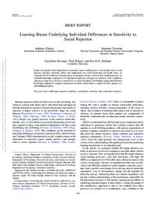
were trained on the task in an fMRI simulator using a parallel set of stimuli to 75% proficiency to minimize contamination by “learning the task” during fMRI data acquisition. While lying prone in the MRI scanner, participants heard a spoken pseudoword in their headphones (normalized to 600 ms duration). Shortly after (200 ms) the onset of the auditory pseudoword, they saw a picture of an object (duration 1 s) followed by a fixation cross (2 s). This sequence constituted one trial. Participants were asked to indicate, via a button press on a response pad, if the words and pictures went together in the new vocabulary. The pairings were sometimes “correct” and sometimes “incorrect.” While participants were informed at the outset that they would be learning a new vocabulary, there was otherwise no feedback given to participants throughout the experiment. The “correct” pairings repeated throughout the experiment, each “incorrect” pairing was presented only once. Thus, the underlying learning principle was a higher cooccurrence of “correct” trials with a 20:1 (correct:incorrect) ratio by the end of both scanning sessions (Figure 1). The lexicon was randomly generated from a matched set of stimuli and different for each participant. Each participant’s lexicon remained the same across the scanning days and, thus, the learning runs were additive. Each day of learning in the scanner involved 5 “learning runs” (approximately 6 min in length), which were each comprised of 120 trials as described above for a total of 1,200 trials over the course of the entire experiment with half being “correct” pairings and the other half “incorrect.”
Study Procedure
The study occurred over a 3-day period. On day 1, participants were assessed for suitability, and the PANSS and RBANS were administered. Participants were trained on the fMRI learning task in an MRI simulator using a parallel set of stimuli. On days 2 and 3, participants completed the fMRI experiment in which they learned a 30-word novel lexicon while undergoing fMRI scanning. The structure of the sessions on days 2 and 3 were identical. The vocabulary was the same for both days and thus day 3 functioned as a “practice session” to consolidate the learning from the prior session. The mean interval between scanning sessions for all participants was 2 days.
Stimuli Selection and Task Design
Participants were asked to learn a novel 30-word vocabulary comprised of auditory English pseudowords arbitrarily paired with pictures of everyday objects while undergoing event-related fMRI scanning. The task was developed by Breitenstein and Knecht (34, 35) for German speakers and modified by us for native English speakers. In our version of the task, participants were informed in advance that they would be learning a new vocabulary over the course of the experiment. All participants
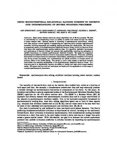
Figure 1 | Vocabulary learning task. Over the course of 10 “learning runs” (five runs on day 1 and five runs on day 2) with 1,200 trials total, each pseudoword (here: skog) appeared 20 times paired with the same picture (here: sun) and only once with other varying pictures. Participants were instructed that they would be learning a new vocabulary, but were not informed of the underlying learning principle and did not receive feedback throughout the experiment. Thus, they intuitively learned that the recurring couplings were the correct pairs. A 30-word picture-word vocabulary was randomly generated for each participant using a normed set of English pseudowords paired with a standardized picture of an everyday object. Each participant’s vocabulary remained the same for both scanning sessions. They indicated their response (“correct” or “incorrect” pairing) via a button press while in the fMRI scanner.
Frontiers in Psychiatry | www.frontiersin.org
3
January 2017 | Volume 7 | Article 212
Korostil et al.
Spatiotemporal Learning Networks in Schizophrenia
fMRI Data Acquisition and Preprocessing
fMRI Analyses
To identify multivariate patterns of relationships between performance and brain activity, we used Behavioral-Partial Least Squares (BPLS) (39). PLS is a multivariate statistical method that identifies maximal covariance [latent variables (LVs)] between sets of independent measures and allows for both spatial and temporal interpretation. It is mathematically similar to canonical correlation analysis. PLS analysis of neuroimaging data has been demonstrated as an effective method in SZ experiments with small samples and multicollinear measures (40, 41). BPLS is a type of PLS that allows for direct identification of multivariate patterns characterizing brain–behavior (in our case brain–accuracy) relationships rather than relying on inference via task performance. In BPLS, PLS solutions are constrained to the part of the covariance structure directly related to behavior. Importantly, the approach allows for analysis of all the relationships between performance on the task across two days of learning for all participants and BOLD signal in all gray matter regions in one model, without the need to restrict analysis to specific regions of interest. A correlation matrix between accuracy for each learning run and the BOLD signal at each voxel for each correct trial across both fMRI scans (10 learning runs) was created for each subject. Given that the hemodynamic response function for each condition lasts for several scans, PLS utilizes a “lag window” (i.e., a short signal segment) to best capture the response of each voxel. The lag-window size was 8 (TR = 2, 16 s), beginning at the offset of the auditory pseudoword (600 ms from beginning of each trial in the learning runs). The event markers were set at the beginning of this lag window to best capture language-related activation (35). The correlation matrix was then decomposed using singular value decomposition to produce LVs that capture the optimal relationship between learning accuracy and BOLD signal across the entire brain. Essentially, behavior-PLS is mathematically identical to a multivariate seed-connectivity analysis wherein accuracy is the “seed” and the results demonstrate how accuracy covaries with brain activity across subjects during the experimental learning runs. Each LV contains the spatial pattern displaying brain regions where BOLD activity is mostly strongly related to learning performance for each group. The LVs consist of the correlation strength (i.e., the “singular value”) and the weighting pattern across all brain voxels that optimally expresses the correlation (i.e., the “brain saliences”). Brain scores, akin to component scores in PCA, are summary measures for each participant that shows the degree to which a participant expresses the multivariate spatial pattern captured by a given latent variable. Significance testing of the resultant multivariate relationship between brain functional patterns and learning accuracy was assessed with 1,000 permutation tests of the singular value (SV) associated with each latent variable. As this operation is done on the entire data structure simultaneously, there is no need to correct for multiple comparisons at the voxel level. Bootstrap resampling (1,000 resamples) was used as a measure of the robustness of the voxel contribution to the effect. Each voxel’s bootstrapped mean salience was divided by its estimated SE to obtain a normalized estimate of robustness [i.e.,
fMRI was performed on a research-dedicated Siemens Trio 3-T MRI scanner. T2* functional images (TE = 30 ms, TR = 2,000 ms, flip angle = 70°, FOV = 200 mm, in-plane voxel size = 3.5 mm × 3.5 mm × 5.0 mm) were acquired using a single shot echo-planar image sequence leading to a BOLD contrast. Each functional sequence consisted of 28 5-mm thick slices in the axial-oblique plane positioned to image the entire brain. A T1-weighted anatomical scan was obtained using a SPoiled Gradient Recalled sequence (TE = 2.6 ms, TR = 2,000 ms, FOV = 256 mm, slice thickness = 1 mm) for coregistration with the functional images and to ensure that there were no significant brain abnormalities in any participants. Participants’ responses were made on an MR-compatible button response pad (Fiber-Optic Response Pad system).1 Visual stimuli were viewed in a head-coil mounted mirror directed toward a rear projection screen at the foot of the scanner. Auditory stimuli were presented using the Silent Scan auditory presentation system (AVOTEC). E-Prime Software2 was used to control stimulus presentation and to collect behavioral responses. The functional data were processed prior to statistical analysis. Slice-timing correction was done using AFNI.3 Motion correction was completed using AIR.4 The motion-corrected functional volumes within each run were averaged to create a mean functional volume per run. This mean functional run was then registered with each participant’s structural volume using a rigid body transformation model. The structural data were spatially normalized to the Common Anatomical Template and the end result was a direct non-linear transform from each initial fMRI volume into the Common Template space (36–38). Given the possible morphologic differences between the two groups, our approach helps protect against distortion that could be caused by the use of a standard space template based only on healthy brains. The functional data were then smoothed using a 7-mm Gaussian kernel. The voxel time series were further adjusted by regressing out motion correction parameters, white matter time series, and CSF time series, as in Garrett et al. (37). Results from data analyses were transformed into MNI space using the FSL/FNIRT registration algorithm to find a non-linear transform between our anatomical template and the MNI152_T1 template provided with FSL software.5 We used SPM56 to further assist with anatomical localization.
Data Analysis
Behavioral Measures
Behavioral performance (accuracy and reaction time) during the fMRI experiments was analyzed using repeated measures ANOVAs. http://www.curdes.com. http://www.pstnet.com. 3 http://afni.nimh.nih.gov/afni. 4 http://bishopw.loni.ucla.edu/AIR5/. 5 www.fmrib.ox.ac.uk/fsl. 6 http://www.fil.ion.ucl.ac.uk/spm/. 1 2
Frontiers in Psychiatry | www.frontiersin.org
4
January 2017 | Volume 7 | Article 212
Korostil et al.
Spatiotemporal Learning Networks in Schizophrenia
Behavioral Data
“bootstrap ratio” (BSR)]. A BSR of 3.00 approximates a 99% confidence interval. Our tables and images were thresholded at a BSR of 3. Correlation matrices for the brain scores for the significant LVs across all 10 learning runs were constructed. High correlations that tracked across a series of learning runs would indicate consistent movement through the learning task for a given individual subject. For example, assuming consistent effort and a clear underlying learning principle of the task, behavioral performance for any given subject on any given run should correlate with performance on subsequent runs. Similarly, one could make the same assumptions for brain activity and deviations from this could indicate a change in brain “strategy” due to changes in the underlying learning processes. The mean of the brainscores for a given series of runs that represented a coherent brain–behavior relationship was calculated for each individual for each significant LV. The mean was then correlated with symptom (PANSS), neuropsychological scores (RBANS), and antipsychotic medication dose (CPZ equivalents) (42) to better understand characteristics of participants with a given brain–behavior pattern. The approach also had the benefit of reducing the data so as to reduce risks of spurious conclusions inherent with multiple correlational analyses.
There was a significant learning effect for both groups across the 10 learning runs [F(4, 89) = 116.43, p = 0.0001] (Figure 2). Both groups showed similar learning rates over the 2 days [F(4, 89) = 0.80, p = 0.53]. The SZ group scored at “chance” in the first run and began to acquire the lexicon in the second run. This behavioral difference set the stage for the remaining acquisition of the lexicon and explained the main group effects [F(1, 22) = 7.82, p = 0.011]. Aside from this initial difference, the curves tracked each other for the duration of the experiment and performance plateaued for both groups at the same time. Reaction time data mirrored the accuracy data with both groups showing an expected decrease in reaction time for correct responses across 10 runs [F(2.8, 61.69) = 11.79, p = 0.0001], main effects of group with the HC group being faster overall [F(1, 22) = 18.43, p = 0.0001], and no interaction of run by group.
Imaging Results
Behavior-PLS Analyses
The initial behavior-PLS analysis was conducted on both groups simultaneously. Three significant LVs emerged that characterized patterns of overlap and differences between brain regions, supporting accurate learning for both groups. Similar to a multivariate factorial ANOVA, these patterns represented group by learning run interactions and showed no main effects of group or learning run. To disentangle these interactions, we performed the analysis within each group, similar to how one would proceed to decompose an interaction effect in ANOVA. Analysis of the HC group data extracted two significant LVs that characterized the brain–accuracy relationship across all 10 learning runs (Figure 3; Tables 2 and 3). There was a clear demarcation for the HC group between the early (runs 2 through 7) and late (runs 8 through 10) brain patterns supporting accurate learning. The first LV emerged as the most statistically robust spatiotemporal pattern of all patterns for both groups [p